Chondrogenesis from stem cells requires fine tuning the culture conditions. Here, we present a magnetic approach for condensing cells, an essential step to initiate chondrogenesis. In addition, we show that dynamic maturation in a bioreactor applies mechanical stimulation to the cellular constructs and enhances cartilaginous extracellular matrix production.
Cartilage engineering remains a challenge due to the difficulties in creating an in vitro functional implant similar to the native tissue. An approach recently explored for the development of autologous replacements involves the differentiation of stem cells into chondrocytes. To initiate this chondrogenesis, a degree of compaction of the stem cells is required; hence, we demonstrated the feasibility of magnetically condensing cells, both within thick scaffolds and scaffold-free, using miniaturized magnetic field sources as cell attractors. This magnetic approach was also used to guide aggregate fusion and to build scaffold-free, organized, three-dimensional (3D) tissues several millimeters in size. In addition to having an enhanced size, the tissue formed by magnetic-driven fusion presented a significant increase in the expression of collagen II, and a similar trend was observed for aggrecan expression. As the native cartilage was subjected to forces that influenced its 3D structure, dynamic maturation was also performed. A bioreactor that provides mechanical stimuli was used to culture the magnetically seeded scaffolds over a 21-day period. Bioreactor maturation largely improved chondrogenesis into the cellularized scaffolds; the extracellular matrix obtained under these conditions was rich in collagen II and aggrecan. This work outlines the innovative potential of magnetic condensation of labeled stem cells and dynamic maturation in a bioreactor for improved chondrogenic differentiation, both scaffold-free and within polysaccharide scaffolds.
Magnetic nanoparticles are already used in the clinic as contrast agents for magnetic resonance imaging (MRI), and their therapeutic applications keep expanding. For example, it has recently been shown that labeled cells can be manipulated in vivo using an external magnetic field and can be directed and/or maintained at a defined site of implantation1,2,3. In regenerative medicine, they can be used to engineer organized tissues in vitro4, including vascular tissue5,6,7, bone8, and cartilage9.
Articular cartilage is immersed in an avascular environment, making repairs of the extracellular matrix components very limited when damages occur. For this reason, research is currently focused on the engineering of hyaline cartilage replacements that can be implanted at the defect site. In order to produce an autologous replacement, some research groups are exploring the use of autologous chondrocytes as a cell source10,11, while others emphasize the capacity of mesenchymal stem cells (MSC) to differentiate into chondrocytes12,13. In previous studies recapitulated here, we selected MSC, as their bone marrow sampling is fairly simple and does not require the sacrifice of healthy chondrocytes, which risk losing their phenotype14.
An early step essential to initiating the chondrogenic differentiation of stem cells is their condensation. Cell aggregates are commonly formed using either centrifugation or micromass culture15; however, these condensation methods neither present the potential to create cell clusters within thick scaffolds nor the potential to control the fusion of aggregates. In this paper, we describe an innovative approach to condensing stem cells using MSC magnetic labeling and magnetic attraction. This technique has been proven to form scaffold-free 3D constructs via the fusion of aggregates with one another to obtain a millimeter-scale cartilaginous tissue9. Magnetic seeding of thick and large scaffolds has also allowed the possibility of increasing the size of the engineered tissue, designing a shape more readily useful for implantation, and diversifying the potential for clinical applications in cartilage repair. Here, we detail the protocol for the magnetic seeding of MSC into porous scaffolds composed of natural polysaccharides, pullulan, and dextran, scaffolds previously used to confine stem cells16,17. Chondrogenic differentiation was finally performed in a bioreactor to ensure continuous nutrient and gas diffusion into the matrix core of the scaffolds seeded with a high density of cells. Besides providing nutrients, chondrogenic growth factors, and gas to the cells, the bioreactor offered mechanical stimulation. Overall, the magnetic technology used to confine stem cells, combined with dynamic maturation in a bioreactor, can markedly improve chondrogenic differentiation.
1. Construction of the Magnetic Devices
NOTE: The devices used for cell seeding vary depending on the application (Figure 1). To form aggregates, the number of cells is limited to 2.5×10⁵/aggregate, so the magnetic tips must be very thin (750 µm in diameter). To seed the 1.8 cm2/7 mm-thick scaffolds, the magnets must be larger (3 mm in diameter) and will ensure cell migration through the pores of the scaffold.
2. Stem Cell Labeling
NOTE: Stem cells were labeled with 0.1 mM magnetic nanoparticles for 30 min (2.6 ± 0.2 pg iron/cell) to form aggregates, while they were labeled with 0.2 mM magnetic nanoparticles for 30 min (5 ± 0.4 pg iron/cell) to seed scaffolds. These nanoparticle concentrations and incubation times have been used previously and published for MSC and other cells18,19, and it has been determined that nanoparticles impacted neither cell viability nor MSC differentiation capacity. The iron mass incorporated by the stem cells was measured via single-cell magnetophoresis19,20.
3. Magnetic Cell Seeding
4. Differentiation into Chondrocytes
NOTE: After 4 days of incubation, remove the magnets and continue the chondrogenic maturation either in a Petri dish (static conditions) or in a bioreactor (dynamic conditions). Negative control samples are matured in static conditions with chondrogenic medium without TGF-β3.
5. RNA Extraction and Gene Expression Analysis
NOTE: Prior to RNA extraction, digest the scaffolds with an enzymatic solution.
6. Histological Analysis
First, aggregates can be individually formed using micro-magnets by depositing 2.5×10⁵ labeled stem cells (Figure 2A). These single aggregates (~0.8 mm in size) can then be fused into larger structures thanks to sequential, magnetically induced fusion. For instance, on day 8 of chondrogenic maturation, aggregates were placed in contact in pairs to form doublets; quadruplets were assembled on day 11 by merging 2 doublets; and finally, on day 15, the 4 quadruplets were fused to form a 3D construct containing 16 of the initially formed aggregates, with a total of 4×10⁶ cells (~4 mm in size) (Figure 2B). Second, the same magnetic attraction technique has been used to form cell aggregates within scaffolds. Scaffolds were seeded over the magnetic device and showed densely condensed cells within the pores of the scaffold, at exact micro-magnet locations (Figure 2C). By contrast, when cells were seeded within a scaffold without magnetic attraction (passive seeding), they were found to be evenly distributed. Cellularized scaffolds were then inserted into cages (Figure 3A) attached to the bioreactor chamber to perform the chondrogenic process under dynamic maturation conditions (Figure 3B). Such a bioreactor improves nutrient and gas exchanges and provides mechanical stimulation by transduction. The rotation speed of both the arm and the chamber was adjusted to 5 rpm, as recommended by the constructor for soft 3D tissue regeneration. A peristaltic pump that provides a continuous supply of medium was set at 10 rpm.
For all conditions of cell organization (fused aggregates and seeding within scaffolds) and tissue maturation (within or without a bioreactor), gene expression was analyzed on day 25. The tissue formed by magnetic fusion showed a significant increase in collagen II expression compared to the pellet obtained by centrifugation (Figure 4A), together with an increased trend of aggrecan expression. For cellularized scaffolds, we obtained an increase in aggrecan and collagen II expression-significant for collagen II-when magnetic seeding was used, as compared to the scaffolds seeded without magnetic forces. In addition, the expression of both genes was much higher (significant for Col II) when magnetic seeding was combined with dynamic differentiation (Figure 4B).
Histological analyses were also performed, for instance using a toluidine blue stain to reveal the glycosaminoglycans (GAG). The sequential magnetic fusion of 16 aggregates exhibited abundant deposition of GAG, as evinced by the blue-purple color (Figure 5A). For the scaffolds, only those magnetically seeded were stained with toluidine blue. GAG content was higher when the scaffolds were differentiated in a bioreactor (Figure 5B) rather than statically (Figure 5C). Taken together, these results demonstrate the potential of magnetic aggregation and magnetic seeding within scaffolds to enhance chondrogenesis. It also indicates that the dynamic maturation conditions within the bioreactor are much more favorable for efficient differentiation.
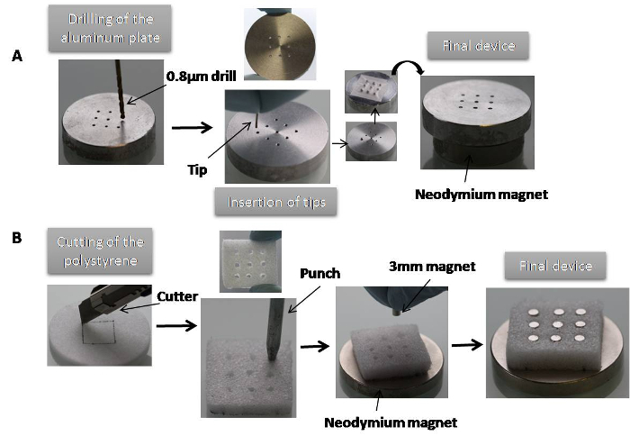
Figure 1. Construction of the magnetic devices. (A) Example of a magnetic device for aggregate formation: the aluminum plate was drilled (0.8 µm-diameter holes) and tips were inserted in each hole and then placed on a permanent neodymium magnet, which ensured magnetization to saturation. (B) Magnetic device to seed the scaffold: hard polystyrene (24 mm2) with 9 manually made holes was placed on a permanent neodymium magnet, which ensured magnetization to saturation. Small magnets (3 mm in diameter) were then inserted into each hole to form the device. Please click here to view a larger version of this figure.
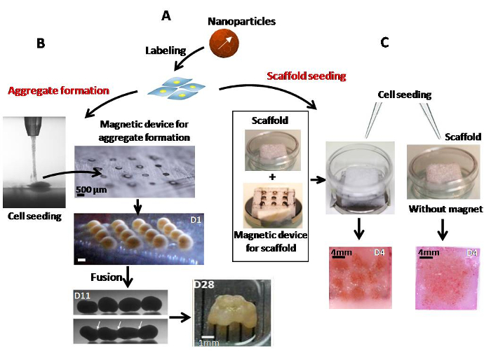
Figure 2. Magnetic labeling of stem cells and seeding. (A) Stem cells were labeled with iron oxide nanoparticles for 30 min at 37 °C. (B) Spheroids were formed from labeled cells attracted by a network of 16 micro-magnets. The aggregates were merged into quadruplets on day 11 by fusion of the doublets formed 3 days before. The quadruplets were then fused on day 15 to construct the final engineered tissue. (C) A scaffold, placed into a glass-bottomed dish, was seeded with or without magnetic forces. On day 4, spots of compacted stem cells were observed in the magnetically seeded scaffold, while the cells appeared uniformly distributed in the scaffold seeded without a magnet. Please click here to view a larger version of this figure.
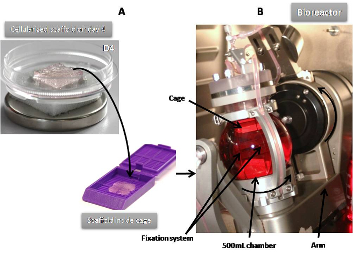
Figure 3. Dynamic maturation of cellularized scaffolds. (A) After magnetic or passive seeding, cellularized scaffolds were put into cages to avoid disruption. (B) Cages, fixed using the needles of the cap, were placed into the vessel of the bioreactor filled with chondrogenic medium. The bioreactor applied biaxial rotation with an independently controlled speed (1-12 rpm and 1-35 rpm for the arm and the chamber, respectively). A peristaltic pump continuously provided medium. Please click here to view a larger version of this figure.
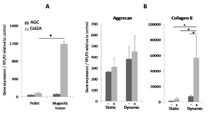
Figure 4. Expression of specific chondrogenic genes on day 25. (A) The magnetically induced fusion of MSC spheroids showed a significant increase in collagen II compared to the pellet formed by centrifugation. *denotes a statistical difference using Student's t-test (p-value < 0.05). (B) The expression of aggrecan and collagen II were clearly increased in scaffolds differentiated with a combination of magnetic seeding and dynamic maturation in a bioreactor. Gene expression was normalized to RPLP0 mRNA and expressed in arbitrary units relative to control (~1 ± SEM). Results are presented as means ± SEM of two to four independent experiments. *denotes a statistical difference using the Kruskal-Wallis test (one-way ANOVA nonparametric test) (p-value < 0.05). (-): seeding without magnet; (+): seeding with magnetic forces. Please click here to view a larger version of this figure.
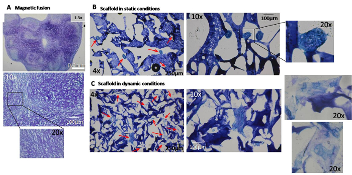
Figure 5. Histological staining of glycosaminoglycans on day 25. Glycosaminoglycan (GAG) deposits are evidenced by blue-purple coloration. (A) A positive toluidine blue stain was observed in the 8-µm cryosections of the final cartilaginous structure obtained from the sequential fusion of 16 aggregates. The 12-µm cryosections of scaffolds magnetically seeded after static (B) or dynamic (C) conditions clearly showed that GAG content was higher in scaffolds differentiated in a bioreactor. Arrows indicate aggregates of differentiated cells. Please click here to view a larger version of this figure.
First, because the techniques presented here rely on the internalization of magnetic nanoparticles, one important issue is the outcome of the nanoparticles once they localize within the cells. It is true that iron nanoparticles may trigger potential toxicity or impaired differentiation capacity depending on their size, coating, and time of exposure19,22. However, several studies have shown no impact on cellular physiology when encapsulated iron nanoparticles were used23 in the form of magnetoferritin, a biological magnetic nanoparticle24, or were used with a simple citrate coating and adequate concentrations18. In addition, when iron oxide nanoparticles were used in similar conditions as those described in this paper (with MSC and for chondrogenesis), we recently demonstrated that a rapid and almost total degradation of the nanoparticles occurs within the endosomes in about 10 days upon cellular incorporation and MSC spheroid formation. Interestingly, this massive degradation was associated with the efficient storage of the free iron within the ferritin protein and resulted in a very limited impact on the cellular iron metabolism, boding well for future clinical applications25.
Another critical point is the requirement of cell compaction to initiate chondrogenesis. Usually, the condensation of cells is achieved through centrifugation; however, this method is limited by a low number of cells (not more than 2.5×10⁵ cells). Past this number, the nutrients and gas cannot diffuse to the center of the aggregates, triggering cell necrosis. Here, the magnetic condensation of labeled stem cells into aggregates appears as a significant method to form 3D constructs for cartilage tissue regeneration. Such a magnetic procedure has been used by other authors to build 3D spheroids: by magnetic levitation with a magnet placed on the top of the plate after the dissociation of cells23 or with iron pins to localize the magnetic field26. However, magnetic levitation does not appear to be suited for further stages of aggregate fusion. By contrast, with the magnetic method proposed here, we can control all fusion steps to obtain a step-by-step cartilage tissue construction. In brief, this multi-step process starts with confinement of stem cells into aggregate building blocks and is followed by the fusion of these blocks into a larger structure. The critical steps of this scaffold-free stem cell aggregation procedure are the following: first, one must form each aggregate with as small a volume of cells as possible and second, one must control the fusion steps to avoid the formation of a single, large aggregate. The tissue obtained here was rich in collagen II and aggrecan. It also presented the advantages of having flexible geometry and size and of being scaffold-free.
The magnetic approach was also used to guide stem cells within thick and large scaffolds; another alternative for the design of various shapes and sizes. The critical step here is to seed the scaffolds with an appropriate volume of cells: neither too little to have an overall homogeneous distribution of cells, nor too much to avoid any cell loss. Magnetic forces were previously used to attract and retain cells within scaffolds and to enhance cell seeding27,28. Here, sufficient cell condensation within the pores of the polysaccharide scaffolds led to successful chondrogenesis. The extracellular matrix production was markedly improved when the magnetically cellularized scaffolds were subjected to the transduction/shear stress stimuli provided by the bioreactor thanks to its bi-axial rotation. It has been shown in other studies that multi-axial loading conditions improved the quality of tissues formed from chondrocytes29. This novel bioreactor concept presents a real gain when compared to the existing techniques, where only compressive forces are applied30,31,32.
In conclusion, for chondrogenic differentiation, the use of magnetic confinement of labeled stem cells to form and manipulate aggregates as well as to seed scaffolds allowed for the creation of millimeter-size cartilage cellular constructs. In addition, combining magnetic cell seeding with dynamic differentiation provides a valuable new tool for regenerative medicine applications.
The authors have nothing to disclose.
The authors would like to acknowledge QuinXell Technologies and CellD, particularly Lothar Grannemann and Dominique Ghozlan for their help with the bioreactor. We thank Catherine Le Visage, who provided us with the pullulan/dextran polysaccharide scaffolds. This work was supported by the European Union (ERC-2014-CoG project MaTissE 648779) and by the AgenceNationalede la Recherche (ANR), France (MagStem project ANR-11 SVSE5).
Iron oxide (maghemite) nanoparticules ( γ-Fe2O3) | PHENIX – University Paris 6 | Made and given by C. Ménager | Mean diameter of 8.1 nm and negative surface charge |
Polysaccharide Pullulan/Dextran scaffolds | LIOAD – University Nantes | Made and given by C. Le Visage | Prepared from a 75:25 mixture of pullulan/dextran in alkaline conditions (10M NaOH). Porosity: 185-205µm; Thickness: 7mm; Surface area: 1.8cm2. |
TisXell Regeneration System | QuinXell Technologies | QX900-002 | Biaxial bioreactor with 500 mL culture chamber |
Cage for scaffolds: Histosette II M492 | VWR | 720-0909 | |
Mesenchymal Stem Cell (MSC) | Lonza | PT-2501 | Three independant batches have been used |
MSCGM BulletKit medium | Lonza | PT-3001 | For the complete medium, add the provided BulletKit (containing serum, glutamine and antibiotics) to the MSCGM medium |
DMEM with Glutamax I | Life Technologies | 31966-021 | No sodium pyruvate, no HEPES |
RPMI medium 1640, no Glutamine | Life Technologies | 31870-025 | No sodium pyruvate, no HEPES |
PBS w/o CaCl2 w/o MgCl2 | Life Technologies | 14190-094 | |
0.05% Trypsin-EDTA (1x) | Life Technologies | 25300-054 | |
Penicillin (10.000U/mL)/Streptomicin (10.000µg/mL) | Life Technologies | 15140-122 | |
ITS Premix Universal Culture Supplement (20x) | Corning | 354352 | |
Sodium pyruvate solution 100mM | Sigma | S8636 | |
L-Ascorbic Acid 2-phosphate | Sigma | A8960 | Prepare the concentrated solution (25 mM) in distilled water extemporaneously |
L-Proline | Sigma | P5607 | Prepare the 175 mM stock solution diluted in distilled water and store at 4°C |
Dexamethasone | Sigma | D4902 | Prepare the 1 mM stock solution diluted in Ethanol 100% and store at -20°C |
TGF-beta 3 protein 10µg | Interchim | 30R-AT028 | |
Tri-sodium citrate | VWR | 33615.268 | Prepare the 1M stock solution diluted in distilled water and store at 4°C |
Pullulanase from Bacillus acidopullulyticus | Sigma | P2986 | |
Dextranase from Chaetomium erraticum | Sigma | D0443 | |
NucleoSpin RNA Extraction Kit | Macherey-Nagel | 740955.5 | |
SuperScript II Reverse Transcriptase | Life Technologies | 18064-014 | |
Random Primer – Hexamer | Promega | C1181 | 500 µg/mL: Use diluted 1/2 and put 1 µL per sample |
Recombinant RNAsin ribonuclease inhibitor | Promega | N2511 | 40 U/µL: put 1 µL per sample |
PCR nucleotide dNTP mix (10mM each) | Roche | 10842321 | |
SyBr Green PCR Master Mix | Life Technologies | 4368708 | |
Step One Plus Real-Time PCR System | Life Technologies | 4381792 | |
Formalin solution 10% neutral buffered | Sigma | HT5012 | |
OCT solution | VWR | 361603E | |
Isopentane | Sigma | M32631 | |
Toluidine blue O | VWR | 1.15930.0025 | |
Ethanol absolute | VWR | 20821.310 | |
Toluene | VWR | 1.08323.1000 | |
Mounting medium Pertex | Histolab | 840 | |
RPLP0 Primer for qPCR | Eurogentec | 5'-TGCATCAGTAC CCCATTCTATCAT-3' ; 5'-AAGGTGTAATC CGTCTCCACAGA-3' |
|
Aggrecan Primer for qPCR | Eurogentec | 5'-TCTACCGCTGCGAGGTGAT-3' ; 3'-TGTAATGGAACACGATGCCTTT-5' | |
Collagen II Primer for qPCR | Eurogentec | 5'-ACTGGATTGACCCCAACCAA-3' ; 3'-TCCATGTTGCAGAAAACCTTCA-5' |