Mantle cell lymphoma (MCL) is a difficult to treat B cell disorder and it is equally difficult to establish a xenograft mouse model of primary MCL to study and develop therapeutics. Here, we describe the successful establishment of MCL xenografts in mice to help understand its underlying biology.
B lymphocytes are key players in immune cell circulation and they mainly home to and reside in lymphoid organs. While normal B cells only proliferate when stimulated by T lymphocytes, oncogenic B cells survive and expand autonomously in undefined organ niches. Mantle cell lymphoma (MCL) is one such B cell disorder, where the median survival rate of patients is 4 – 5 years. This calls for the need of effective mechanisms by which the homing and engraftment of these cells are blocked in order to increase the survival and longevity of patients. Therefore, the effort to develop a xenograft mouse model to study the efficacy of MCL therapeutics by blocking the homing mechanism in vivo is of utmost importance. Development of animal recipients for human cell xenotransplantation to test early stage drugs have long been pursued, as relevant preclinical mouse models are crucial to screen new therapeutic agents. This animal model is developed to avoid human graft rejection and to establish a model for human diseases, and it may be an extremely useful tool to study disease progression of different lymphoma types and to perform preclinical testing of candidate drugs for hematologic malignancies, like MCL. We established a xenograft mouse model that will serve as an excellent resource to study and develop novel therapeutic approaches for MCL.
Lymphocytes by nature play a major role in immune surveillance, and lymphocyte trafficking is a critical step in mounting antigen specific immunity1,2. This process includes migration of naïve T lymphocytes from the thymus to the blood stream, and from there to secondary lymphoid organs, including lymph nodes, Peyer's patches, or spleen, where they meet cognate antigens. The B lymphocytes differentiate in the bone marrow and migrate as naïve cells into follicles of secondary lymphoid organs3. Some of these B cells bind antigen with their receptor and are activated by specific T cells. Proliferation and differentiation of these B cells pushes the non-activated, naive B cells into the mantle zone of the follicle. Activated cells can then differentiate into memory B cells, which patrol the body, or mature into immunoglobulin secreting plasma cells that migrate to the bone marrow4.
MCL occurs when naïve B lymphocytes in the mantle zone transform into a tumor. These lymphoma cells reside in the microenvironment of the lymphoid organs and proliferate independently of specific T lymphocyte control. However, at a certain stage of density they escape from this niche and recirculate in the bloodstream in search for niches in other organs. Considering the complexity of adhesion molecules and the promiscuity of chemokines and their receptors, the mechanism of this cellular trafficking in vivo is poorly understood and therefore hampers therapy. Novel methods are needed to effectively block this migration process to prevent the lymphoma B cells from reaching new microenvironments.
MCL is one of the most difficult to treat B cell malignancies. The development of a neoplastic phenotype of MCL is the result of a multistep cascade, characterized by the acquisition of unique biologic properties. At the time of diagnosis, most patients (70%) already present with a disseminated disease, with a majority of cases exhibiting extranodal involvement in spleen, bone marrow, and/or the gastrointestinal tract5,6. In treated patients, relapse by resistant tumors within a few years is common, even though conventional chemotherapy induces high remission rates at short term7,8. Here we present a new disease model that can help understand MCL dissemination and its underlying biology: we established a human MCL xenograft mouse model that originated from primary tumor cells of patients. We hope that this model will help develop therapeutic strategies against MCL dissemination, and possibly provide new clinical perspectives for optimal diagnosis and treatment of relapsed patients.
The human blood samples were used according to procedures approved by the local ethics and human experimentation committees of the Geneva University Hospital.
Animal procedures were performed in accordance with the Institutional Ethical Committee of Animal Care in Geneva, Switzerland and the Cantonal Veterinary Office (authorization number: GE/26/15).
1. Preparation of Primary Peripheral Blood Mononuclear Cells (PBMCs) by Density Gradient Separation
NOTE: 3 – 5 mL of peripheral blood was obtained from patients presenting with MCL in a leukemic phase. The diagnosis was established according to standard diagnostic criteria for flow cytometry (CD5+, CD23−, CD200+, monoclonal B cell population), and subsequently confirmed by presence of the chromosomal translocation t(11;14) and overexpression of cyclin D1. In all cases, monoclonal B cells constituted to >90% of the total B cell population. The remaining cells were mainly monocytes and some T cells.
2. Enrichment of B Cells by B Cell Negative Selection
3. Development of the Xenograft Mouse Model
4. Chimerism Analysis of Tumor Cell Engrafted in Different Organs
The manuscript describes an optimized protocol for the successful development of a xenograft mouse model for engraftment of MCL cells. Preparation of a pure cell population (in this case MCL cells), is very critical to develop successful MCL xenografts. Figure 1 represents the preparative steps for mononuclear cell isolation from MCL patient's blood by density gradient separation. The mononuclear cells are further processed to obtain pure B cells using a negative B cell enrichment kit to obtain a pure cell population for xenograft injection into mice. Care should be taken to obtain maximum purity in order to have successful MCL engraftment. The purity obtained using this method is usually >90%. The remaining cells were mainly monocytes and some T cells (data not shown).
Figure 2 represents the different steps of the B cell purification protocol as described in the methods section. The enriched cells are further analyzed for their purity by flow cytometer using different markers (CD45, CD19, CD20, CD23, CD200, CD5, kappa, and lambda). Sequential gating as shown in Figure 3 allows characterizing of MCL cells: CD45+, CD19+, CD20+, CD5+, CD23−, and CD200− are selected. Compensation for multicolor staining was carried out by using single stained controls for each of the fluorochromes used, according to standard cytometry set-up.
Figure 3E, G represents the typical dot plot of B cells before and after enrichment. In this case, >95% of the enriched cells using this kit are B cells. The cells are suspended in PBS at a concentration of 40 – 60 ×106 cells in 150 – 200 µL PBS as mentioned in the protocol. They were immediately injected i.v. to NSG mice. After 10 weeks, >90% of the injected animals developed lymphoma shows signs of terminal illness (weight loss, ruffled hair, decreased activity, etc.). After sacrifice, spleen and liver are removed and processed to obtain single cell suspensions by mechanical disruption (Figure 4D, F); blood is drawn carefully by cardiac heart puncture and the bone marrow is processed by cutting both ends of the femur and flushing it using a 2 mL syringe filled with RPMI medium or PBS (Figure 4C). The cells are further processed by flow cytometry using human B cell specific markers like CD19/CD20 and CD45. We used human B cell specific markers here because the recipient mouse strain (NSG) lacks B Cells. Figure 4B–F represents the collection of organs after sacrifice and the steps of further processing. The degree of engraftment of MCL cells derived from two patients is represented in Figure 4G. The engraftment pattern in different organs and between patients can be compared following this technique.
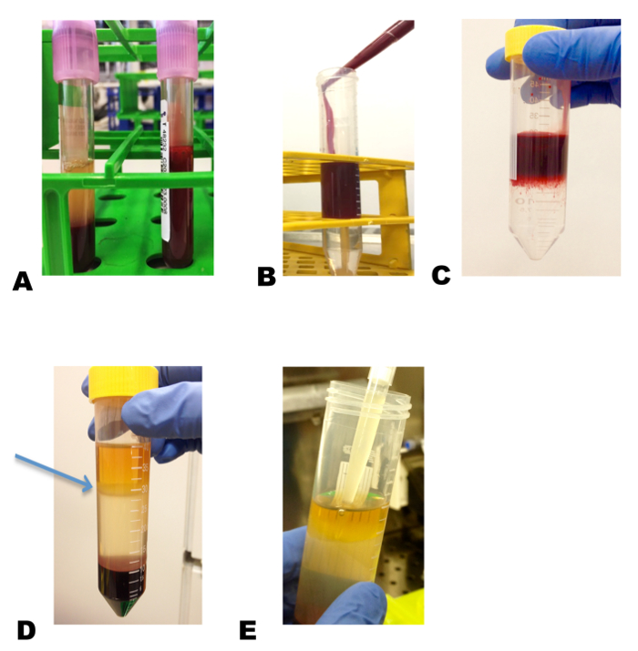
Figure 1: Isolation of PBMCs from whole blood. Blood drawn from a MCL patient is diluted 1:1 in RPMI medium (A), and placed carefully on top of the density gradient media layer (B) without mixing the blood into this density gradient media (C). Centrifugation at 400 x g for 45 min separates the mononuclear cells, which appear as whitish ring (arrow indicates the mononuclear cell layer) (D). This layer is pipetted gently without mixing with other layers into a clean tube for further processing (E). Please click here to view a larger version of this figure.
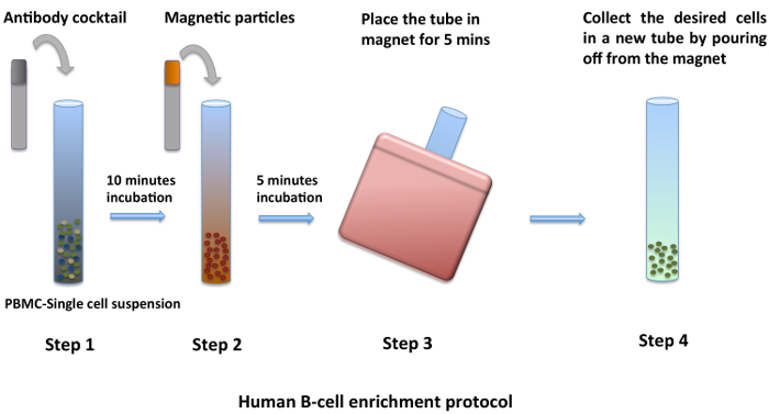
Figure 2: Enrichment of B cells from PBMCs. PBMCs isolated by density gradient centrifugation are washed twice with PBS. Using the negative B cell enrichment kit and by following the manufacturer's protocol, a pure population of B cells is obtained (1–4). Please click here to view a larger version of this figure.
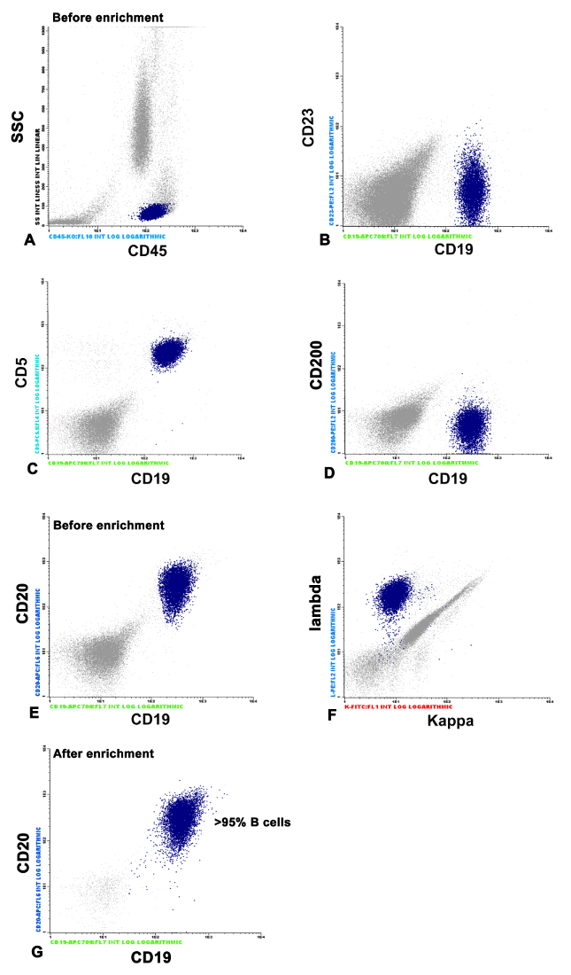
Figure 3: FACS analysis of MCL cells and purity of B cells before and after enrichment. The mononuclear MCL cells are characterized by FACS analysis using different markers like CD45, CD19, CD20, CD23, CD200, CD5, kappa, and lambda. Cells that are positive for CD45, CD19, CD20, CD5, and negative for CD23 and CD200 are selected (A–F). G represents the cell population after enrichment using the negative selection kit compared to E, which is before enrichment. Please click here to view a larger version of this figure.
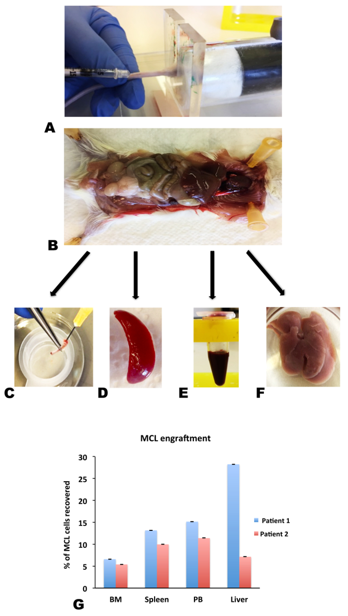
Figure 4: NSG mice as a xenograft model for engraftment of MCL cells. B cells derived from MCL patients were injected i.v. via the lateral tail vein of NSG mice (A). After allowing several days for the tumor to engraft, the mice were sacrificed, and dissected (B) to collect different organs like bone marrow (C), spleen (D), peripheral blood (E) (drawn by cardiac heart puncture), and liver (F). These organs were processed further and analyzed by flow cytometry for the presence of B cells. G represents the engraftment pattern of MCL cells in different organs derived from MCL injected NSG mice. Results from two patients are shown. Data are shown as mean ± SEM, n = 3. Please click here to view a larger version of this figure.
Clinical trials are possible for drugs that are in an advanced stage of development but cannot be used for drug discovery. Efforts to develop animal recipients for human cell xenotransplantation in order to test early stage drugs have long been pursued. Here we present an animal model that avoids human graft rejection and can establish a model for human diseases, such as MCL. This is at present a state of the art xenograft model to study the mechanisms of human tumor engraftment and tumor growth. Here we use NSG mice, one of the most immune deficient mouse models to date in order to achieve greater success of lymphoma engraftment. NSG mice lack mature T cells, B cells, and NK cells. They also have impaired cytokine signaling and have defects in innate immune response.
Lymphocyte preparation from the primary MCL sample by density gradient is an important step to remove other cell types like red blood cells and platelets. Traditionally, a density gradient centrifugation allows successful isolation of lymphocytes. Further processing of these lymphocytes to enrich the B cell population is attained by the use of a B cell enrichment kit following the protocol as described within this manuscript. Care should be taken to attain a high purity of B lymphocyte populations to reach optimal engraftment of the lymphoma cells. The MCL cells are injected intravenously (at least 40 – 60 x 106 cells/mouse) through the lateral tail vein, and mice were allowed to develop lymphoma for up to 10 weeks. The mice were closely examined every day for symptoms of illness including, weight loss, ruffled hair, decreased activity, hind limb paralysis, etc. Analysis of different organs like spleen, liver, bone marrow, and blood by flow cytometry revealed MCL engraftment. Recently, it has been shown that primary MCL cells engraft in bone marrow and spleen of irradiated NSG mice at 20 weeks of injection9. Independent of this finding, we have successfully developed our own xenograft model of primary MCL with more rapid tumor formation.
This xenograft mouse model may become an extremely useful tool for the study of disease progression of different lymphoma types. Xenograft models also provide powerful tools to perform preclinical testing of candidate drugs for hematologic malignancies like MCL, however, with certain limitations for example, the dominant clone present at relapse in a patient is not necessarily the clone emerging on xenotransplantation10. This could be due to the different selective pressure that the xenograft undergoes in different host systems. The hematopoietic composition in this strain does not fully recapitulate human hematopoiesis, thus, limiting the long-term maintenance of human cells.The advantages in using human tumor xenografts overrides the limitations as results are obtained in a few weeks from a human tumor biopsy regarding response to therapy. Drug responses do not often correlate with clinical activities in patients11 when human cell lines instead of primary tumor cells are used as xenografts, but they help form the groundwork for possible therapeutic responses. The use of primary tumors as an orthotropic xenograft has a stronger predictive response value, particularly when a clinically relevant drug dosage is used11,12,13. Homing of lymphoma cells to their niches is an important pre-requisite in order to survive and to establish tumor engraftment. This process is majorly regulated via the lymphatic system, and the entry of circulating lymphocytes through the high endothelial venules maintains the lymphocyte homeostasis in lymph nodes.
Circulation of B lymphocytes through lymph nodes requires crossing endothelial barriers and chemoattractant-triggered cell migration by coordinated interaction with different adhesion molecules. Therefore, targeting the molecules that control the homing of lymphocytes to their survival niches could constitute a new treatment strategy for B cell lymphomas as they behave similar to normal lymphocytes. Our aim was to target the molecule JAM-C, a junctional adhesion molecule, which is present on MCL. This xenograft model will be used to study the therapeutic effects of JAM-C antibody on homing and engraftment of primary lymphoma cells to their survival niches. We have recently shown an effect of anti-JAM-C antibody in mice that received a MCL cell line, Jeko-114. This antibody had a blocking effect on homing of Jeko-1 cells and also when administered in regular intervals, it efficiently prevented the engraftment of Jeko-1 cells. In addition to its effect on homing, the antibody treatment eradicated the lymphoma engrafted in most of the organs tested in this mice14.
In the light of the scientific and clinical question being addressed, these xenografted tumors represent at present, an interesting model to study the therapeutic outcome of MCL.
The authors have nothing to disclose.
This work was supported by the Ligue Genevoise contre le Cancer, Fondation Dr. Dubois Ferriere Dinu-Lipatti, Oncosuisse KPS-OCS, OCS-02260-08-2008 and 2914-02-2012, and Swiss National Science Foundation Grant 31003A_156760 and 310030-153456.
Ficoll-paque media | GE Healthcare | 17-1440-02 | for separation of mononuclear cells |
RPMI Medium 1640 | Gibco-Life technologies | 61870-010 | for dilution of blood sample |
Phosphate Buffered Saline (PBS) | Sigma-Aldrich | D8537 | washing of cells |
Bovine Serum Albumin (BSA) | Sigma-Aldrich | A8412 | for preparation of PBS with 1% BSA used in washing cells during isolation |
CD19-APC700 | Beckman Coulter | B49212 | human pan-B cell marker |
CD20-APC | Beckman Coulter | A21693 | human pan-B cell marker |
CD20-ECD | Beckman Coulter | IM3607 | human pan-B cell marker |
CD5-PC 5.5 | Beckman Coulter | PN A70203 | human T cell marker |
CD23-PE | Pharmingen | 555711 | Cell surface protein typically absent in MCL |
CD45 KO | Beckman Coulter | B36294 | Pan-leucocyte marker |
CD200-PE | Pharmingen | 552475 | Cell surface protein typically absent in MCL |
NOD scid gamma (NSG) mice | Charles River Laboratories | 5557 | used to develop MCL xenografts in this study |
Easy sep Human B cell enrichment kit | Stem cell technologies | 19054 | used to enrich B cells to obtain pure cells for injecting into mice |
FACS | Beckman Coulter | Navios | used to characterize MCL sample and to study the organs for MCL engraftment |
1X ammonium chloride potassium buffer | red blood lysis buffer (NH4Cl 8,024 mg/l; KHCO3 1,001 mg/l; EDTA.Na2·2H2O 3.722 mg/l ) |