Using human primary prostate epithelial cells, we report a novel biomarker-free method of functional characterization of stem-like cells by a spheroid-based label-retention assay. A step-by-step protocol is described for BrdU, CFSE, or Far Red 2D cell labeling; three-dimensional spheroid formation; label-retaining stem-like cell identification by immunocytochemistry; and isolation by FACS.
Despite advances in adult stem cell research, identification and isolation of stem cells from tissue specimens remains a major challenge. While resident stem cells are relatively quiescent with niche restraints in adult tissues, they enter the cell cycle in anchor-free three-dimensional (3D) culture and undergo both symmetric and asymmetric cell division, giving rise to both stem and progenitor cells. The latter proliferate rapidly and are the major cell population at various stages of lineage commitment, forming heterogeneous spheroids. Using primary normal human prostate epithelial cells (HPrEC), a spheroid-based, label-retention assay was developed that permits the identification and functional isolation of the spheroid-initiating stem cells at a single cell resolution.
HPrEC or cell lines are two-dimensionally (2D) cultured with BrdU for 10 days to permit its incorporation into the DNA of all dividing cells, including self-renewing stem cells. Wash out commences upon transfer to the 3D culture for 5 days, during which stem cells self-renew through asymmetric division and initiate spheroid formation. While relatively quiescent daughter stem cells retain BrdU-labeled parental DNA, the daughter progenitors rapidly proliferate, losing the BrdU label. BrdU can be substituted with CFSE or Far Red pro-dyes, which permit live stem cell isolation by FACS. Stem cell characteristics are confirmed by in vitro spheroid formation, in vivo tissue regeneration assays, and by documenting their symmetric/asymmetric cell divisions. The isolated label-retaining stem cells can be rigorously interrogated by downstream molecular and biologic studies, including RNA-seq, ChIP-seq, single cell capture, metabolic activity, proteome profiling, immunocytochemistry, organoid formation, and in vivo tissue regeneration. Importantly, this marker-free functional stem cell isolation approach identifies stem-like cells from fresh cancer specimens and cancer cell lines from multiple organs, suggesting wide applicability. It can be used to identify cancer stem-like cell biomarkers, screen pharmaceuticals targeting cancer stem-like cells, and discover novel therapeutic targets in cancers.
The human prostate gland contains luminal epithelium with secretory function and basal cells underlying it along with an unusual neuroendocrine cell component. The epithelial cells, in this case, are generated from a rare population of prostate stem cells that are relatively quiescent in vivo and act as a repair system to maintain glandular homeostasis throughout life1. Despite many advances, the identification and functional isolation of prostate stem cells remains a major challenge in the field. Stem cell biomarkers, including cell surface marker-based methodologies combined with flow cytometry are commonly used for stem cell research2,3,4. However, results for enrichment and isolation vary widely as a function of marker combinations and antibody specificity5,6, raising questions about the identity of the isolated cells. Another widely used approach for stem-like cell enrichment is three-dimensional (3D) spheroid culture2,3,4. While resident stem cells are relatively quiescent in vivo with niche restraints, they undergo cell division in 3D matrix culture (both symmetric and asymmetric), generating both stem and progenitor cells that rapidly reproduce toward the lineage commitment7,8. The formed spheroids are a heterogeneous mixture containing both stem cells and progenitor cells at various stages of lineage commitment, including early and late stage progenitor cells. Thus, assays using the whole spheroids are not stem cell exclusive, making the identification of unique stem cell properties inconclusive. Therefore, it is critical to create assays to identify and separate prostate stem cells from their daughter progenitors. Towards this end, the goal of the current protocol is to establish an assay system that allows for the efficient identification and isolation of stem cells from human prostate tissues followed by robust downstream analysis of their biological functions.
Long-term 5-bromo-2′-deoxyuridine (BrdU) label-retention is widely used for in vivo and in vitro lineage tracing of stem cells based on their prolonged doubling time9,10. The current approach for prostate stem cell identification and isolation described herein is based on their relative quiescent characteristic and label-retention properties within a mixed epithelial population. Furthermore, based on the immortal strand DNA hypothesis, only stem cells can undergo asymmetric cell division. The stem cell represents the daughter cell that contains the older parental DNA while the progenitor cell, which is a committed daughter cell, receives the newly synthesized DNA. The unique stem cell property described above is exploited to perform BrdU labeling in parental stem cells in primary cultures and then track their label following BrdU-washout upon transfer to 3D anchor-free spheroid culture. While the majority of primary prostate epithelial cells retain a basal and transit amplifying phenotype in 2D culture, there is also a rare population of multipotent stem cells replenishing and maintaining the epithelial homeostasis as evidenced by formation of spheroids or fully differentiated organoids with corresponding culture media upon transfer to 3D systems3,12. In our current protocol, by using HPrEC prostate spheroids or prostasphere-based BrdU, CFSE, or Far Red retention assays followed by fluorescence activated cell sorting (FACS), we identify label-retaining stem cells in spheroids at a single cell level13.
Importantly, we further confirmed the stem cell characteristics of label-retaining cells within early-stage spheroids compared to the progenitor cells with lineage commitment. These include stem cell asymmetric division, in vitro spheroid formation ability and in vivo tissue regeneration capacity, elevated autophagy activity, augmented ribosome biogenesis and decreased metabolic activity. Subsequently, RNAseq analysis was performed. Differentially expressed genes in label-retaining spheroid cells were observed that may serve as novel biomarkers for human prostate stem cells. This spheroid-based label-retaining approach can apply to cancer specimens to similarly identify a small number of cancer stem-like cells, thus providing translational opportunities to manage the therapeutic resistant populations13. Presented below is the prostasphere-based label-retention assay using human primary prostate epithelial cells (HPrEC) as an example.
All cell handling and media preparations should be performed with aseptic technique in a Class II biological safety cabinet (BSC).
1. Culture and maintenance of HPrEC Cells in 2D
2. Labeling HPrEC with BrdU, CFSE, or Far Red pro-dyes
NOTE: The cells can proceed either to step 2.1 or step 2.2 followed by the transfer to 3D prostasphere culture as described in step 3.1.
3. Prostasphere formation in the 3D basement membrane culture system
4. Identification of BrdU-retaining prostate stem cells by immunofluorescent staining
5. Isolation of the CFSE label-retaining prostate stem cells by FACS sorting
Primary normal human prostate epithelial cells are placed into fibronectin-coated culture dishes and cell growth is maintained in 2D culture (Figure 1a). Upon transfer into 3D culture with a basement membrane matrix, differentiated epithelial cells slowly die out. Only prostate stem cells can survive in an anchor-free culture and form spheroids in 5 days (Figure 1b).
Dual labeling of prostate epithelial cells in 2D culture followed by spheroid formation in 3D culture indicates the colocalization of BrdU, CFSE, and Far Red in the same label-retaining cells (Figure 2a-i).
Label-retaining cells show stem cell characteristics in Day 5 spheroids. Dual immunostaining shows that label-retaining cells exhibit lower levels of cytokeratin protein KRT14; decreased cell junction protein E-cadherin14; increased stem cell early marker proteins Wnt10B13,15,16 and ALDH1A1; increased autophagy protein LC3, an indicator of autophagy flux activity17; and increased myosin IIB (Figure 3a-f).
A spheroid-based label-retention assay also successfully detects cancer stem-like cells in prostate cancer specimens (Figure 4). This will enable the discovery of true biomarkers for cancer stem-like cells and has the potential to identify novel therapeutic targets for prostate cancer.
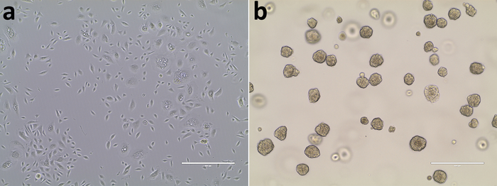
Figure 1: Maintenance of HPrEC in 2D culture and spheroid formation in 3D culture. (a) A 2D primary culture of HPrEC was BrdU-labeled and transferred to 3D culture with prostasphere formation on Day 5 (b). Scale bars = 400 µm. Please click here to view a larger version of this figure.
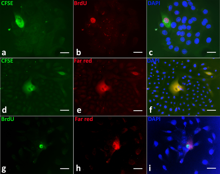
Figure 2: Identification of long-term label-retaining cells in primary prostaspheres. Double labeling of BrdU (red) and CFSE (green); CFSE (green) and Far Red (red); BrdU (green) and Far Red (red) identified the same stem-like cells with retention of parental DNA. Any BrdU, CFSE, or Far Red labels in rapidly dividing progenitor cells (DAPI, blue) were diluted and lost (a-i). Representative images show BrdU/CFSE (a-c) (upper panel), CFSE/Far Red (d-f) (middle panel), and BrdU/Far Red (g-i) (lower panel) co-labeling in single prostasphere (PS) cells. Scale bars = 50 µm. Please click here to view a larger version of this figure.
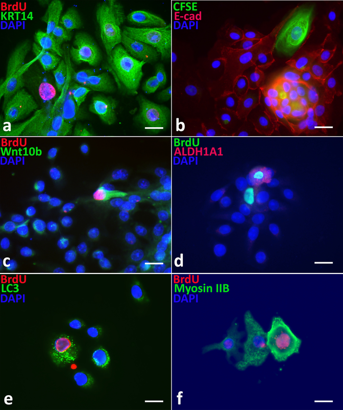
Figure 3: Label-retaining PS cells exhibiting stem cell properties. As compared to non-label-retaining progenitor cells, BrdU or CFSE label-retaining stem cells exhibit (a) lower levels of cytokeratin 14 (KRT 14), (b) decreased levels of E-cadherin, (c) elevated levels of Wnt10B, (d) higher levels of ALDH1A1, (e) increased LC3, and (f) increased myosin IIB proteins. Scale bars = 50 µm. Please click here to view a larger version of this figure.
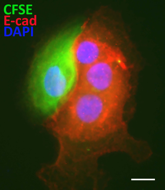
Figure 4: Using the sphere-based label-retaining assay for identification of cancer stem-like cells. CFSE label-retaining cancer stem-like cells in spheroids derived from human prostate cancer specimens exhibited reduced E-cadherin protein relative to the non-labeled progenitor cells. Scale bars = 50 µm. Please click here to view a larger version of this figure.
Flow cytometry using multiple stem cell surface markers is a commonly used approach for stem cell research despite lacking both specificity and selectivity1,5,6. While spheroid formation in a 3D culture system is another useful method in enriching the rare stem cell population from primary epithelial cells, including HPrEC, the resulting spheroids are still a heterogeneous mixture of stem and progenitor cells2,3,4. In prostaspheres, compared to the rapidly proliferating progenitor cells, the relatively quiescent character of stem cells allows them to be identified by long-term label-retention assay.
The methods summarized in this paper describe the labeling of HPrEC using BrdU, CFSE, and/or Far Red pro-dyes13. While BrdU, CFSE, and/or Far Red pro-dyes in rapidly proliferating cells are all quickly diluted by each cell division, there is an additional mechanism that accounts for the loss of BrdU known as immortal strand DNA segregation11. This is when the stem cell undergoes asymmetric division, giving rise to one daughter stem cell and a progenitor cell. The daughter stem cell retains all the old parental DNA with BrdU label, while the daughter progenitor cell receives the newly synthesized DNA without the BrdU label. Therefore, a label-retention assay using BrdU allows for clearer and easier identification of label-retaining stem cells by immunofluorescent staining. This is especially useful to confirm stem cell biomarkers by IF double staining using two different antibodies. One major limitation of BrdU labeling is that cells must be fixed for IF staining, thus preventing further functional studies following BrdU labeling and cell fixation.
To solve this issue, two fluorescent pro-dyes for live HPrEC labeling were tested in label-retaining assays. Results indicated that there was a complete overlap of BrdU, CFSE, and Far Red labeled sphere cells. BrdU label could be substituted with CFSE or Far Red in sphere-based label-retaining assays to allow visualization of live cells for imaging and separation by FACS, promoting functional characterization of isolated live stem cells using both in vitro cell culture assays and in vivo xenograft assays13. CFSE or Far Red label-based FACS sorted live stem and progenitor cells could also be used for RNA-seq including single cell RNA-seq, which is very powerful in identifying novel stem cell gene markers and signaling pathways as well as epithelial cell lineage hierarchy.
The novel biomarker-free method of functional characterization of stem cells by a spheroid-based label-retention assay presented here can identify cancer stem-like cells from primary patient specimens and cancer cell lines. Thus, it provides an avenue for discovering novel biomarkers of cancer stem-like cells and developing effective therapeutics targeting cancer13.
The authors have nothing to disclose.
This study was supported by grants from the National Cancer Institute R01-CA172220 (GSP, WYH), R01-ES02207 (GSP, WYH). We thank the Flow Cytometry Core at the University of Illinois at Chicago for assistance on cell sorting.
0.05% Trypsin-EDTA | Gibco | 25300-054 | |
1 mL tuberculin syringes | Bectin Dickinson | BD 309625 | |
1.5 mL microcentrifuge tubes, sterile | |||
100 mm culture dishes | Corning/Falcon | 353003 | |
12-well culture plate | Corning/Falcon | 353043 | |
15 mL centrifuge tubes | Corning/Falcon | 352097 | |
22 x 22 mm coverslips, sq | Corning | 284522 | For MatTek 35 mm culture dish |
24 x 50 mm coverslips | Corning | 2975245 | |
26G x 1.5 inch hypodermic needle | Monoject | 1188826112 | |
2N HCl | |||
35 mm culture dish with cover glass bottom | MatTek Corp | P35G-0-10-C | Glass bottom No. 0, uncoated, irradiated |
40 µm pore nylon cell strainer | Corning | 352340 | |
5% CO2 culture incubator, 37 °C | Forma | ||
50 mL centrifuge tubes | Corning/Falcon | 352098 | |
5mL Polystyrene Round-Bottom Tube with strainer snap cap | Corning | 352235 | 35 µm nylon mesh |
6-well culture plates | Corning | 353046 | |
8-well chamber slides | Millipore Sigma | PEZGS0816 | |
Aqueous mounting medium containing DAPI | Vector Laboratories | H-1200 | A nuclear fluorescent dye |
Biological safety cabinet, Level 2 certified | |||
BrdU (5-bromo-2′-deoxyuridine) | Sigma-Aldrich | B5002 | 1 mM stock solution in DMSO |
Centrifuge for 1.5 mL microcentrifuge tubes | Eppendorf | ||
Centrifuge for 15 mL tubes | Beckman Coulter | Allegra 6 | |
CFSE (carboxyfluorescein succinimidyl ester) | Thermo Fisher Scientific | C34554 | 5 mM stock solution in DMSO |
cytochalasin D | Thermo Fisher Scientific | PHZ1063 | |
Dispase 1U/mL | StemCell Technologies | 07923 | |
FACS CellSorter MoFlo XDP | Beckman Coulter | s | |
Far-Red pro-dye | Thermo Fisher | C34564 | 5 mM stock solution in DMSO |
Fetal Bovine Serum (FBS) | |||
Fibronectin | Sigma-Aldrich | F0895 | For coating 100 mm culture dishes |
Fluorescent microscope with color digital camera | Carl Zeiss | Axioskop 20 fluorescent microscope; color digital Axiocamera | |
Goat anti-Mouse IgG (H+L) Highly Cross-Adsorbed Secondary Antibody, Alexa Fluor 488 | Thermo Fisher | A-11029 | |
HPrEC (Primary normal human prostate epithelial cells) | Lifeline Cell Technology | FC-0038 | Pooled from 3 young (19-21yr od) disease-free organ donors; 1 x 10⁵ cells/mL; stored in liquid nitrogen |
ice bucket and ice | |||
Inverted microsope with digital camera | |||
Matrigel, low growth factor, phenol-red free | Corning | 356239 | |
Methanol | Corning | A452-4 | |
Mouse anti-BrdU antibody | Cell Signaling | 5292S | |
Mouse IgG antibody (negative control) | Santa Cruz Biotechnology | sc-2025 | |
Normal goat serum | Vector Laboratories | S-1000 | |
Phosphate Buffered Saline (PBS), pH 7.4 | Sigma-Aldrich | P5368-10PAK | |
Pipettors and tips, various sizes | |||
PrEGM (ProstaLife Epithelial Cell Growth Medium) | Lifeline Cell Technology | LL-0041 | |
Propidium Iodide (PI) | R & D Systems | 5135/10 | 10 μg/mL PI in PBS stored at 4 °C in the dark |
Serological pipets, various sizes | |||
Software for sphere counting and size measurements | |||
Software: 3D images using Imaris an imageing software with freeform drawing capabilities | |||
Triton X-100 | Millipore Sigma | T8787 | |
Water bath, 37 °C | |||
z-stack images using a transmitted light inverted fluorescent confocal microscope |