In this manuscript, we demonstrate the experimental techniques to encapsulate the F-actin cytoskeleton into giant unilamellar lipid vesicles (also called liposomes), and the method to form a cortex-biomimicking F-actin layer at the inner leaflet of the liposome membrane.
The actin cytoskeleton, the principal mechanical machinery in the cell, mediates numerous essential physical cellular activities, including cell deformation, division, migration, and adhesion. However, studying the dynamics and structure of the actin network in vivo is complicated by the biochemical and genetic regulation within live cells. To build a minimal model devoid of intracellular biochemical regulation, actin is encapsulated inside giant unilamellar vesicles (GUVs, also called liposomes). The biomimetic liposomes are cell-sized and facilitate a quantitative insight into the mechanical and dynamical properties of the cytoskeleton network, opening a viable route for bottom-up synthetic biology. To generate liposomes for encapsulation, the inverted emulsion method (also referred to as the emulsion transfer method) is utilized, which is one of the most successful techniques for encapsulating complex solutions into liposomes to prepare various cell-mimicking systems. With this method, a mixture of proteins of interest is added to the inner buffer, which is later emulsified in a phospholipid-containing mineral oil solution to form monolayer lipid droplets. The desired liposomes are generated from monolayer lipid droplets crossing a lipid/oil-water interface. This method enables the encapsulation of concentrated actin polymers into the liposomes with desired lipid components, paving the way for in vitro reconstitution of a biomimicking cytoskeleton network.
The actin cytoskeleton plays a fundamental role in constructing the intracellular architecture of the cell by coordinating molecular-level contractility and force generation1,2,3. As a result, it mediates numerous essential cellular activities, including cell deformation4,5, division6, migration7,8, and adhesion9. The in vitro reconstitution of actin networks has gained tremendous attention in recent years10,11,12,13,14,15,16,17. The goal of reconstitution is to build a minimal model of the cell devoid of the complex biochemical regulation that exists within live cells. This offers a controllable environment to probe specific intracellular activities and facilitates the identification and analysis of different components of the actin cytoskeleton18,19. Further, the encapsulation of in vitro actin networks inside phospholipid giant unilamellar vesicles (GUVs, liposomes) provides a confined but deformable space with a semi-permeable boundary. It mimics the physiological and mechanical microenvironment of the actin machinery within the cell9,20,21,22.
Among various methods to prepare liposomes, the lipid film hydration method (also known as the swelling method) is one of the earliest techniques23. The dry lipid film hydrates with the addition of buffers, forming membranous bubbles that eventually become vesicles24. To produce larger vesicles with a higher yield, an improved method advancing from the film hydration method, known as the electroformation method25, applies an AC electric field to efficiently promote the hydration process26. The major limitations of these hydration-based methods for actin encapsulation are that it has low encapsulation efficiency of highly concentrated proteins, and it is only compatible with specific lipid compositions24. The inverted emulsion technique, in comparison, has fewer limitations for lipid components and protein concentrations20,27,28,29. In this method, a mixture of proteins for encapsulation is added to the inner aqueous buffer, which is later emulsified in a lipid-containing mineral oil solution, forming lipid-monolayer droplets. The monolayer lipid droplets then cross through another lipid/oil-water interface through centrifugation to form bilayer lipid vesicles (liposomes). This technique has proven to be one of the most successful strategies for actin encapsulation24,30. Separately, there are some microfluidic device methods, including pulsed jetting31,32, transient membrane ejection33, and the cDICE method34. The similarities between the inverted emulsion method and the microfluidic method are the lipid solvent (oil) that is utilized and the introduction of lipid/oil-water interface for the formation of the outer leaflet of liposomes. By contrast, the generation of liposomes by the microfluidic method requires a set-up of microfluidic devices and is accompanied by oil trapped between the two leaflets of the bilayer, which requires an extra step for oil removal35.
In this manuscript, we used the inverted emulsion technique to prepare liposomes encapsulating a polymerized F-actin network as used previously22. The protein mixture for encapsulation was first placed in a buffer with nonpolymerizing conditions to maintain actin in its globular (G) form. The whole process was carried out at 4 °C to prevent early actin polymerization, which was later triggered by allowing the sample to warm to room temperature. Once at room temperature, the actin polymerizes into its filamentous (F) form. A variety of actin-binding proteins can be added to the inner aqueous buffer solution to study protein functionalities and properties, thus, further providing insights into its interaction with the actin network and membrane surface. This method can also be applied to the encapsulation of various proteins of interest36 and large objects (microparticles, self-propelled microswimmers, etc.) close to the size of the final liposomes28,37.
1. Preparation of buffers and protein solutions
2. Preparation of liposomes based on the inverted emulsion techniques
3. Microscopy observation
The preparation of liposomes based on the inverted emulsion technique is illustrated graphically and schematically in Figure 1.
First, empty (bare) liposomes (~5-50 µm in diameter) that were composed of phospholipid (EPC) and fluorescent lipid (DHPE) were prepared. A bright, far-red fluorescent dye was encapsulated within bare liposomes as a control experiment. Whether a lipid monolayer has successfully formed in the peripheral of the droplet could be determined by observing monolayer lipid droplets incorporating fluorescent dye in the emulsion, as shown in Figure 2A. Successful liposome generation could be verified through the visualization of the thin circular ring, which is the green-fluorescent lipid bilayer (conjugation of lipids with DHPE), under 488 nm laser using confocal microscopy (Figure 2A, right-most panel). To confirm encapsulation of the fluorescent dye, the internal environment of the liposomes should be uniformly fluorescent under a 647 nm laser (Figure 2A; overlay image) due to the far-red fluorescent dye (Table of Materials).
Different forms of encapsulated actin inside the monolayer lipid droplet are shown in Figure 2B. Images from left to right show globular actin (G-actin), filamentous actin (F-actin), actin forming a thin F-actin layer with the addition of VCA-His to the final buffer and Nickel-lipid to the membrane, and actin forming a thin F-actin layer with the addition of VCA-His, cofilin, and gelsolin to the final buffer and Nickel-lipid to the membrane.
Next, purified G-actin and associated F-actin binding proteins were encapsulated within the liposome. Composed of the same membrane components as above (EPC mixed with DHPE), liposomes here were ~5-50 µm in diameter. The formation of liposomes could be visualized by the thin green circular rings shown in Figure 3A,B. Actin polymerization was triggered by allowing the sample to warm to room temperature. As shown in Figure 3A, the reconstituted F-actin networks (with 20% fluorescently labeled actin) inside liposomes are heterogeneous, manifesting as branched network structures of actin filaments. The branched architecture was triggered by the introduction of the Arp2/3 complex, which simultaneously controls the nucleation and branching of actin filaments, along with VCA-His40,41,42,43,44,45.
Finally, an F-actin cortex-biomimicking system was created. A thin, but densely branched F-actin layer was created at the inner leaflet of the bilayer of liposomes22, which could be visualized as a fluorescent shell (Figure 3C). In this case, additionally, VCA-His, cofilin, and gelsolin were encapsulated into liposomes. Nickel-lipids were required in the component of the lipid membrane. VCA-His, a WASP fragment, carries a histidine tag in interaction with the Nickel-lipids of the lipid membrane. In the meantime, it recruits the Arp2/3 complex20,46,47,48. As a result, actin nucleated by the Arp2/3 complex generates an F-actin layer coated along the inner layer of the membrane in the presence of cofilin and gelsolin.
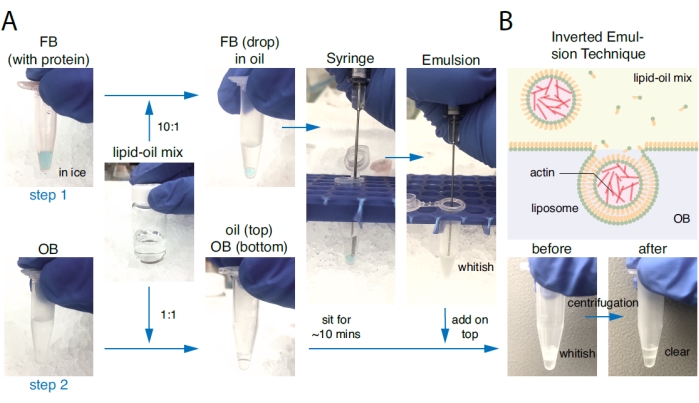
Figure 1: Preparation of liposomes based on the inverted emulsion technique. (A) Liposome preparation consists of two steps. Step one: A droplet of 10 µL of Final Buffer (FB) carrying proteins of interest was added to 100 µL of the phospholipid-oil mixture at a ratio of 1:10 and suspended by gently aspirating up and down with a glass syringe. Solutions containing monolayer lipid droplets became whitish after the back-and-forth aspiration. Step two: In a separate plastic tube, a few microliters of the lipid-oil mixture were placed on top of the same amount of OB and allowed to sit for ~10 min to develop a lipid monolayer at OB/oil interface. The emulsion from step one was poured on top of the oil phase from step two and was centrifuged (100 x g; 15 mins) at 4 °C. After centrifugation, the top oil solution should be clear, and the bottom OB solution (containing liposomes) should be slightly cloudy. (B) Schematics of the liposome preparation from an inverted emulsion. The whitish emulsion on the top contained monolayer lipid droplets that incorporated a branched actin network inside. During centrifugation, monolayer lipid droplets passed through the OB/oil interface to form an outer leaflet such that liposomes were created and accumulated at the bottom of the plastic tube. Please click here to view a larger version of this figure.
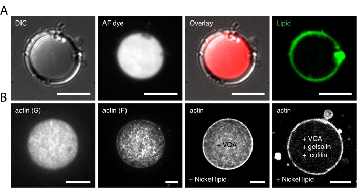
Figure 2: Microscopic images of the monolayer lipid droplet. (A) The monolayer lipid droplet incorporating fluorescent dye in the emulsion. Images from left to right show a monolayer lipid droplet under the DIC channel, fluorescent 640 nm channel, overlay of the DIC and 640 nm channel, and fluorescent 488 nm channel, respectively. (B) Different forms of encapsulated actin inside the monolayer lipid droplet under a fluorescent 561 nm channel. Images from left to right show globular actin (G-actin), filamentous actin (F-actin), actin forming a thin F-actin layer with the addition of VCA-His to the Final Buffer and Nickel-lipid to the membrane, and actin forming a thin F-actin layer with the addition of VCA-His, cofilin, and gelsolin to the Final Buffer and Nickel-lipid to the membrane. Scale bars are 10 µm at 63x magnification. Please click here to view a larger version of this figure.
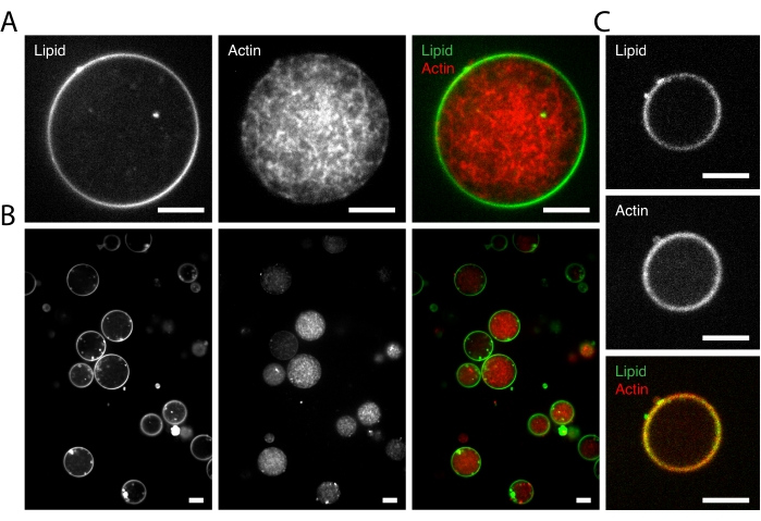
Figure 3: Microscopic images of the encapsulation of actin networks inside liposomes. (A) A liposome encapsulating polymerized branched (Arp 2/3 nucleated) F-actin network. Left to right: fluorescent 488 nm channel (left), fluorescent 561 nm channel (center), overlay (right). (B) A representative result of suspensions of liposomes encapsulating polymerized branched (Arp 2/3 nucleated) F-actin networks. Left to right: fluorescent 488 nm channel (left), fluorescent 561 nm channel (center), and overlay (right). (C) The appearance of the formation of a thin but dense F-actin layer coated along the inner layer of the lipid membrane of a liposome. Top to bottom: fluorescent 488 nm channel (top), fluorescent 561 nm channel (center), overlay (bottom). Scale bars are 10 µm at 63x magnification. Please click here to view a larger version of this figure.
Several key steps determine the success of a high yield of liposomes during the preparation process. To completely dissolve the lipid film in the oil, the sample must be sonicated until the lipid film at the bottom of the glass vial disappears completely. After the sonication, the lipid-oil mixture must be stored overnight at room temperature under dark conditions for the lipid molecules to disperse further29. The mixture can be stored at 4 °C for up to a week. When preparing an FB/oil emulsion with monolayer lipid droplets containing protein of interest, the lipid-oil-FB mixture must be gently pumped back and forth through a glass syringe to avoid introducing air bubbles. During this process, one big droplet is sheared off at the tip of the glass syringe into many smaller droplets (~5-100 µm in diameter) after several repetitions. The mixture must be whitish after the process30. To check the quality of phospholipids, and whether a lipid monolayer had successfully formed in the peripheral of the FB containing droplet, monolayer lipid droplets incorporating fluorescent dye were examined in the emulsion, as shown in Figure 2A. To ensure the passage of the monolayer lipid droplets through the OB/oil interface during centrifugation, and allow the collected liposomes in OB to settle on the glass surface, the density of FB should be slightly higher than the OB. Therefore, sucrose and glucose were chosen as the main component of the inner (FB) and outer (OB) aqueous solutions, respectively. In the previous work, dextran was used to adjust the density of the inner aqueous solution20, which is not included in this protocol. Moreover, the osmolarity of the OB was adjusted with glucose such that the osmotic pressure of the OB is slightly larger than that of FB (20-60 mOsm) as reported in the previous work20,22. Large osmolarity difference led to the shrinkage (when the osmolarity of the OB is greater than that of FB) or rupture (when the osmolarity of the FB is greater than that of OB) of the liposomes. An osmometer was used to check the osmolarities of the buffers. In the protocol, β-casein was added in the OB to passivate both plastic tube surfaces and glass surfaces of the imaging chamber and minimize sticking between the liposomes20,49. Mineral oil was used as the oil solution (i.e., the lipid solvent). Different lipid solvents have been reported elsewhere, including dodecane50, liquid paraffin30, squalane51, etc. Accordingly, different centrifugation speeds and durations are used for various lipid solvents because of their viscosity differences and the influence they bring on the liposome surface tension. The duration of step 2.3 needs to be extended for charged lipids because, in the monolayer, these charged molecules repel each other, thus requiring more time to diffuse and organize well at the interface between the lipid/oil mixture and OB38. For lipids incorporated with proteins of interest, an extension of this step is also recommended to ensure better monolayer formation52. For the liposome yield of liposomes encapsulating polymerized F-actin network, the number of liposomes per field of view (100 µm x 100 µm) is around 5-10 with an average diameter of 20 µm. A zoomed-out image of a typical microscopy sample is shown in Figure 3B. More zoomed-out confocal images of the liposomes produced through the inverted emulsion method can be found in a recent study52 where various parameters (density difference, centrifugation speed/time, lipid concentration, pH, temperature, etc.) have been optimized for high-yield production of liposomes.
For the reconstitution of the F-actin network, it is essential to choose an appropriate concentration of the adenosine triphosphate (ATP), dithiothreitol (DTT), the associated salts, and the pH condition. An intermediate checkpoint of the reconstituted F-actin network can be done by observing monolayer lipid droplets with encapsulated actin inside. Different forms of encapsulated actin inside the monolayer lipid droplet are shown in Figure 2B. Actin polymerization occurs at >20 mM K+ salt and in >0.2 mM Mg2+ salt with the pH stabilized between 6.5 and 8.5, as reported previously53,54,55. Before the imaging of liposomes, all solutions were kept on ice to prevent early actin polymerization. The samples were imaged at room temperature to trigger actin polymerization. The architecture of the reconstituted F-actin network is predominantly regulated by the activity of the Arp2/3 complex. The Arp 2/3 complex nucleates and branches actin filaments from the side of an existing mother filament to generate dendritic architectures56,57. The Nickel-lipid (DOGS-NTA-Ni) and the VCA-His are critical for the formation of the F-actin layer. VCA-His serves as a nucleation-promoting factor, which is linked to Nickel-lipids at the membrane22,58. A branched F-actin network is then nucleated from VCA-His facilitating the Arp2/3 complex, and as a result, a dense F-actin shell is formed at the inner leaflet of the lipid membrane. The concentration of VCA-His within the liposome determines the thickness of the F-actin layer. The addition of the actin regulatory proteins (cofilin and gelsolin) also promotes the formation of the F-actin layer. The cofilin severs actin filaments to increase the number of filament ends, while the gelsolin binds to the barbed ends of actin filaments to limit their growth. Thereby, in the presence of cofilin and gelsolin, a larger number of filaments can be nucleated by Arp2/3, and then recruited by VCA-His to form the F-actin layer.
Similar papers based on the emulsion transfer method for protein encapsulation have been published in recent years28,30,34. One major difference between the protocol here and the protocols in these papers is the choice of the primary lipid for liposomes. In this work, we use the L-α-phosphatidylcholine (EPC, a mixture of different phosphatidylcholine species containing approximately 60% POPC59) as the main component of the liposome membrane. To form an F-actin layer, a mixture of the EPC and the Nickel-lipid (DOGS-NTA-Ni) was used at a ratio of 10:1. In a previous work, liposomes were spread on polyhistidine coated glass, and EPC, cholesterol, and Nickel-lipid were mixed at a ratio of 53:37:1020. In comparison, Natsume et al. and Bashirzadeh et al. chose the dioleoyl-phosphocholine (DOPC, a single type of phospholipid) to encapsulate microspheres28 and fascin-actin bundles34, respectively, while Fujii et al. recommended 1-Palmitoyl-2-oleoyl-sn-glycero-3-phosphocholine (POPC, a pure synthetic phospholipid) as the primary lipid to establish a liposomes-based membrane protein synthesis technology30. The choice of the primary lipids affects the physical properties of liposomes. For example, as the degree of unsaturation of the acyl chain increases (i.e., POPC< EPC< DOPC60), the permeability properties of various molecules through the bilayer decrease61. The choice also depends on the lipids of interest being mixed. For instance, the addition of the cholesterol makes EPC liposomes more stable, while it destabilizes the bilayer structure of DOPC/DOPE mixture61.
The similarities between this work and a recent publication from Bashirzadeh et al.34 fall on the same topic of actin encapsulation. Meanwhile, there are clear distinctions. Bashirzadeh et al. reported a modified cDICE approach utilizing a 3D-printed rotating chamber. Here, we adopt the simple, traditional inverted emulsion transfer method using a syringe and a centrifuge machine. Both of these two methods generate a high yield of liposomes and have high encapsulation efficiencies for large biomolecules24. Besides the difference in the lipid components as mentioned above, the architectures of the encapsulated actin network alter as well. Bashirzadeh et al. encapsulated fascin-actin bundles within the liposome, while in this work, we encapsulated a branched network triggered by the Arp2/3 complex. In addition, the formation of an F-actin layer to create a cortex-biomimicking system in the presence VCA-His has been elaborated here.
The method reported here has two limitations. One is that the size of the liposomes that incorporate in vitro reconstituted actin networks cannot be precisely manipulated (varying from 5 to 50 µm in diameter), and the yield is low compared to the electroformation method. The other limitation of a water-oil-based method is that trace amounts of oil are trapped between the bilayer leaflets24. Although it does not significantly affect the membrane thickness or the biocompatibility of these bio-mimicking liposomes24,35,62,63, the membrane dynamics of the oil-inserted liposomes may be altered62. In this method, the actin nucleator Arp2/3 is incorporated. Future studies may include other cytoskeleton proteins64,65,66,67, nucleators, such as formin68, or molecular motors such as myosin II69.
The authors have nothing to disclose.
We acknowledge funding ARO MURI W911NF-14-1-0403 to M.P.M., the National Institutes of Health (NIH) R01 1R01GM126256 to M.P.M., the National Institutes of Health (NIH) U54 CA209992, NIH RO1 GM126256, NIH U54 CA209992, University of Michigan / Genentech, SUBK00016255 and Human Frontiers Science Program (HFSP) grant number RGY0073/2018 to M.P.M. Any opinion, findings, conclusions, or recommendations expressed in this material are those of the authors(s) and do not necessarily reflect the views of the ARO, NIH, or HFSP. S.C. acknowledges fruitful discussions with V. Yadav, C. Muresan, and S. Amiri.
1,2-dioleoyl-sn-glycero-3-{[n(5-amino-1-carboxypentyl)iminodiacetic acid]succinyl} nickel salt (DOGS-NTA-Ni) | Avanti Polar Lipids Inc. | 231615773 | Nickel Lipid |
1,4-Diazabicyclo[2.2.2]octane | Sigma | D27802-25G | DABCO |
Actin protein (>99% pure): rabbit skeletal muscle | Cytoskeleton, Inc | AKL99-D | non-fluorescent G-actin |
Actin protein (rhodamine): rabbit skeletal muscle | Cytoskeleton, Inc | AR05 | fluorescently labeled actin |
Adenosine 5′-triphosphate disodium salt hydrate | Sigma | A2383-10G | ATP |
Alexa Fluor 647 dye | ThermoFisher | fluorescent dye | |
Andor iQ3 | Andor Technologies | control and acquisition software for confocal microscope | |
Arp2/3 Protein Complex: Porcine Brain | Cytoskeleton, Inc | RP01P-A | Arp 2/3 |
Calcium chloride dihydrate | Sigma | 10035048 | CaCl2 |
Chamlide Chambers (4-well for 12 mm round coverslip) | Quorum Technologies | incubation chamber | |
Cofilin protein: human recombinant | Cytoskeleton, Inc | CF01-C | cofilin |
Confocal Microscope (63× oil-immersion objective) | Andor Technologies | LEICA DMi8 | |
D-(+)-GLUCOSE BIOXTRA | Sigma | G7528 | glucose |
Dithiothreitol | DOT Scientific | DSD11000-10 | DTT |
Gelsolin Protein: Homo Sapiens Recombinant | Cytoskeleton, Inc | HPG6 | gelsolin |
Hamilton 1750 Gastight Syringe, 500 µL, cemented needle, 22 G, 2" conical tip | Cole-Parmer | UX-07940-53 | glass syringe |
HEPES | AmericanBio | 7365-45-9 | |
ImageJ/Fiji | https://imagej.net/tutorials/ | ||
L-alpha-Phosphatidylcholine | Avanti Polar Lipids Inc. | 97281442 | EPC |
Magnesium chloride | Sigma | 7786303 | MgCl2 |
Mineral oil, BioReagent, for molecular biology, light oil | Sigma | 8042475 | mineral oil |
N-WASP fragment WWA (aa400–501, VCA-His) | VCA-His is purified using lab protocol. The protocol can be provided upon reasonable requests | ||
Oregon Green 488 1,2-Dihexadecanoyl-sn-Glycero-3-Phosphoethanolamine (Oregon Green 488 DHPE) | Thermo Fisher | O12650 | DHPE |
Potassium chloride | Sigma | 7447407 | KCl |
Sucrose | Sigma | 57-50-1 | sucrose |
β-Casein from bovine milk | Sigma | C6905-250MG |