The Miniature Pig: A Large Animal Model for Cochlear Implant Research
Summary
Miniature pigs (mini-pigs) are an ideal large animal model for research into cochlear implants. Cochlear implantation surgery in mini-pigs can be utilized to provide initial evidence of the safety and potential performance of novel electrode arrays and surgical approaches in a living system similar to human beings.
Abstract
Cochlear implants (CI) are the most effective method to treat people with severe-to-profound sensorineural hearing loss. Although CIs are used worldwide, no standard model exists for investigating the electrophysiology and histopathology in patients or animal models with a CI or for evaluating new models of electrode arrays. A large animal model with cochlea characteristics similar to those of humans may provide a research and evaluation platform for advanced and modified arrays before their use in humans.
To this end, we established standard CI methods with Bama mini-pigs, whose inner ear anatomy is highly similar to that of humans. Arrays designed for human use were implanted into the mini pig cochlea through a round window membrane, and a surgical approach followed that was similar to that used for human CI recipients. Array insertion was followed by evoked compound action potential (ECAP) measurements to evaluate the function of the auditory nerve. This study describes the preparation of the animal, surgical steps, array insertion, and intraoperative electrophysiological measurements.
The results indicated that the same CI used for humans could be easily implanted in mini-pigs via a standardized surgical approach and yielded similar electrophysiological outcomes as measured in human CI recipients. Mini-pigs could be a valuable animal model to provide initial evidence of the safety and potential performance of novel electrode arrays and surgical approaches before applying them to human beings.
Introduction
According to the World Health Organization (WHO), over 1 billion people are at risk of hearing loss globally, and it is estimated that, by 2050, one out of four people will suffer hearing loss1. Over the last 2 decades, CIs have been the most effective intervention for people with permanent severe and profound sensorineural hearing loss (SNHL). A CI converts physical signals of sound into bioelectrical signals that stimulate the spiral ganglion neurons (SGNs), bypassing hair cells. Over time, the indications for a CI have been broadened so that they now include people with residual hearing, unilateral hearing loss, and very old or young people2,3,4. Meanwhile, totally implantable CIs and advanced arrays have been developed5. There is, however, no economically feasible large animal model for investigating the electrophysiology and histopathology of the inner ear with a CI. This lack of a large animal model limits research seeking to improve CIs and gain insights into the electrophysiological impact of CIs on the inner ear.
Several rodent animal models have been applied in CI research, such as mouse6, gerbil7, rat8, and guinea pig9; however, the characteristics of morphology and electrophysiological responses are different from that in humans. Cochlear structures of animal models traditionally used for CI studies, such as cats, guinea pigs, and other animals, differ greatly from those of human cochlear structures10. Although array insertion has been conducted on cats11 and rabbits12, because of their smaller cochleae, this was done with arrays that were not designed for use in humans. Several large animal models have also been explored for CI. Lambs are well suited as a training model for atraumatic cochlear implantation, but the smaller size of the cochlea makes full array insertion impossible13. Primates might be the most suitable animals for CI research because of their anatomical similarity to humans14,15; however, the sexual maturity of monkeys is delayed (4-5 years), the gestation period is up to about 165 days, and each female usually produces only one offspring per year16. These reasons, and the expensive cost, hinder the extensive application of primates in CI research.
In contrast, pigs reach sexual maturity at 5-8 months and have a gestation period of ~114 days, making pigs more accessible for CI research as a large animal model16. Bama mini pigs (mini-pigs) originated from a small-sized pig species in China in 1985, whose genetic background is well understood. They are characterized by an inherent small size, early sexual maturity, rapid breeding, and ease of management17. The mini-pig is an ideal model for otology and audiology because of its similarity to humans in morphology and electrophysiology18. The scala tympani length of a Bama mini-pig is 38.58 mm, which is close to the 36 mm length in humans10. The mini-pig cochlea has 3.5 turns, which is similar to the 2.5-3 turns seen in humans10. In addition to morphology, the electrophysiology of Bama mini-pigs is also highly similar to that of humans18. Therefore, in the present study, we inserted arrays designed for human use into the mini-pig cochlea via the round window membrane and followed a similar surgical approach to that used in human CI recipients. Intraoperative ECAP measurements were applied to evaluate the procedure. The process we describe herein could be used both for preclinical translational research associated with CIs and as a platform for resident training.
Protocol
All procedures and animal surgeries were conducted according to the guidelines of the Ethics Committee of the PLA General Hospital and were approved.
1. Anesthesia and surgical preparation
- Inject the pig (male, 2 months old, 5 kg) muscularly with tiletamine and zolazepam with a dosage of 10-15 mg/kg and intubate it with a 5.5-French endotracheal tube. Maintain anesthesia by ventilator-assisted respiration with isofluorane inhalation. Monitor the oxygen saturation (>90%), breathing (15-20/min), and heart rate (60-120 beats/min) using the pulse oximetry clamp of an ECG monitor, which is connected to the pig's tongue.
- Put the mini-pig in a left lateral position (when the right side is to be implanted) on a thermostatically regulated heating pad to prevent hypothermia. Confirm the pig is adequately anesthetized using various stimuli. Ensure the absence of all responses (e.g., toe pinch reflex). Apply artificial tear ointment to the eyes of the minipig to keep the cornea from drying. Keep the eyes closed using a medical patch.
- Shave the surgical area around the earlobe, keeping it 10 cm in diameter (Figure 1) and disinfect it with three alternating swabs of iodine and alcohol in a circular motion from the center toward the outside. Cover the surgical area with sterile surgical drapes.
- Cover the microscope with a sterile plastic sleeve and remove the parts covering the eyepieces and objective.
2. Surgical procedure
- Locate the surface projection site of the cochlea 1 cm behind the posterior auricular sulcus at the level of the earlobe. Make a postauricular incision about 5 cm long with the projection site as the center using a #15 scalpel. Divide the subcutaneous tissue, parotid gland, and sternocleidomastoid muscle with micro-scissors to expose the surface of the mastoid bone (Figure 2A). Use bipolar cautery when necessary to minimize bleeding.
- Cortical mastoidectomy
- Drill the mastoid at the surface projection of the cochlea on the mastoid bone (Figure 2B) to the external auditory canal (EAC), which is dense and pale blue (Figure 2C). Be careful not to damage the pale or reddish vertical segment of the facial nerve dorsal to the EAC to avoid bleeding (Figure 2C).
NOTE: If the facial nerve is damaged, bipolar cautery is a good choice to stop bleeding.
- Drill the mastoid at the surface projection of the cochlea on the mastoid bone (Figure 2B) to the external auditory canal (EAC), which is dense and pale blue (Figure 2C). Be careful not to damage the pale or reddish vertical segment of the facial nerve dorsal to the EAC to avoid bleeding (Figure 2C).
- Expose the tympanum by drilling the bone surrounding the posterior bony EAC (Figure 2D). Separate the skin of the EAC and the facial nerve with a hypodermic needle to avoid damaging the facial nerve. Carefully push the skin of the EAC away to expose the tympanum (including the ossicular chain) and round window niche (Figure 3A).
- Expose the round window membrane. Remove the round window niche with a small diamond burr, and keep continuous suction-irrigation to expose the basal turn of the cochlea and round window membrane (Figure 3B).
- Fix the receiver package. Separate the cranial parietal muscle to form a pocket just large enough for the receiver. Place the internal receiver package in the muscular pocket and fix it with a fixation suture.
- Insert the electrode array, which is connected to a receiver fixed in a muscular pocket, by opening the round window membrane with a sharp microsurgical knife and inserting the array using micro forceps slowly, steadily, and continuously in relationship to the modiolus of the cochlea (Figure 3C). Suture the surgical incision with an absorbable suture 2-0.
- ECAP measurements
NOTE: The setup is composed of a PC with MAESTRO Software connected to the patient's cochlear implant (CI) through the stimulation device (MAX Programming Interface) and the CI coil.- Magnetically connect the CI coil to the CI receiver through the skin. Confirm the integrity of the CI and check the electrode impedance for all channels before ECAP measurements using the telemetry function of the CI, which is conducted by MAESTRO Software automatically (Figure 4A,B).
- Conduct the ECAP measurements as described previously19. Select the ECAP module, choose all 12 electrodes for stimulation, and wait for the ECAP tests of the electrodes to be completed. See the Table of Materials for the software and stimulation device used to measure ECAP responses. Stimulate all 12 electrodes for ECAP measurements using biphasic stimuli of 30 µs phase duration, with an alternating-polarity paradigm, averaging over 25 iterations, and a stimulation rate of 45.1 pulses/s.
3. Postoperative care
- Keep monitoring the mini-pig to avoid harm due to unconscious movement until it has regained sufficient consciousness to maintain sternal recumbency. Put the minipig on the thermostatically regulated heating pad to prevent hypothermia.
- Place the mini-pig back into its home cage alone.
- Inject antibiotics to prevent postoperative infection for 7 days.
- Check the mini-pig for symptoms of vestibular injury such as nystagmus, circling, or rolling over.
4. Postoperative CT scan
- Administer intramuscular injection of 3% sodium pentobarbital 1 ml/kg, and 0.1 ml/kg of Sulforaphane to minipig to induce anesthesia. Use a 37℃ warming plate to keep it warm. After 3 or 5 minutes, a CT scan can be conducted.
- To confirm the correct position of the electrode array, narcotize the mini-pig with tiletamine and zolazepam 1 week after the operation. Perform the CT scan and 3D reconstruction20 using the 3D slicer image computing platform (see the Table of Materials). Import DICOM data of the CT and conduct the Volume rendering module to achieve 3D images of the CI.
Representative Results
The integrity (Figure 4A) and impedances (Figure 4B) of the CI were confirmed by MAESTRO Software. ECAP results showed that all 12 electrodes demonstrated good neural responses (Figure 4C), meaning the electrode array was well attached to the cochlear axis and stimulated the auditory nerve. Figure 5 demonstrates postoperative 3D reconstructed electrode coils in the right cochlea. The array did not fold or dislocate. The electrode array was coiled in the basal turn of the cochlea (Figure 5A), and the electrodes are rendered in green (Figure 5B). 3D reconstruction demonstrates that the electrode array was spirally coiled in the cochlea (Figure 5C).
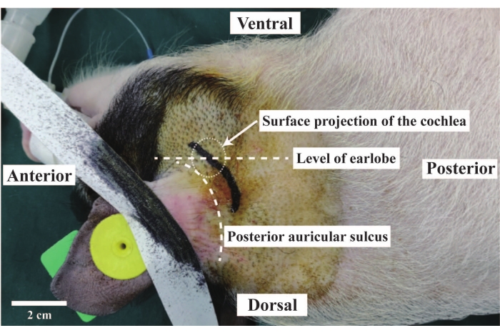
Figure 1: The surgical position and surface projection of the cochlea. The anesthetized pig was in a left lateral position. The white dashed circle shows the surface projection of the cochlea: 1 cm behind the posterior auricular sulcus at the level of the earlobe. Scale bar = 2 cm. Please click here to view a larger version of this figure.
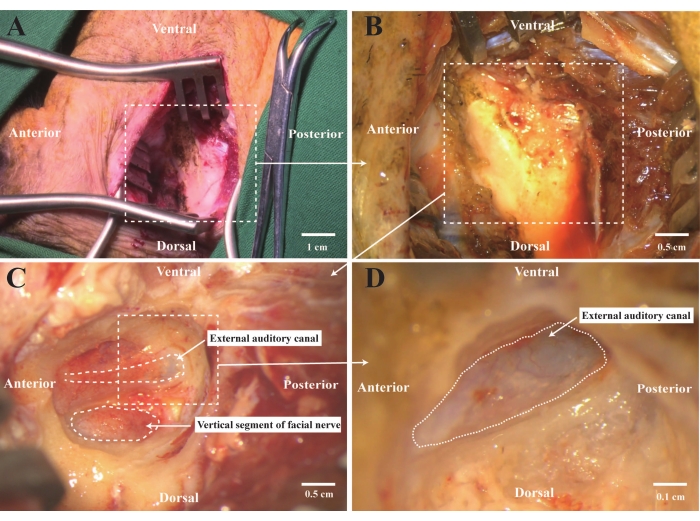
Figure 2: Cortical mastoidectomy. (A) Make a postauricular incision and divide the subcutaneous tissue, parotid gland, and sternocleidomastoid muscle to expose the surface of the mastoid bone. (B) Drill the mastoid at the surface projection of the cochlea on the mastoid bone. (C) Expose the EAC and the vertical segment of the facial nerve. (D) Drill the bone surrounding the posterior EAC to reveal the skin of the EAC. Scale bars = (A) 1 cm, (B,C) 0.5 cm, (D) 0.1 cm. Abbreviation: EAC = external auditory canal. Please click here to view a larger version of this figure.
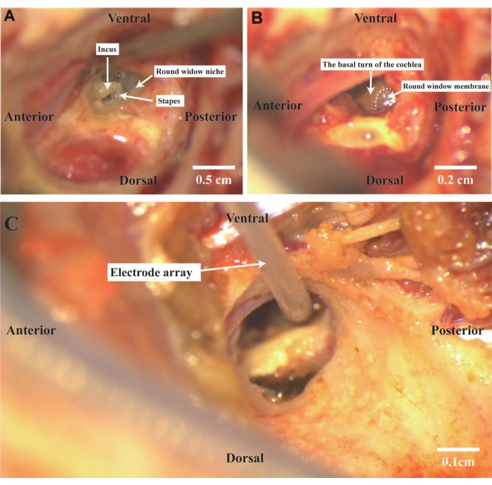
Figure 3: Exposing the tympanum. (A) Push the skin of the EAC forward and expose the tympanum. The landmarks of the middle ear, the incus, stapes, as well as round the window niche, must be clearly visible. (B) Remove the round window niche and expose the round window membrane. (C) Insert intraoperative electrodes through the round window membrane. Scale bars = (A) 0.5 cm, (B) 0.2 cm, (C) 0.1 cm. Abbreviation: EAC = external auditory canal. Please click here to view a larger version of this figure.
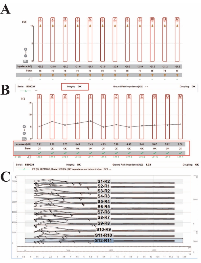
Figure 4: Telemetry of the CI and ECAP results of 12 electrodes. (A) Integrity test of the CI. (B) Impedance tests on electrodes. (C) ECAP results of all 12 electrodes. Abbreviations: CI = cochlear implant; ECAP = evoked compound action potential. Please click here to view a larger version of this figure.
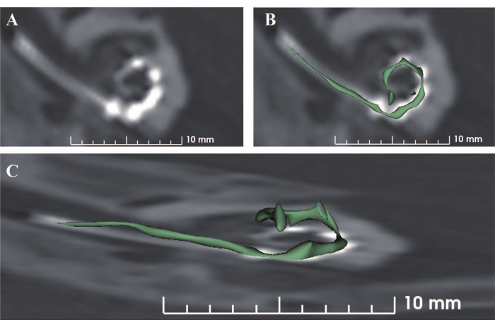
Figure 5: Postoperative CT 3D reconstruction of electrodes (Electrodes of Concerto F28). (A) The electrode array is coiled in the basal turn of the cochlea. (B) The electrodes are rendered in green. (C) 3D reconstruction demonstrates that the electrode array is spirally coiled in the cochlea. Scale bar = 10 mm. Please click here to view a larger version of this figure.
Discussion
Around 15% of the world’s population have some degree of hearing loss, and over 5% have disabling hearing loss21. CI provision is the most efficient treatment for both adult and pediatric patients with severe and profound sensorineural hearing loss. As the first successful implantable cranial nerve stimulator, over the past 2 decades, CIs have offered thousands of people with hearing loss the opportunity to return to the world of sound and (re)integrate into mainstream society. Even though CIs are now very different from their original appearance and function, CI research still lacks a large animal model similar to humans. An economical and accessible large animal model would provide important electrophysiological and histopathological information that is not easy or ethical to obtain directly from humans.
Several bioelectrical treatments depending on CI are currently being investigated. Guo et al.22 found that electric-acoustic stimulation via a CI could promote neural stem cells to proliferate and differentiate into neurons. Furthermore, several factors such as growth hormone23, glial cell line-derived neurotrophic factor (GDNF)24, and brain-derived neurotrophic factor (BDNF)25 have been proven to promote neurite extension or increase the survival rate of SGNs. These results may give hope to patients with SGN degeneration, who may not benefit from CI use.
Nevertheless, the promising research that may improve CI performance mentioned above is performed in vitro or on small animal models. Experiments must be conducted on large animal models before conducting them on living humans. Thus, the protocol described herein demonstrates how to perform cochlear implantation in a Bama mini-pig. The great advantage of using this animal model is that the same devices can be studied in animals as are used in humans, i.e., the devices or dosages do not need to be scaled up or down.
Unlike cochlear implantation in guinea pigs or mice, where general anesthesia in a spontaneously breathing animal is sufficient, cochlear implantation in Bama mini-pigs is similar to that in humans in terms of operation time and protocols. Tiletamine and zolazepam were injected intramuscularly with a dosage of 10-15 mg/kg. After the successful induction of anesthesia, endotracheal intubation and ventilator-assisted respiration with isofluorane were essential to ensure the depth of anesthesia intraoperatively.
There are two key steps to successfully exposing the round window. The first is the position of the animal during surgery. Placing a cushion under the animal’s neck in a lateral position helps to clearly expose the mastoid bone. The second is to determine the projection area of the cochlea on the surface of the mastoid, which is located 1 cm behind the posterior auricular sulcus at the level of the earlobe (Figure 1). Drilling the mastoid at this site enables easy access to the EAC and facial nerve.
Two important anatomical landmarks, the EAC and the vertical segment of the facial nerve, are helpful to identify the middle ear. The facial nerve appears reddish or pale, while the skin of the EAC appears bluish (Figure 2D). One should remove the posterior bony EAC and carefully push the skin of the EAC forward to reveal the tympanum. The round window niche shelters the round window membrane (Figure 3A). Removing the niche with a drill exposes the membrane (Figure 3B). The facial nerve may block the round window membrane, in which case the facial nerve must be cut to expose the membrane. Cutting the facial nerve results in massive bleeding and obscures the surgical view. Bipolar coagulation should be used to stop bleeding. Unlike cochlear implantation surgery in humans, in which the implant is fixed in a bone groove on the skull, we fixed the implant in a muscle pocket because the skull of a mini-pig is thinner than that of a human. Fixing the receiver on the top of the skull should avoid damage due to collision on both sides, because pigs often rub their cage with the sides of their heads.
The procedure described herein could be applied to research into new types of arrays and into biotherapy and gene therapy combined with CI. Due to the normal hearing of the pigs used in this research, it is difficult to observe the postoperative responses toward sound (e.g., a whistle for food). As a topic of future research, we aim to establish a series of methods to observe the pigs’ responses to sound transmitted by the CI.
Divulgaciones
The authors have nothing to disclose.
Acknowledgements
This study was funded by grants from the National Natural Science Foundation of China (Nos. 81970890) and the Chongqing Scientific Research Institution Performance incentive project (Nos. 19540). We thank Anandhan Dhanasingh and Zhi Shu from the MED-EL company for their support.
Materials
| 0.5 mm diamond burr | |||
| 1 mm diamond burr | |||
| 5 mm diamond burr | |||
| 2-0 suture silk | |||
| 3D Slicer image computing platform | 3D reconstruction of CT image | ||
| Alcohol | |||
| Bipolar cautery | |||
| Bipolar electrocoagulation | Stop bleeding | ||
| CI designed for human use (CONCERTO FLEX28) | MED-EL | Concerto F28 | |
| Dressing forceps | |||
| ECG monitor | |||
| Iodine tincture | |||
| Isoflurane | 3.6 mL/h | ||
| Laryngoscope | |||
| MAESTRO Software | MED-EL | Measure ECAP responses | |
| Micro forceps | |||
| Micro spatula | |||
| Mosquito forceps | |||
| Needle holder | |||
| Needle probe | |||
| Negative pressure suction device | |||
| Otological surgical instruments | |||
| Respiratory Anesthesia Machine | |||
| Scalpel with blade No. 15 | |||
| Scissors | |||
| Shaver | |||
| Stimulation device (MAX Programming Interface) | MED-EL | Measure ECAP responses | |
| Surgery microscope | Leica | ||
| Surgical drill | |||
| Surgical Power Device | |||
| Tiletamine and zolazepan | 10-15 mg/kg | ||
| Tissue forceps | |||
| Trachea cannula |
Referencias
- World report on hearing. World Health Organization Available from: https://www.who.int/publications/i/item/world-report-on-hearing (2021)
- Lee, S. Y., et al. Natural course of residual hearing preservation with a slim, modiolar cochlear implant electrode array. American Journal of Otolaryngology. 43 (2), 103382 (2022).
- Lorens, A., et al. Binaural advantages in using a cochlear implant for adults with profound unilateral hearing loss. Acta Oto-Laryngologica. 139 (2), 153-161 (2019).
- Lally, J. W., Adams, J. K., Wilkerson, B. J. The use of cochlear implantation in the elderly. Current Opinion in Otolaryngology & Head and Neck Surgery. 27 (5), 387-391 (2019).
- Rhodes, R. M., Tsai Do, B. S. Future of implantable auditory devices. Otolaryngologic Clinics of North America. 52 (2), 363-378 (2019).
- Colesa, D. J., et al. Development of a chronically-implanted mouse model for studies of cochlear health and implant function. Hearing Research. 404, 108216 (2021).
- Toulemonde, P., et al. Evaluation of the efficacy of dexamethasone-eluting electrode array on the post-implant cochlear fibrotic reaction by three-dimensional immunofluorescence analysis in Mongolian gerbil cochlea. Journal of Clinic Medicine. 10 (15), 3315 (2021).
- King, J., Shehu, I., Roland, J. T., Svirsky, M. A., Froemke, R. C. A physiological and behavioral system for hearing restoration with cochlear implants. Journal of Neurophysiology. 116 (2), 844-858 (2016).
- Chen, M., Min, S., Zhang, C., Hu, X., Li, S. Using extracochlear multichannel electrical stimulation to relieve tinnitus and reverse tinnitus-related auditory-somatosensory plasticity in the cochlear nucleus. Neuromodulation. , (2021).
- Yi, H., et al. Miniature pigs: A large animal model of cochlear implantation. American Journal of Translational Research. 8 (12), 5494-5502 (2016).
- Vollmer, M., Beitel, R. E., Schreiner, C. E., Leake, P. A. Passive stimulation and behavioral training differentially transform temporal processing in the inferior colliculus and primary auditory cortex. Journal of Neurophysiology. 117 (1), 47-64 (2017).
- Sunwoo, W., Delgutte, B., Chung, Y. Chronic bilateral cochlear implant stimulation partially restores neural binaural sensitivity in neonatally-deaf rabbits. The Journal of Neuroscience. 41 (16), 3651-3664 (2021).
- Mantokoudis, G., et al. Lamb temporal bone as a surgical training model of round window cochlear implant electrode insertion. Otology & Neurotology. 37 (1), 52-56 (2016).
- de Abajo, J., et al. Effects of implantation and reimplantation of cochlear implant electrodes in an in vivo animal experimental model (Macaca fascicularis). Ear and Hearing. 38 (1), 57-68 (2017).
- Johnson, L. A., Della Santina, C. C., Wang, X. Temporal bone characterization and cochlear implant feasibility in the common marmoset (Callithrix jacchus). Hearing Research. 290 (1-2), 37-44 (2012).
- Yin, P., Li, S., Li, X. J., Yang, W. New pathogenic insights from large animal models of neurodegenerative diseases. Protein & Cell. , (2022).
- Yu, S. M., Wang, C. W., Zhao, D. M., Zhang, Q. C., Pei, D. Z. Raising and pathogen purification of Chinese experimental mini-pig. Laboratory Animal Science and Administration. 20, 44-46 (2003).
- Guo, W., et al. The morphology and electrophysiology of the cochlea of the miniature pig. The Anatomical Record. 298 (3), 494-500 (2015).
- Christov, F., et al. Electric compound action potentials (ECAPs) and impedances in an open and closed operative site during cochlear implantation. Cochlear Implants International. 20 (1), 23-30 (2019).
- Zhong, L. L., et al. Inner ear structure of miniature pigs measured by multi-planar reconstruction techniques. American Journal of Translational Research. 10 (3), 709-717 (2018).
- The Lancet. Hearing loss: An important global health concern. The Lancet. 387 (10036), 2351 (2016).
- Guo, R., et al. Cochlear implant-based electric-acoustic stimulation modulates neural stem cell-derived neural regeneration. Journal of Materials Chemistry B. 9 (37), 7793-7804 (2021).
- Gabrielpillai, J., Geissler, C., Stock, B., Stöver, T., Diensthuber, M. Growth hormone promotes neurite growth of spiral ganglion neurons. Neuroreport. 29 (8), 637-642 (2018).
- Li, H., et al. Guided growth of auditory neurons: Bioactive particles towards gapless neural – electrode interface. Biomaterials. 122, 1-9 (2017).
- Wille, I., et al. Development of neuronal guidance fibers for stimulating electrodes: Basic construction and delivery of a growth factor. Frontiers in Bioengineering and Biotechnology. 10, 776890 (2022).
