$$\rightleftharpoonup{xx}$$
$$\longleftharp{xx}$$,
$$\longrightharp{xx}$$,
All procedures involving animal models have been reviewed by the local institutional animal care committee and the JoVE veterinary review board.
1. Laparotomy
- Anesthetize animals with xylazine (10 mg/kg) and ketamine solution (80 mg/kg), subcutaneously (0.1 mL/10g of body weight) using a 1 mL syringe and 13 x 0.45 mm needle 26 G ½. Check for sufficient anesthesia depth by pinching the toe. Control body temperature using heated pads. Ensure that all surgical materials are sterile.
- Clean the abdominal area with 5% povidone-iodine solution and use a trimmer to remove hair between the chest and lower abdomen (approximately 2 cm2). Clean the surgical area with 70% alcohol.
- Immobilize the animal on the surgical board using surgical tape. Use scissors to cut 5 mm of the skin horizontally, on the upper part of the abdomen and 1 cm below the xiphoid process. Repeat the cut on the peritoneum. This will result in a laparotomy with minimal exposure of the cavity.
2. Locating and Exposing the Pancreas
- With the aid of a retractor, pull the liver towards the mouse's head, ~1 cm from the intestine.
- Locate the region of the pancreas that will be injected with sodium taurocholate (pancreas head). Locate the duodenum with reference to the liver—below the liver, on the right side (on the left as the mouse is viewed). The duodenum is the first part of the small intestine and is connected to the final portion of the stomach.
- With the aid of forceps, lift the liver towards the animal head, and gently pull the small intestine portion. Fix the two lateral ends of the small intestine with a 6-0 polypropylene suture to better view the distal portion of the common bile duct.
3. Severe Acute Pancreatitis Induction
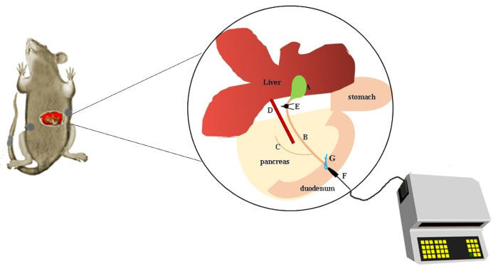
- Temporarily occlude the proximal common bile duct with a microvessel clip to prevent retrograde infusion from leaking into the liver. The common bile duct can be seen on the liver side of the duodenum and its junction with the duodenum will appear white. Expose the organ out of the abdominal cavity.
- Puncture the periampullary region (whitish part of the small intestine's wall) to access the common bile duct with a 0.4 mm needle connected to a 0.54 mm polyethylene tube.
- Make a temporary occlusion of the distal common bile duct with 8-0 suture to prevent the sodium taurocholate solution from leaking into the duodenum.
- Start the infusion pump and program a 2.5% sodium taurocholate solution (diluted in 0.9% saline) infusion at a constant speed of 10 µL/10 g body weight for 3 min.
- After the infusion, remove the microvessel clip, the temporary 8-0 suture, and the injection needle from the bile pancreatic duct to reconstitute the physiological flow of the bile.
- At the end, suture the abdomen with 6-0 nonabsorbent monofilament polypropylene suture. The time between the laparotomy and the end suture should be a maximum of 30 min (see Figure 1).
- After the surgery, house the animals in polyethylene boxes lined with wood shavings and water and food ad libitum.
- Treat control mice in the same manner as the experimental mice but ensure that the infusate consists of saline only. Perform the surgical procedure and the infusion of saline solution (10 mL/min, for 3 min) in a control group (SHAM) to eliminate the inflammatory bias caused by surgery and cannulation.
- Use tramadol 12.5 mg/kg subcutaneously every 8 hours, starting after post-surgical recovery.