The use of fluorophores for in vivo imaging can be greatly limited by opsonization, rapid clearance, low detection sensitivity and cytotoxic effects on the host. Encapsulation of fluorophores in liposomes by film hydration and extrusion leads to fluorescence quenching and protection which enables in vivo imaging with high detection sensitivity.
Optical imaging offers a wide range of diagnostic modalities and has attracted a lot of interest as a tool for biomedical imaging. Despite the enormous number of imaging techniques currently available and the progress in instrumentation, there is still a need for highly sensitive probes that are suitable for in vivo imaging. One typical problem of available preclinical fluorescent probes is their rapid clearance in vivo, which reduces their imaging sensitivity. To circumvent rapid clearance, increase number of dye molecules at the target site, and thereby reduce background autofluorescence, encapsulation of the near-infrared fluorescent dye, DY-676-COOH in liposomes and verification of its potential for in vivo imaging of inflammation was done. DY-676 is known for its ability to self-quench at high concentrations. We first determined the concentration suitable for self-quenching, and then encapsulated this quenching concentration into the aqueous interior of PEGylated liposomes. To substantiate the quenching and activation potential of the liposomes we use a harsh freezing method which leads to damage of liposomal membranes without affecting the encapsulated dye. The liposomes characterized by a high level of fluorescence quenching were termed Lip-Q. We show by experiments with different cell lines that uptake of Lip-Q is predominantly by phagocytosis which in turn enabled the characterization of its potential as a tool for in vivo imaging of inflammation in mice models. Furthermore, we use a zymosan-induced edema model in mice to substantiate the potential of Lip-Q in optical imaging of inflammation in vivo. Considering possible uptake due to inflammation-induced enhanced permeability and retention (EPR) effect, an always-on liposome formulation with low, non-quenched concentration of DY-676-COOH (termed Lip-dQ) and the free DY-676-COOH were compared with Lip-Q in animal trials.
Liposomes have been intensively investigated and serve as one of the most biocompatible biomedical drug delivery systems for clinical applications1,2. They are mainly composed of phospholipids and cholesterol, both of which are biocompatible compounds mimicking parts of natural cell membranes. Whereas hydrophilic substances can be entrapped in the aqueous interior, lipophilic agents can be incorporated within the liposomal phospholipid bilayer3. Encapsulation of substances within the aqueous interior of liposomes grants protection against degradation in vivo and also prevents the host system from toxic effects of cytotoxic drugs used for the therapy of diseases, for example chemotherapeutics aimed at destroying tumor cells. The modification of the liposomal surface with polymers like polyethylenglycol (PEGylation) further extends the liposomal blood circulation time in vivo due to sterical stabilization4. Moreover, liposomes can sequester high concentrations of several substances such as proteins5,6, hydrophilic substances7,8 and enzymes9. They therefore serve as reliable clinical therapeutic and diagnostic tools which merit their approval for delivery of cytotoxic drugs such as doxorubicin for cancer therapy4. Due to their flexibility, liposomes can also be loaded with fluorochromes for diagnostic and image-guided surgical purposes.
Fluorescence imaging provides a cost-effective and non-invasive in vivo diagnostic tool which however, demands some basic requirements. It could be demonstrated that fluorochromes which suit best for in vivo imaging have characteristic absorption and emission maxima in the range where light dispersion and scattering as well as tissue autofluorescence originating from water and hemoglobin is low. Thus, such probes have their abs/em maxima between 650 and 900 nm10. Besides this, the stability of fluorochromes both in vitro and in vivo is critical, as opsonization and rapid clearance can greatly limit their application for in vivo imaging11. Other effects such as poor stability and low sensitivity or cytotoxic effects on target organs as seen with indocyanine green (ICG)12-16, are unwanted and must be taken into consideration when designing probes for in vivo imaging. These observations have led to the active development of several preclinical NIR fluorochromes, nanoparticles as well as new techniques for the in vivo imaging of inflammatory processes, cancer and for image-guided surgery17-20. Despite the stability of most preclinical NIRF (near-infrared fluorescence) dyes in vitro, their rapid perfusion and clearance through the liver and kidney impede their use in the in vivo optical imaging of diseases and inflammatory processes.
We therefore present a protocol for the encapsulation of fluorochromes such as the well characterized near-infrared fluorescent dye DY-676-COOH, known for its tendency to self-quench at relatively high concentrations21 in liposomes. At high concentrations H-dimer formation and/or pi-stacking interactions between fluorophore molecules located within each other’s Förster radius result in Förster resonance energy transfer (FRET) between the fluorochrome molecules. At low concentration the space between the fluorophore molecules increases, thereby preventing pi-stacking interaction and H-dimer formation and resulting in high fluorescence emission. The switch between high and low concentration and the accompanying fluorescence quenching and activation is a promising strategy that can be exploited for optical imaging22. In this respect, encapsulation of high concentrations of the NIRF dye DY-676-COOH in the aqueous interior of liposomes is more favorable for in vivo imaging than the free dye. The challenge of the method lies first of all in the correct encapsulation and secondly, in the validation of the benefits resulting from encapsulating high concentrations of the dye. Comparing the imaging properties of quenched liposomes with that of the free dye and also with a non-quenched liposome formulation with low concentrations of the dye is indispensable. We show by a simple, but highly effective film hydration and extrusion protocol combined with alternate freeze and thaw cycles that encapsulation of quenching concentrations of DY-676-COOH in liposomes is feasible. Other methods used to prepare liposomes such as the reversed phase evaporation method23 as well as the ethanol injection method24 enable liposome preparation with high encapsulation efficiencies for many hydrophilic substances. However, the nature of the substance to be encapsulated can influence the encapsulation efficiency. In effect, the film hydration and extrusion protocol presented here revealed the highest efficiency for encapsulation of DY-676-COOH. To illustrate the benefits of liposomal encapsulation of DY-676-COOH, a zymosan-induced edema model, which permits the study of inflammatory processes within a few hours, was used. Here, it is demonstrated that liposomes with high concentrations of the encapsulated DY-676-COOH are more suitable for whole body in vivo optical imaging of inflammatory processes than the free dye or the non-quenched liposomal formulation with low dye concentrations. Thus the underlying protocol provides a simple and fast method to produce quenched fluorescent liposomes and the validation of their activation and imaging potential both in vitro and in vivo.
NOTE: All procedures are approved by the regional animal committee and in accordance with international guidelines on the ethical use of animals.
1. Preparation of Materials and Instruments
2. Validation of Fluorescence-quenching and Activation of Prepared Liposomes
3. Liposome-based In Vivo Fluorescence Imaging of Inflammation
The encapsulation of high concentrations of fluorescent dyes such as the NIRF dye DY676-COOH used here in the aqueous interior of liposomes leads to a high level of fluorescence quenching. Fluorescence quenching, a phenomenon seen with many fluorophores at high concentration, can be exploited in several in vivo imaging applications where a high sensitivity and reliable detection of the target area are demanded. The use of liposomes also provides protection of the dye which is indispensable for in vivo applications. A thorough characterization of the liposomes is necessary and includes several factors such as the level of dye encapsulated, stability and size of the liposomes, fluorescence quenching and activity of encapsulated dye in vitro and also applicability for in vivo imaging purposes. A comparison of the free dye, DY-676-COOH and quenched liposomes (Lip-Q) as well as a non-quenched liposome (Lip-dQ) with very low concentration of the encapsulated dye is therefore critical especially for in vivo characterizations.
Liposomes prepared by using the film hydration and extrusion technique with successive freeze and thaw cycles before extrusion contain residual free dye molecules which can be successfully separated from the liposomes due to their longer retention in the gel filtration matrix, compared to the liposomes which elute faster (Figure 1B). Depending on the level of dilution after gel filtration, an optional ultracentrifugation step enables the concentration of the liposomes as illustrated (Figure 1C). Based on the absorption and emission properties of the encapsulated dye, the concentration of encapsulated dye is determined with the help of a calibration curve of the free dye (Figure 1D). Besides the concentration of the encapsulated dye, it is important to determine the size and homogeneity of the liposomes after preparation. As seen in Figure 1E, electron micrographs of liposomes prepared by the underlying method reveal a mostly unilamellar morphology of the liposomal vesicles, containing intravesicular DY-676-COOH either within the quenching concentration range (Lip-Q; 606–846 µM DY-676) or at non-quenched dye concentration (Lip-dQ; 25 µM DY-676). Furthermore, they reveal a homogenous size distribution of about 120 nm and polydispersity indices far below 1 (Table 1). Owing to fluorescence quenching, Lip-Q shows two absorption maxima in aqueous buffer, whereby one peak is characterized by a shift towards the blue wavelengths. In line with this, the fluorescence emission is very low compared to the free dye (Figure 2A and B, right). Freeze-damage of the liposomes results in release of the dye, which gets diluted in the surrounding solution. The blue-shifted absorption peak therefore disappears, resulting in a single absorption peak of Lip-Q. Corresponding to this, an increase in fluorescence intensity of freeze-damaged Lip-Q is seen, which indicates that fluorescence activation of released dye molecules took place. The free dye reveals only a single absorption maximum and high fluorescence intensity which remain at the same level, irrespective of freezing (Figure 2A and B, left). This finding suggests that encapsulation of the dye in liposomes, as in Lip-Q would protect the dye from the environment, retain high concentration and the associated fluorescence quenching and if activated by target triggers would enable detection due to increase in fluorescence.
Liposomal probes with the underlying lipid composition reveal a predominant phagocytic uptake which is inhibited by energy depletion. This can be seen via the uptake of Lip-Q by the highly phagocytic murine macrophage cell line J774A.1, and the mildly phagocytic human glioblastoma cell line U-118MG at 37 °C and inhibition at 4 °C (Figure 3A and B). The free dye DY-676-COOH reveals uptake in the phagocytic cell lines both at 37 °C and at 4 °C which indicates that the liposomal probe Lip-Q contains no residual free dye in the solution and can only undergo active uptake. Confocal laser scanning microscopic images further substantiate the uptake and activation of Lip-Q in phagocytic cells (Figure 3C). Furthermore, the lack of fluorescence in the non-phagocytic human fibrosarcoma cell line, HT-1080 indicates that the uptake of Lip-Q is predominantly by phagocytosis and thus, would be suitable for imaging of inflammation where phagocytic monocytes/macrophages are involved.
Consistent with the phagocytic uptake of liposomes seen in cultured cell lines, and owing to fluorescence quenching intravenous injection of Lip-Q leads to a time-dependent increase in fluorescence intensity of edema in mice models (Figure 4A, Lip-Q), with very low background fluorescence. The maximum fluorescence intensity of edema is detected 8-10 hr post injection of Lip-Q. Contrarily, relatively strong NIR-fluorescence of the whole mouse is seen after application of the free DY-676-COOH (Figure 4A, DY-676-COOH) or the always-on liposome, Lip-dQ (Figure 4A, Lip-dQ). Compared to Lip-Q, rapid perfusion and clearance of the free DY-676-COOH as seen from 0-4 hr post injection, interferes with imaging, so that reliable detection of edema is not possible (Figure 4A, DY-676-COOH). Furthermore, the non-quenched liposome, Lip-dQ reveals a maximum fluorescence of edema within 2-4 hr post injection which remains almost constant till 8 hr, then gradually decreases similar to the quenched Lip-Q-based edema fluorescence. Performing semi-quantitative analyses, whereby regions of interest (ROIs) are set for edema versus background, one can make conclusions about the different levels of detection with different probes. According to semi-quantitative analysis of 5 animals per group (probe), edema can be more significantly (P = 0.001) detected with Lip-Q than with the free DY-676-COOH or the non-quenched, Lip-dQ (Figure 4B).
Imaging organs of mice euthanized 24 hr post injection of probes reveal mild ex vivo fluorescence of the liver/gall bladder and kidneys and a very low or no fluorescence of the spleen, lungs and heart (Figure 5), which serves as evidence for the elimination of the probes through the hepatobiliary route.
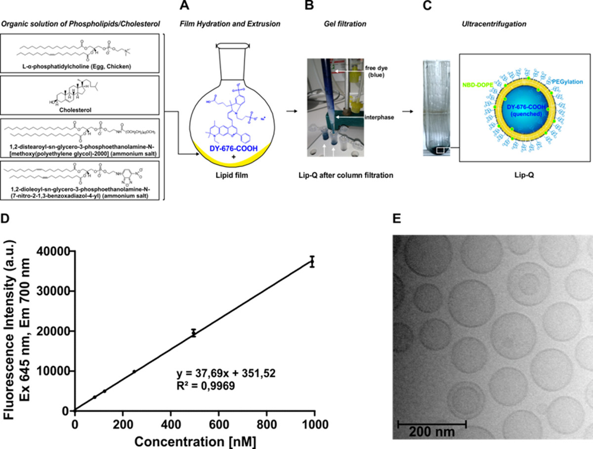
Figure 1: Preparation of DY-676-COOH-loaded liposomes. (A-C) Schematic overview of the synthesis steps involved. (A) Setup of the film hydration and extrusion with the near-infrared dye DY-676-COOH. (B) Picture of the self-made gel filtration setup showing the interphase between non-encapsulated (free) dye and liposomes. Liposomes elute first and appear blue-green due to the encapsulated DY-676-COOH (blue) and the incorporated green phospholipid, NBD-DOPE. (C) Representative image of liposome-sediment (Lip-Q) after concentration by ultracentrifugation. (D) Representative calibration curve of DY-676-COOH in 10 mM Tris pH 7.4 (containing 1% Triton X100) used for the quantification of liposomal dye. (E) Cryo-transmission electron micrograph of Lip-Q. Please click here to view a larger version of the figure.
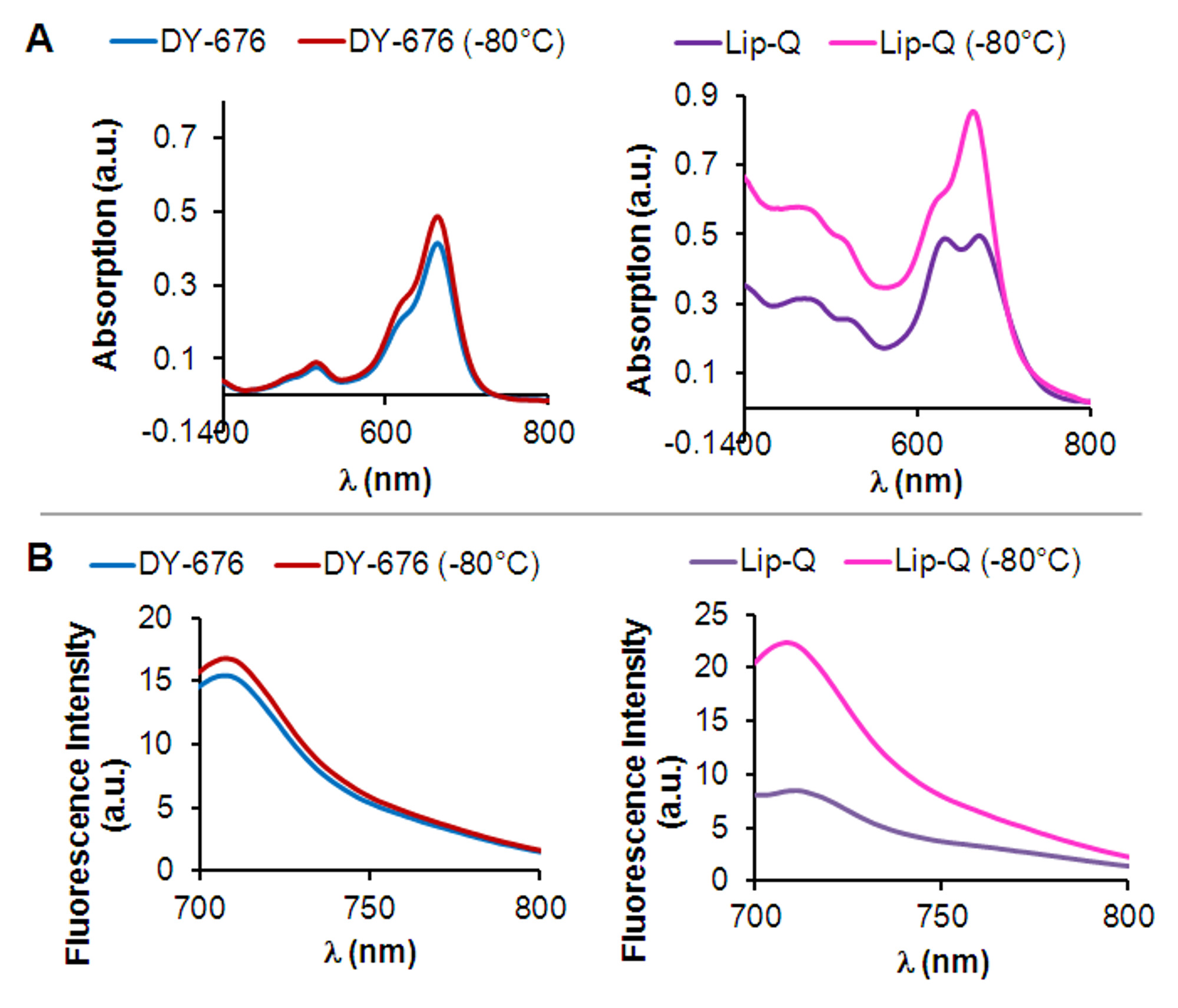
Figure 2: Physicochemical determination of fluorescence quenching and activation in vitro. (A) Absorption spectra of Lip-Q (right) and free DY-676-COOH (left) measured in 10 mM Tris buffer pH 7.4, before or after freezing at -80 °C. Note the characteristic double peak of Lip-Q compared to free DY-676-COOH and the disappearance of the blue-shifted peak after freeze-damage of Lip-Q. (B) Corresponding fluorescence emission spectra of Lip-Q (right) and free DY-676-COOH (left) measured in 10 mM Tris buffer pH 7.4 before or after freezing at -80 °C.
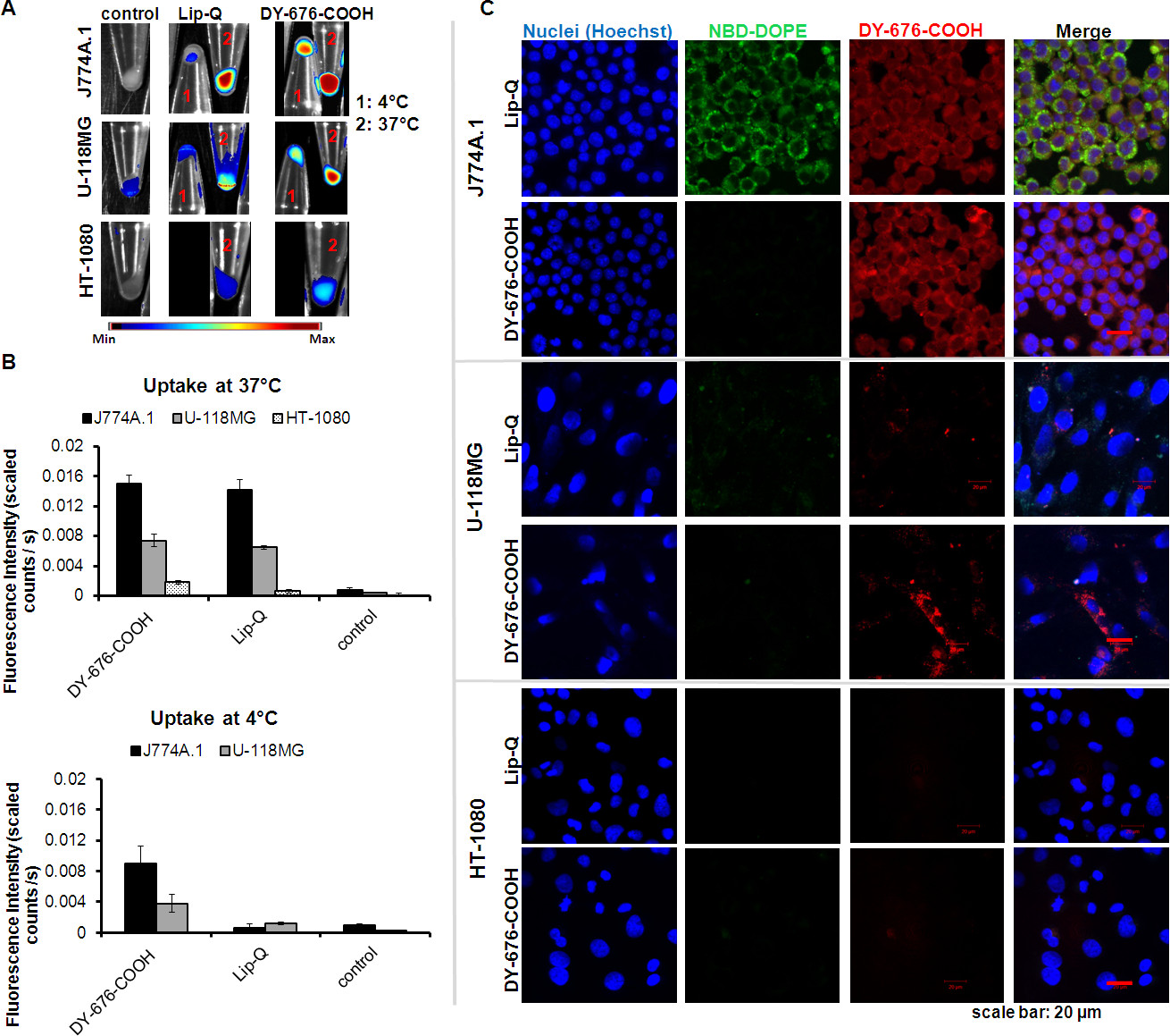
Figure 3: Cellular uptake and fluorescence activation of liposomes. The images in (A) were prepared by NIR fluorescence imaging of cell pellets after exposure to the corresponding probes for 24 hr at the indicated temperatures. The HT-1080 cells did not survive the 24 hr incubation periods at 4 °C. The bar diagrams in (B) represent the semi-quantitative levels of fluorescence signals got by assigning ROIs to the cell pellets in A. Each bar denotes the average intensities of n = 3 experiments ± SD. Images in (C) were acquired by confocal laser scanning microscopy of cells exposed to probes on culture chamber slides for 24 hr at 37 °C. Note the high level of fluorescence in the high phagocytic murine macrophage cell line J774A.1 and the moderate fluorescence in the mild phagocytic human glioblastoma cell line U-118MG. The non-phagocytic human fibrosarcoma cell line HT-1080 shows no fluorescence of the probes. NBD-DOPE: green fluorescent phospholipid. Please click here to view a larger version of the figure.
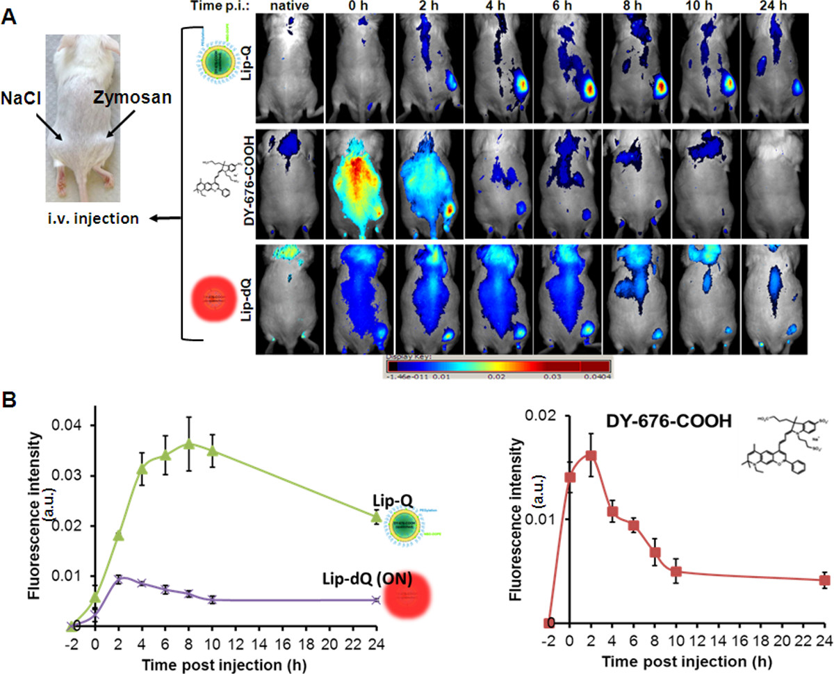
Figure 4. In vivo optical imaging of zymosan-induced edema in mice. (A) The mouse picture on the left shows the positions of subcutaneously applied zymosan-A (500 µg in 50 µl saline) and the control saline solution (50 µl) on the left flank. Intravenous injection of the indicated probes and whole body NIR fluorescence imaging at the indicated time points reveal gradual, but high increase in fluorescence signals of edema and low background signals of Lip-Q (upper panel) with a maximum fluorescence at 8 hr post injection. The free dye reveals perfusion and rapid clearance within 4 hr post injection (middle panel), whereas Lip-dQ reveals detection of edema with low signal intensities and an overall higher background fluorescence. The graphical presentation in (B) reiterate the observed fluorescence signals of edema detected with each probe compared to the control region (saline) in n=5 animals per group. Each plot represents the mean fluorescence signals of (n=5) ± SEM. With the graphs, the maximum fluorescence signal of edema detected with each probe is easily distinguished (Lip-Q, 8 hr; Lip-dQ 2–4 hr and free DY-676-COOH, 2 hr post injection). There is a significantly (P = 0.001) higher fluorescence intensity of edema with Lip-Q versus Lip-dQ at t = 0–24 hr, and with Lip-Q versus free DY-676-COOH at t = 4–24 hr. Please click here to view a larger version of the figure.
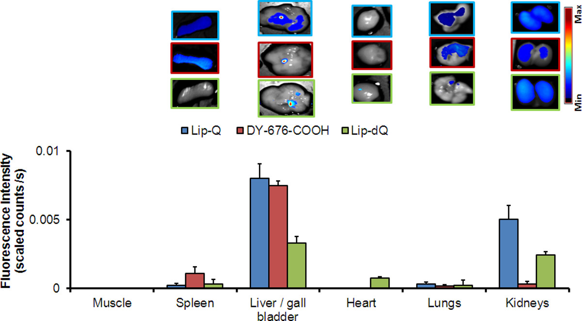
Figure 5: Bio-optical ex vivo images of organs from mice 24 hr post probe application, and corresponding semi-quantitative analysis of fluorescence intensities of the organs. Each bar represents the mean of fluorescence intensities (n = 4) ± SEM. Please click here to view a larger version of the figure.
Liposome formulation | Size [nm] | Polydispersity Index (PI) | Zeta Potential [mV] |
Lip-dQ (dequenched) | 123.4±0.6 | 0.055±0.02 | -10.6±0.4 |
Lip-Q (quenched) | 118.5±0.7 | 0.04±0.02 | -9±2 |
Lip-NBD (w/o DY-676-COOH) | 123.0±1.4 | 0.04±0.03 | -11±1 |
Table 1: Characterization of liposomes by dynamic light scattering.
Since liposomes can also serve as delivery systems for fluorescent dyes, they enable imaging of target diseases. The encapsulation of high concentrations of fluorescent dyes such as the NIRF dye, DY676-COOH used here, leads to a high level of fluorescence quenching of the entrapped dye. Fluorescence quenching, a phenomenon seen with many fluorophores at high concentration can be exploited in several in vivo imaging applications, where a high sensitivity and reliable detection of the target area is demanded. The use of liposomes also provides protection of the dye which is indispensable for in vivo applications.
The film hydration and extrusion technique is a widely used method which enables successful preparation of liposomes with different size ranges depending on need, and permits modifications such as encapsulation of a multitude of different substances25. Thus, it is suitable for the preparation of liposomes encapsulated with the NIR fluorescent dye, DY-676-COOH for imaging purposes. The method yields liposomes with 600–840 µM intravesicular dye concentrations, which are within the quenched concentration of the dye. The LiposoFast-Basic hand extruder used for the homogenization of spontaneously formed vesicle dispersion is suitable for liposome preparation in small lab scale, due to the compatibility of the device, with syringes up to 1 ml volume. For large scale preparations the use of larger high pressure homogenizers which are able to homogenize vesicle dispersions with a capacity of 1,000 L per hour is recommended. The gel filtration (size exclusion) chromatography is a crucial step which ensures separation of the encapsulated liposomal dye from non-encapsulated (free) dye molecules26. The length of the gel filtration column is vital for efficient separation of the liposomes from free dye. Thus, it is necessary to prepare a column of at least 28 cm length to successfully separate the liposomes from the free DY-676-COOH used here. Interestingly, this length is two times as long as that used to purify carboxyfluorescein (CF) loaded liposomes from free carboxyfluorescein. The major drawback of gel filtration is a five to six-fold dilution of the purified samples. This can be compensated by ultracentrifugation, if highly concentrated liposomes are needed. During ultracentrifugation the liposomes sediment and the supernatant can be removed easily27. Other ways to concentrate liposomes such as by dialysis28 are more time consuming than ultracentrifugation.
Besides the film hydration and extrusion method, the reversed phase evaporation method23 as well as the ethanol injection method24 enable liposome preparation with high encapsulation efficiency for many hydrophilic substances. However, our investigations revealed the film hydration combined with the freeze and thaw cycles to be the most suitable method for a sufficient intraliposomal encapsulation of DY-676-COOH. Increasing the starting DY-676-COOH concentration used for film hydration increases the efficacy and the concentration of the intraliposomal dye, when a fixed lipid concentration of 30 mM is used. Despite this the encapsulation efficacy with the freeze and thaw method is below 10% of the starting dye concentration used, but however is sufficient for encapsulation of the quenching concentration required for imaging. Furthermore, the free dye separated from liposomes by gel filtration can be recycled by desalting and dehydration according to manufacturer instructions, making re-encapsulation possible and an overall minimal dye loss. The encapsulation of the dye by the underlying protocol has no influence on the size and morphology of the liposomes as seen by the polydispersity indices and the electron micrograph of Lip-Q.
Several simple methods can be used to evaluate the activity of prepared fluorescent liposomes.DY-676-COOH has a high tendency to self-quench at high concentrations21,29 probably resulting from H-dimer formation and pi-stacking interactions between dye molecules. These interactions, which occur due to nearness of the Förster radii of dye molecules at high concentrations, can be annihilated by dilution30. Therefore, encapsulation of high concentrations of DY-676-COOH does not only protect the dye from opsonization in vivo, but also from the surrounding buffer, thereby maintaining its high concentration and retaining fluorescence quenching which, can be detected as a blue-shifted absorption peak and low fluorescence emission as seen in Figure 2. Freezing liposomes gradually at -80 °C leads to formation of ice crystals within the aqueous interior31 which causes damage to liposomal membrane when rashly thawed at 30 °C. The release, dilution and fluorescence activation of the intra-liposomal DY-676-COOH in buffer after freezing is revealed in a single absorption peak and almost 2.5-fold increase in fluorescence intensity (Figure 2), which indicates that Lip-Q thus sequesters a high, quenching concentration of DY-676-COOH and is activatable. Other methods to damage liposomal lipid bilayer such as the use of detergent or organic solvents do not reveal clear-cut differences between the free dye and the liposomal encapsulated dye, since they both influence the spectral properties of many fluorescent dyes32. The slow freezing and harsh thawing method reported here therefore serves as a more reliable and promising method to validate the high encapsulation of fluorophores in liposomes and a consequent fluorescence quenching. From the absorption and emission spectra of intact Lip-Q (Figure 2), some level of residual fluorescence can be detected. This can result from non-quenched dye monomers within the liposomes and also from electrostatic interaction and influence of encapsulated dye by phospholipid polar head groups33.
As can be seen in Figure 3, a distinct accumulation of Lip-Q in the highly phagocytic murine macrophage and mild phagocytic human glioblastoma cell line, U-118MG34, but not in the non-phagocytic human fibrosarcoma cell line HT-1080, indicates that Lip-Q- based imaging of inflammation would be favorable, since phagocytes are the key players of inflammatory processes. The fact that energy depletion abolishes uptake of Lip-Q but not uptake of the free DY-676-COOH substantiates the specificity of phagocytic uptake of Lip-Q and reveals that Lip-Q remains intact during the experiment, else release of the dye and energy-independent uptake would take place leading to fluorescence detection. Embedding the green fluorescent phospholipid, NBD-DOPE in the liposome bilayer enables microscopic imaging and discrimination between non-degraded from cellular degraded liposomes especially if time-dependent uptake experiments are done as reported earlier32. The microscopic images reveal NIR-fluorescence of DY-676-COOH which, correlates with the levels of fluorescence intensities of cell pellets, as determined by semi-quantitative analysis after incubation at 37 °C. In accordance with this, the murine macrophage cell lines show the highest fluorescence, whereas the human glioblastoma reveal lower fluorescence of both DY-676-COOH and NBD-DOPE than the former. As expected the human fibrosarcoma cell line, HT-1080 reveal no fluorescence of either liposomal dyes or the free DY-676-COOH, which strengthens the fact that Lip-Q uptake, is predominantly by phagocytosis.
To investigate the potential of Lip-Q-based phagocytosis-driven in vivo imaging of inflammation, several controls were considered. Considering that the free DY-676-COOH can be taken up by phagocytosis, opsonization of Lip-Q may lead to release of the dye, which may be taken up by phagocytes. To avoid this, Lip-Q was prepared with 5 mol% PEGylation. Furthermore, it is necessary to distinguish between uptake due to inflammation-based EPR effect and active uptake and fluorescence activation. To address this, the evaluation and comparison of three different probes, namely the always-on, Lip-dQ, the quenched Lip-Q and the free DY-676-COOH in mice bearing zymosan-induced edema was necessary. Zymosan-A which is prepared from the cell walls of Saccharomyces cerevisiae and Candida albicans is a natural stimulant of cytokine secretion via the dectin-1 and the toll-like receptor 2/6 (TLR 2/6). Secreted cytokines in turn induce the activation of downstream cascades which result in vascular leakages, thereby easing the intravasation/extravasation of monocytes/macrophages from a splenic reservoir35 as well as neutrophils, and their migration to inflammation sites (edema)36-39. The zymosan-based monocytes/macrophage extravasation and migration process needs only 4.5–6 hr which makes zymosan a strategic tool for studying inflammatory processes36-39. Due to the vascular leakages that result during inflammation, intravenously injected probes can either be taken up by monocytes/macrophages (phagocytes) during their migration to the inflammation site or extravasate and be taken up at the edema site (EPR effect)40. The use of the non-quenched Lip-dQ reveals an overall stronger background to edema signals than Lip-Q, and a fluorescence increase of edema which reflects the migration of monocytes and macrophages. In effect, the maximum fluorescence of edema is seen already 2-4 hr post application of Lip-dQ and remains almost constant till 8 hr. Opposed to Lip-dQ and Lip-Q, the free DY-676-COOH undergoes rapid perfusion after injection and is cleared within 4 hr, so that distinct imaging of edema is not possible. Interestingly, the use of Lip-Q results in persistent increase in fluorescence intensity of edema and very low background signals. This persistent increase in fluorescence intensities with Lip-Q is attributed to fluorescence activation of the liposomal released dye. Taken together, it can be concluded that the contribution of EPR-effect in Lip-Q based imaging is minimal since Lip-dQ reveal a maximum fluorescence (EPR effect and monocyte migration) at 4 hr post injection. Thus, liposomal encapsulation provides protection and distinct delivery of DY-676-COOH, which in turn enables a more reliable in vivo imaging of edema, exclusively after internalization and degradation (fluorescence activation) by phagocytic cells. So far, the use of zymosan-induced hind leg edema to validate the imaging properties of fluorescent liposomes is new. The protocol reported herein can be expanded both by encapsulation of different fluorescent dyes and by imaging the effect of different inhibitory drugs on the induction of inflammation, and therefore represents a useful tool for preparation and characterization of probes suitable for biomedical imaging.
Another crucial step towards defining suitable imaging probes is the verification of their pharmacological properties and elimination routes. The distribution of probes in organs which play vital roles in excretion, such as the liver and kidneys as well as their short retention and suitable elimination from these organs is usually an indication, that the probes will much likely show no adverse effects on the patient. In accordance with this, the organs of mice prepared 24 hr post injection of Lip-Q or the free DY-676-COOH reveal only mild fluorescence of the liver/gall bladder and the kidneys, which signifies a preferred elimination of the liposomal fluorophore through the hepatobiliary route. The short accommodation of the probes in these organs and their efficient elimination is substantiated by a 7-fold higher fluorescence signals of organs prepared 6 hr post injection of Lip-Q or the free DY-676-COOH32. These observations are in accordance with the elimination of liposomes41 and backs up the importance of including biodistribution studies when characterizing probes. Although adverse side effects, such as skin irritations and complement activation42,43 have been reported for liposomal formulations used in clinical applications, such effects were not detected with the underlying liposomes. Furthermore, observation of immune deficient mice for two weeks after probe injection led to complete clearance of the probes from the mice organs (not shown).
The authors have nothing to disclose.
This work was supported by the Deutsche Forschungsgemeinschaft grants HI-698/10-1 and RU-1652/1-1. We thank Doreen May for excellent technical assistance and the company DYOMICS GmbH, Jena for their kind support.
Name of Material/ Equipment | Company | Catalog Number | Comments/Description |
Materials and equipments for preparation of liposomes | |||
egg phospahtidylcholine | Avanti Polar Lipids | 840051P | Dissolve in Chloroform and store in glass vials (214 mg/ml) |
cholesterol | Sigma | C8667 | Dissolve in Chloroform and store in glass vials (134 mg/ml) |
1,2-distearoyl-sn-glycero-3-phosphoethanolamine-N-[methoxy(polyethylene glycol)-2000] (ammonium salt) | Avanti Polar Lipids | 880120P | Dissolve in Chloroform and store in glass vials (122 mg/ml) |
1,2-dioleoyl-snglycero-3-phosphoethanolamine-N-(7-nitro-2-1,3-benzoxadiazol-4-yl) (ammonium salt) | Avanti Polar Lipids | 810145P | Dissolve in Chloroform and store in glass vials (2mg/ml) |
Sartorius MC1 (d = 0.01 mg) | Sartorius AG | Research RC 210 P | used for weighing the phospholipids |
Rotavapor | Büchi Labortechnik AG | R-114 | used for hydration of phospholipid film |
Waterbath | Büchi Labortechnik AG | R-481 | used for hydration of phospholipid film |
Vacuum Controller | Büchi Labortechnik AG | B-720 | used for hydration of phospholipid film |
Vacobox | Büchi Labortechnik AG | B-177 | used for hydration of phospholipid film |
Circulation Chiller | LAUDA DR. R. WOBSER GMBH & CO. KG |
WKL 230 | used for hydration of phospholipid film |
DY-676-COOH | Dyomics GmbH | 676-00 | Dissolve in 10 mM Tris and store stock at -20°C |
Tris-(Hydroxymethyl)-aminomethan | Applichem | A1086 | buffer 10 mM, pH 7.4 |
Trichlormethan | Carl Roth GmbH + Co. KG | Y015.2 | used for liposome preparation |
Sonicator | Merck Eurolab GmbH | USR 170 H | used for liposome preparation |
Vortex Genie 2 (Pop-off Cup, No. 146-3011-00) | Scientific Industries Inc. | SI-0256 | used for liposome preparation |
Sephadex G25 medium | GE Healthcare Europe GmbH | 17-0033-01 | used for liposome purification |
Triton X100 | Ferak Berlin GmbH | 505002 | used to destruct liposomes for dye quantification |
LiposoFast-Basic | Avestin Inc. | used for the extrusion of liposomes | |
Polycarbonate filter membrane, 100 nm (Whatman Nucleopore Trans Etch Membrane, NUCLEPR PC 19 MM, 0.1 U) | VWR | used for the extrusion of liposomes via LiposoFast-Basic | |
Fluostar Optima | BMG Labtech | used for dye quantification | |
Zetasizer Nano ZS | Malvern | used for the determination of liposome size and zetapotential | |
Ultracentrifuge | Beckmann Coulter GmbH | XL 80 | used for concentration of the samples |
Rotor | Beckmann Coulter GmbH | SW 55 TI | used for concentration of the samples |
Materials and equipments for the evaluation of liposome and optical imaging | |||
Zymosan-A from Saccharomyces cereviciae | Sigma | Z4250-250MG | used for induction of inflammation |
Isotonic Saline (0.9) | Fresenius GmbH | PZN-2159621 | used for the dilution of Zymosan-A |
Isoflurane vaporizer | Ohmeda Isotec 4 | used for anesthesizing animals | |
Isoflurane | Actavis GmbH | PZN-7253744 | anesthesia |
Thermo Mat Pro 20 W | Lucky Reptile | 61202-HTP-20 | used to keep animals warm during anesthesia |
Omnican-F (1 ml injection) | Braun | PZN-3115465 | used for subcutaneous and intravenous application of probes |
Panthenol eye cream | Jenapharm | PZN-3524531 | used to prevent dryness of the eyes of animals during anesthesia |
Hanks buffered saline solution | PAA Laboratories /Biochrom AG | L2045 | w/o Mg2+, Ca2+ and phenol red. For dilution of probes and for washing of cells |
8-Well chamber slides | BD Biosciences | 354108 | used for cell culture followed by microscopy |
Cell culture flasks | Greiner BioOne | ||
Cell culture media | Gibco (life technologies GmbH) | ||
Fetal calf serum | Invitrogen | ||
Poly-L-Lysine solution (0,01% – 50 ml) | Sigma | P4832 | used to coat cell culture chamber slides |
Mountant Permafluor | ThermoScientific | S21022-3 | Mounting solution for microscopy |
Hoechst-33258 | AppliChem | DNA stain for microscopy | |
Hera-Safe | Heraeus Instruments | sterile work bench used for cell culture | |
HERA cell | Heraeus Instruments | Incubator used for cell culture | |
LSM510-Meta | Zeiss | used for confocal microscopy | |
Maestro-TM in vivo fluorescence imaging system | CRi, Woburn | used for whole body fluorescence imaging of small animals | |
Spectrophotometer (Ultrospec 4300 pro UV) | GE Healthcare | used for measurement of absorption | |
Spectrofluorometer (Jasco FP-6200) | Jasco | used for measurement of fluorescence emission | |
Animals | |||
NMRI mice (8-12 weeks old, male) | Elevage Janvier, France | used for inflammation trials |