Summary

The Sol Braiding Method for Handling Thick Hair During Transcranial Magnetic Stimulation: An Address for Potential Bias in Brain Stimulation

Published: August 09, 2024
doi:

Summary

Hair type commonly seen in historically underrepresented minorities appears to interfere with transcranial magnetic stimulation (TMS). Here we describe a hair braiding method (The Sol Braiding Technique) that improves TMS.

Abstract

Transcranial Magnetic Stimulation (TMS) is a technique that is frequently utilized in neuroscience for both therapeutic and research purposes. TMS offers critical medical services like treating major depression and is vital in almost every research facility. Because TMS relies on scalp placement, hair is thought to affect efficacy because it varies the distance to the target site. Further, it is presumed that the hair textures and length that are predominantly seen in minoritized persons might pose significant challenges to collecting high-quality data. Here, we present preliminary data demonstrating that TMS may be influenced by hair, particularly in historically underrepresented minoritized groups.

The Sol braiding approach is introduced here as an easy-to-learn, quick-to-implement technique that reduces variability in TMS. Compared across nine participants, it was found that the Sol method significantly increased motor evoked potential (MEP) strength and consistency (p < 0.05). By removing the physical hair barrier that impedes direct coil-to-scalp contact, the Sol approach enhances TMS delivery. The MEP peak amplitude and the MEP area under the curve (AUC) were shown to increase as a result. While preliminary, these data are an important step in addressing diversity in neuroscience. These procedures are explained for non-braiding experts.

Introduction

Neuroscience research, by its very nature, involves paradigm shifts and innovations for understanding brain function, neurological disabilities, and psychiatric disorders1. Despite much progress, the discipline of neuroscience has fallen short in some aspects. For example, there exist racial disparities, both in the number of researchers, but also in the representation of subjects and patients in research. Numerous underrepresented persons from minority groups are absent from experiments and clinical studies2. Only 5 publications out of 81 peer-reviewed scalp-based EEG articles from September to October 2019 specifically indicated having a sample that included minoritized individuals. Furthermore, recent studies demonstrated that individuals from underrepresented minority groups were often misdiagnosed or failed to trust the researchers. Assari et al. found that the healthcare community, specifically half of White medical students and residents, believed that African Americans have thicker skin than Whites, which influenced their medical judgment and treatment strategies3,4. Owing to the absence of data from minority participants, research findings are less generalizable and show disparities for minority populations. To ensure that the trial population is representative of the patients who will use the medication or medicinal product and that the results are generalizable, clinical trials must include a diverse group of participants5.

Of interest to scalp-based neuroscience is the distinct shape, thickness, styling, and density that is often seen in underrepresented minority hair. The follicle shape, for example, is one feature that makes African hair distinctive. African hair comes from smaller, more elliptical, and flat follicles while Caucasian and Asian hair follicles are more circular and large6. When minorities wash their hair, it curls, causing difficulties for researchers in their experiments. Minority groups are sometimes advised to wash and straighten their hair using hair products before coming in for scalp-based imaging, but doing so can have an impact on the accuracy of the data. Data are skewed because fewer participants of minority groups would volunteer and data from them might be discarded for being a lower quality. Moreover, due to their typical hairstyles (such as cornrows and braids), minoritized individuals are sometimes perceived as being difficult to recruit and retain2. Rosen et al. studied a man of African descent who wore dreadlocks, a style worn by underrepresented minoritized individuals, and presented with disfluency in spontaneous speech7. He wanted to receive treatment using scalp-based imaging since it had emerging evidence for efficacy and was tolerable.

One of the scalp-based imaging techniques that is used widely is transcranial magnetic stimulation (TMS). TMS is a surface-based imaging technique that is used in a non-invasive way to induce localized increases in brain activity. The ability to control neuronal activity in the human brain makes TMS a crucial tool for both experimental and therapeutic neuroscience8. To set standard safety recommendations, when represented as a percentage of the motor threshold (MT), TMS intensity provides a generalizable indicator of applied stimulation that may be used with any coil shape or kind of stimulator9. The motor evoked potential (MEPs) used in determining MT can also be a measure of cortico-excitability, which is elicited by TMS over the human motor cortex10,11,12,1,3,14,15,16. TMS is delivered to the motor cortex, which causes activation in the contra-lateral regions. Typically, regions of the hand are targeted as the stimulating target is not difficult to find on the motor cortex, and attaching electrodes or visually monitoring hand/digit responses is simple. The mechanisms governing motor output can be more fully comprehended using MEPs. Since MEPs are used to measure individual differences in MT, they are now a part of practically every TMS application. In general, it is dangerous to utilize TMS without gauging some aspect of MT. If TMS is delivered above the appropriate MT, seizures may result. If TMS is delivered below MT, results may be reduced or absent (i.e., targeted neurons may not be depolarized). Accurate MT reporting is also critical in comparing studies. For example, many of the studies in our lab use a 90% value, which tells other researchers that a 110% application may result in a greater effect.

Stokes et al. examined different distances between the target region and the stimulating coil and subsequently found a direct linear relationship between the distance and the individuals' MT8,17. Therefore, minority groups, some with thicker natural hair might have less accurate MTs/MEP measurements. In a survey targeting the TMS community of published authors,we found when we asked open-ended questions such as "does hair play a role in impedance?" experts in the field replied: "It increases thresholds. Moving hair aside, compressing it, etc.;" We try to use gel to bridge that contact, but not a lot that can be done;" " Thick hair also makes contact difficult; same as above"; " More hair makes stimulation more difficult-especially if it prevents good scalp contact with the coil18. Dense hair growth makes it challenging to achieve contact between the TMS coil and the scalp, leaving minimal to no contact and impeding the signal. Previous research has shown that braiding thick, coarse hair reduces impedances in scalp-based imaging6. Using the characteristics of coarse or curly hair, Etienne et al. found that braiding a participant's hair into cornrows maintains signal integrity when using EEG.

We are introducing the Sol "Sun" method to offer a solution to manage the hair in underrepresented minorities. Due to the thickness and coarseness of their hair, we predicted that hair that is typically seen in underrepresented minorities will respond better to this procedure since it will preserve the hair (i.e., no shaving) and allow for long-term measurement. These methods are easy to teach, learn, and perform; require no additional equipment; do not increase safety risks; honor and respect the participants' natural hair; and promote pride in the participant (and researchers) who may have felt previously discouraged by scalp-based techniques.

Protocol

The research presented here was approved by the Institutional Review Board (IRB) committee of Montclair State University initiated in 2001 and updated yearly through 2023. All participants were treated within the ethical guidelines of the American Psychological Association. Typical safety procedures were followed. For example, we recruited nine adults from the general Montclair State University population using flyers and word of mouth. All subjects were screened in person employing TMS guidelines set forth by Wasserman19. Participants were compensated $25 for their enrollment in the study, and all subjects were treated within the standards put forth by the local Institutional Review Board (IRB) and in accordance with the Helsinki Declaration. Written informed consent was obtained from all participants and all subjects identified as being either Hispanic or African-American.

1. Background and 10/20 transfer

NOTE: For TMS, no additional equipment of any note will be needed (i.e., labs should easily have all these supplies).

  1. Participants
    1. Before starting any procedures, be sure to obtain an IRB Committee Review. If this is an initial protocol, consult experienced researchers and prepare all forms such as Consent, Screening (Figure 1), and Side Effects (Figure 2) and practice their presentation.
      NOTE: Forms should supplement and not substitute direct interviews with participants regarding safety.
    2. Ensure that all team members are trained in their roles.
      NOTE: Training courses are available and almost all TMS laboratories freely share their best safety practices.
    3. Recruit participants through social media and flyers.
      NOTE: Recruitment provides a monetary incentive and a brief description of risks.
    4. Administer the Screening Checklist before enrollment (Figure 1).
    5. Pay all participants for their participation ($25 in this study) and treat them in accordance with the guidelines of the Internal Review Board and the American Psychological Association.
  2. Perform TMS using the parameters appropriate for the institution (see the discussion).
  3. Participant safety and comfort are critical so ask and monitor the participants both verbally and visually throughout the procedure. Be sure to look for nervousness, which can be the norm and, in some cases, leads to more difficult outcomes.

2.TMS equipment handling

  1. Ensure that the 70 mm figure-of-eight coil used for this experiment never reaches dangerous/shut-off temperatures. Keep backup coils ready in case they are needed as replacements.
  2. Use visual inspection (5/10 evoked Abductor Pollicis Brevis) or electromyography (EMG) to determine the motor threshold.

3. Motor threshold on non-braided hair

  1. Mark region CZ nasion/inion line and the midpoint taken using an erasable magic marker. Measure earlobe or pre-auricular points and mark those points as well.
  2. Using the right hemisphere line, go one-third down (from dorsal to ventral) and find the optimal location for the Abductor Pollicis Brevis (APB) activation. Trigger the TMS device via the coil trigger and the footswitch and disengage the safety from the front of the machine.
  3. Orient the TMS coil at 45° for all searches and TMS deliveries.
  4. Use the dial on the front of the machine to start the stimulation output at 30% total machine output and increase in 2% increments until a movement is noted. Be sure to move the location with increasing stimulation intensity due to the careful interplay between coil movement and stimulation intensity.
  5. Once the site providing the maximum APB response is identified, determine the MT.
  6. To facilitate accurate placement before starting MT determination, mark the coil tip site on the swim cap and trace the entire anterior portion of the coil using a magic marker.
  7. For the visual inspection method, use the dial to start at 20% of the machine's intensity ad increase to deliver approximately 20 pulses to identify the stimulation level that results in 5/10 (50%) APB responses. Move the dial based on increased or decreased finger movements. Once 5/10 responses have been obtained, record the intensity displayed in the machine as the individual's MT.
  8. For the (preferred) MEP method, place disposable electrodes on the APB and the tendon of the thumb and a ground (around the back of the wrist), and instead of using visual inspection, confirm that a positive MEP is observed on the recording unit.
    1. Define a positive MEP response as an MEP with ≥50 µV peak-to-peak amplitude.
    2. Similar to visual inspection, stimulate until 5/10 positive MEPs are observed. Ensure that the MEPs are greater than 50 µV. Record the MT when 50% of the MEPs are above (and 50% below) 50 µV.

4. Sol

  1. Practice with different brushes, combs, and paints, as researchers feel differently about different implements.
    NOTE: The length of time to perfect the technique varies based on experience. Although it is not essential to document participant hair20, we include them here (Figure 3).
  2. Be consistent in terms of criteria to braid. There are no universal standards regarding which individuals should be braided prior to TMS. However, be sure to state the intention of using a determinant such as hair texture prior to any study or clinical application.
  3. Describe the methods to the participant and use photographs during consenting. Because valuing the participant should be a priority, use images and describe the braiding as part of the technique.
    NOTE: One potential inducement (i.e., compensation) is the inclusion of braiding the entire head following the experiment.
  4. Know the target points before braiding begins.
  5. Transfer 10/20 coordinates (motor-related target points) to the scalp to provide target braiding points. To achieve this with a standard ERP/EEG cap, load a typical gel syringe and blunt needle with 1-5 mm of Green Florescent Paint (GFP). Apply the cap and, at the target points, insert the syringe as if applying gel and dab ~0.05 mm of GFP (Figure 4A – E).
    NOTE: The amount of GFP will vary depending on the number of electrodes that need marking.
  6. Let the points dry for at least 2 min.
  7. Starting from the marked point on the head, pull the hair away. Ensure a clear patch of skin is seen around the mark. Try to clear the area of hair with fingers or a pick.
  8. Before starting, visualize first. To make 6-8 cornrows that will end up looking like a cartoon sun with the target point in the middle, make each braid, thinking of a clock face: cornrow 6 o'clock first, then cornrow 12 o'clock, and then cornrow 8 and 10 o'clock. End with cornrowing 2 and 4 o'clock. This will result in six cornrows.
  9. If eight cornrows are needed, start with 6 o'clock and 12 o'clock; then, cornrow 9 o'clock, between 7 and 8 o'clock, and between 10 and 11 o'clock. Finalize with all missing rows.
    NOTE: The main difference between cornrowing and braiding is that you will keep adding hair to your initial 'clump'. You will 'adhere' the braid to the scalp, thus reducing the height and awkwardness of the brain. Visualize this before starting cornrowing.
  10. To initiate the first conrow, from any side of the marked point, part a small section. Try to estimate an amount of hair that would be 1/8 or 1/6 of the hair needed to cornrow. This should be a small amount that is easy to manipulate-the initial clump.
  11. Use both hands to divide this initial clump into three sections. Make the sections equal in terms of the amount of hair.
  12. With the left section in the left hand, and the right section in the right hand, hold down the middle section with either, one, or both index fingers.
  13. If done correctly, the right section is now in the middle and what was the middle section is now on the right.
  14. Take the leftmost section and lift it. Put it in between the middle and right sections. If you did it correctly, this section is in the middle and the section that was in the middle is now on the left.
  15. Now take the right section and repeat step 2.6 while adding some new, additional hair from the scalp that is under the piece.
    NOTE: Adding the hair will feel like a sweeping motion. This is what will make cornrows rather than braids.
  16. Continue grabbing one piece at a time while adding in hair going down the section.
  17. Continue cornrowing until the end of that clump of hair.
  18. Do this 6-8x to create a 'sun' shape.

5. Motor threshold on braided hair

  1. Apply TMS now. Ensure that the coil surface at the maximal stimulation point lies flush now to the skin.
  2. Using a coil holder, mounting system, or by hand, press firmly against the skin with the plastic of the coil as per TMS norms.
    NOTE: These methods are appropriate for a 70 mm figure-8 coil.
  3. Measure MEPs using electrodes and visual inspection. Targeted fingers should visibly move.
  4. Cornrow any additional target areas (e.g., Left Dorsal Lateral Prefrontal Cortex) in a similar manner.
    NOTE: You can have multiple sites prepared simultaneously by using more than one trained braider.

Representative Results

A TMS single-pulse device with a 70 mm figure-8 coil was employed for all stimulation sessions. MEPs were acquired using standard amplifiers and software installed on a local computer. All MEPs were obtained by attaching three electrodes targeting the Abductor Pollicis Brevis muscle (APB). The main hypothesis tested was that the Sol method would produce larger amplitudes and AUC compared to unbraided hair. To do this, we used ANOVAs using separate 2 x 9 (Pre/Post x Subject) tests. We predicted larger amplitudes and AUC following braiding compared to no braiding. We also examined variability across the 30 trials in each participant and predicted that the Sol method would decrease variability.

We first ran a 2 x 9 Repeated-Measures ANOVA using Pre/Post and Subject as factors over the 30 TMS pulses sampling at 1,000 Hz for 100 ms post TMS trigger. It is noted that the sample is small and caution must be taken in interpreting these results. It was found using a Kolgoromov-Smirnov test that the data were normally distributed (all p's > .05). For Peak Amplitude, it was found that there was no overall interaction (F(8,232) = 1.82, p = 0.08). We then examined the Main Effect of Pre/Post and found that there was a significant difference (F(1,29) = 8.70, p = 0.006) with the Post Amplitude being 116.99% higher than the Pre. While Subject was significant (F(8,232) = 2.41, p = 0.016), we were interested in the number of subjects in which braiding made a difference. It was found that Amplitude increased in 7/9 Subjects, and in 3, the Pre/Post difference was significant (t(8), p's per Subject= 0.01, 0.01, 0.02, 0.12, 0.55, 0.60, 0.71, 0.76, 0.81; Figure 5).

We ran a similar 2 x 9 ANOVA for AUC (Figure 5). There was no interaction (F(8,232) = 1.30, p = 0.24). There was a significant Pre/Post difference (F(1,29) = 7.39, p = 0.01). The Post AUC was 108.12% greater than Pre AUC. There was also a Subject significant difference (F(8,232) = 2.47, p = 0.01). In 7/9, there was an increase in AUC, and in 2, the Pre/Post difference was significant (t(8), p's per Subject = 0.01, 0.04, 0.10, 0.11, 0.48, 0.71, 0.86, 0.87, 0.96).

Analyzing the variability in Amplitude, it was found that there was non-significant interaction (F(8,232) = 1.41, p = 0.19; Figure 5). There was a trend for less variability looking at Pre/Post scores (F(1,29) = 2.81 p = 0.10). Following braiding, variability was reduced 10.36%. There was no significant effect for Subject (F(8,232) = 1.26, p =. 27). AUC was analyzed for variability and there was no significant interaction (F(8,232) = 1.28,p = 0.25). Pre/Post (F(1,29) = 0.98, p = 0.33) and Subjects (F(8,232) = 1.06, p = 0.39) were non-significant as well. These data indicate a number of important significant findings and trends. The increased amplitude and AUC demonstrate that Motor Threshold (i.e., the level of TMS needed to induce an MEP) would likely be reduced with appropriate braiding. Cornrowing also increased reliability as variability was decreased (though not significantly). Finally, signal strength is increased, which may make clinical applications more efficacious, though this claim is highly speculative.

Figure 1
Figure 1: Screening. Typical screening used to ensure patient safety. Please click here to view a larger version of this figure.

Figure 2
Figure 2: Side Effects. Monitor potential problems before and after TMS. Please click here to view a larger version of this figure.

Figure 3
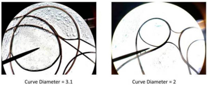
Figure 3: Comparison of differences in curves. The left sample represents a wider curve-longer diameter. This results in less overall hair density. On the right, the 'tighter; curve tends to result in an overall thicker head of hair. Please click here to view a larger version of this figure.

Figure 4
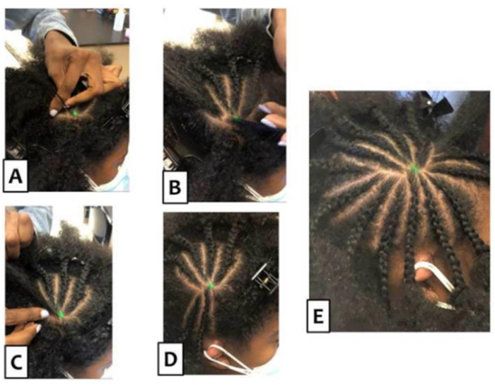
Figure 4: The Sol method. (A) Manageable strands are pulled away from the target point. (BD) Progressive cornrowing is presented so that a clear pattern emerges. To achieve better motor evoked potentials and (E) a vacated target, it is vital to braid so that the hair is flat on the scalp (this is a cornrow). Starting from the mark point on any side of the hair, separate a clump of hair. Using that section, part the hair 'clump' three more times vertically and begin to cornrow. To cornrow, place the left hair section in the left hand and the right section in the right hand. Let the middle section lay flat on the scalp but using the index finger(s) of one or both hands, press down on the middle section. grab the piece of hair on the right section and go over the middle section, and place it in between the middle and left sections. Then, do the same with the left section and go over the middle section. Continue grabbing sections of the hair one at a time, going over the middle section. While doing so, also add pieces of hair next to the parted section going down the scalp. Continue to do so until the section of the hair is done, ensuring the hair will be adhered to the scalp. Continue the same method of parting the section of hair and cornrowing, going around the marked point of hair until a "Sun" is created. Please click here to view a larger version of this figure.

Figure 5
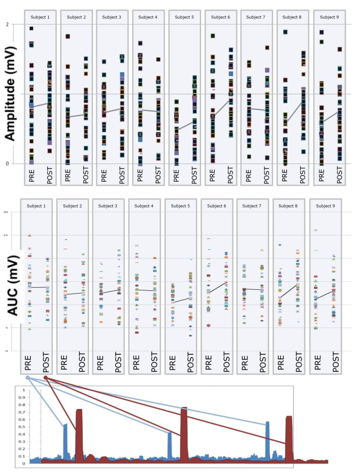
Figure 5: Amplitude, area under the curve, and three sample pre/post motor evoked potentials. The data for all nine participants are presented before and after braiding with the Sol method. Presented are the 30 TMS pulses delivered to M1. The line connecting Pre/Post is the mean of each sample. Abbreviation: TMS = transcranial magnetic simulation. Please click here to view a larger version of this figure.

Figure 6
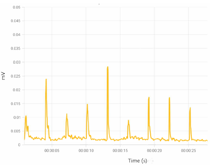
Figure 6: Sample MEPs. Following braiding, MEPs should be consistent and robust. Abbreviation: MEPs = motor evoked potentials. Please click here to view a larger version of this figure.

Discussion

Cornrows should not interfere with the angle (e.g., 45°) of the TMS coil. If they do, one of the cornrows may need to be redone to alleviate this issue. If done correctly, MEPs should be consistent (Figure 6).

By utilizing the characteristics of curly or coarse hair, this method of braiding maintains the integrity of the TMS signal. In this study, we were able to significantly increase the MEP size and found an increased trend for consistency. These data have implications and we believe that it will assist in future braiding studies. As with most novel techniques, we believe and encourage advances from others in the field.

It is possible that fewer underrepresented minoritized individuals participate in clinical research because of their hair, which can affect the neuromodulation signal. Further, those receiving TMS with thicker hair may be getting an inappropriate level of TMS. Not only does this invoke safety concerns, but efficacy issues are raised as well.

Though data were not collected, we note that we have found that participants appreciate the respect that they are shown and comments included, "Thank you for 'honoring the hair' ". Thus, ancillary benefits include an improved research or clinical environment. Practice will reduce times to about 3-6 min per region. Thus, in a typical clinical Major Depressive Disorder session where two areas are isolated (the target region and the motor threshold region-M1), 6-12 min would be added to the treatment.

The current study further identified a potential benefit for persons from racial and ethnic minority groups, and we believe it should serve as a motivation to pursue further evidence-based approaches to diversify the field. These future studies would hopefully include re-examining other technologies so that we may increase diversity and improve the rigor of our scientific research.

Future studies should examine different types of TMS (e.g., rTMS, paired pulse), EEG, or any brain stimulation technique and how they can be employed now in underrepresented populations. Future studies should include control populations and employ neuronavigation and larger samples. By creating a braiding method that exposes the scalp, we hope we have improved these brain stimulation techniques and technologies. We additionally hope to prompt researchers to look at how these same techniques failed underrepresented minorities21. While we acknowledge our work is preliminary, we believe the data are important for rapid dissemination.

Divulgaciones

The authors have nothing to disclose.

Acknowledgements

LSAMP (Louis Stokes Alliance for Minority Participation), Wehner, and The Crawford Foundation, the Kessler Foundation are all thanked for their support.

Materials

Android Samsung Tablet (for MEPs)
Cloth Measuring Tape
COVID Appropriate Sanitizers and Safety Masks/Gloves
Figure of 8 Copper TMS Coil
Lenovo T490 Laptop
Magstim 200 Single Pulse
Magstim Standard Coil Holder
Speedo Swim Caps
Testable.Org Account and Software
Trigno 2 Lead Sensor (for MEPs)
Trigno Base and Plot Software (for MEPs)

Referencias

  1. Yeung, A. W. K., Goto, T. K., Leung, W. K. The changing landscape of neuroscience research, 2006-2015: a bibliometric study. Frontiers in Neuroscience. 11, 120-120 (2017).
  2. Choy, T., Baker, E., Stavropoulos, K. Systemic racism in EEG research: considerations and potential solutions. Affective Science. 3 (1), 14-20 (2021).
  3. Assari, S., Moghani Lankarani, M., Caldwell, C. H. Discrimination increases suicidal ideation in black adolescents regardless of ethnicity and gender. Behavioral Sciences. 7 (4), 75 (2017).
  4. Bailey, R. K., Mokonogho, J., Kumar, A. Racial and ethnic differences in depression: current perspectives. Neuropsychiatric Disease and Treatment. 15, 603-609 (2019).
  5. Clark, L. T., et al. Increasing diversity in clinical trials: overcoming critical barriers. Current Problems in Cardiology. 44 (5), 148-172 (2019).
  6. Etienne, A., et al. Novel electrodes for reliable EEG recordings on coarse and curly hair. Annual International Conference of the IEEE Engineering in Medicine and Biology Society. , 6151-6154 (2020).
  7. Rosen, A. C., et al. TDCS in a patient with dreadlocks: Improvements in COVID-19 related verbal fluency dysfunction. Brain Stimulation. 15 (1), 254-256 (2022).
  8. Stokes, M. G., et al. Distance-adjusted motor threshold for transcranial magnetic stimulation. Clinical Neurophysiology. 118 (7), 1617-1625 (2007).
  9. Wassermann, E. M. Risk and safety of repetitive transcranial magnetic stimulation: report and suggested guidelines from the International Workshop on the Safety of Repetitive Transcranial Magnetic Stimulation, June 5-7, 1996. Electoencephalography and Clincal Neurophysiology/Evoked Potentials Section. 108 (1), 1-16 (1998).
  10. Bestmann, S. Functional modulation of primary motor cortex during action selection. Cortical Connectivity.: Brain stimulation for assessing and modulating cortical connectivity and function. , 183-205 (2012).
  11. Bestmann, S., Krakauer, J. W. The uses and interpretations of the motor-evoked potential for understanding behaviour. Experimental Brain Research. 233 (3), 679-689 (2015).
  12. Chen, R., et al. The clinical diagnostic utility of transcranial magnetic stimulation: Report of an IFCN committee. Clinical Neurophysiology. 119 (3), 504-532 (2008).
  13. Di Lazzaro, V., et al. Theta-burst repetitive transcranial magnetic stimulation suppresses specific excitatory circuits in the human motor cortex. The Journal of Physiology. 565, 945-950 (2005).
  14. George, S., Duran, N., Norris, K. A systematic review of barriers and facilitators to minority research participation among African Americans, Latinos, Asian Americans, and Pacific Islanders. American Journal of Public Health. 104 (2), e16-e31 (2014).
  15. Rothwell, J. C. Techniques and mechanisms of action of transcranial stimulation of the human motor cortex. Journal of Neuroscience Methods. 74 (2), 113-122 (1997).
  16. Terao, Y., et al. Input-output organization in the hand area of the human motor cortex. Electroencephalography and Clinical Neurophysiology/Electromyography and Motor Control. 97 (6), 375-381 (1995).
  17. Stokes, M. G., et al. Simple metric for scaling motor threshold based on scalp-cortex distance: application to studies using transcranial magnetic stimulation. Journal of Neurophysiology. 94 (6), 4520-4527 (2005).
  18. Keenan, J. P., Archer, Q., Duran, G., Chavarria, K., Brenya, J. Preventing potential racial biasing employing transcranial magnetic stimulation. Annual Meeting of the Eastern Psychological Association. , 73 (2023).
  19. Wassermann, E. M. Risk and safety of repetitive transcranial magnetic stimulation: report and suggested guidelines from the International Workshop on the Safety of Repetitive Transcranial Magnetic Stimulation, June 5-7, 1996. Electroencephalography and Clinical Neurophysiology. 108 (1), 1-16 (1998).
  20. Dela Mettrie, R., et al. Shape variability and classification of human hair: a worldwide approach. Human Biology. 79 (3), 265-281 (2007).
  21. Peebles, I. S., Phillips, T. O., Hamilton, R. H. Toward more diverse, inclusive, and equitable neuromodulation. Brain Stimulation. 16 (3), 737-741 (2023).
check_url/es/66001?article_type=t

Play Video

Citar este artículo
Archer, Q., Brenya, J., Chavaria, K., Friest, A., Ahmad, N., Zorns, S., Vaidya, S., Shelanskey, T., Sierra, S., Ash, S., Balugus, B., Alvarez, A., Pardillo, M., Hamilton, R., Keenan, J. P. The Sol Braiding Method for Handling Thick Hair During Transcranial Magnetic Stimulation: An Address for Potential Bias in Brain Stimulation. J. Vis. Exp. (210), e66001, doi:10.3791/66001 (2024).

View Video