The presence of stable microRNAs (miRNAs) in exosomes has generated immense interest as a novel mode of intercellular communication, for their potential utility as biomarkers and as a route for therapeutic intervention. Here we demonstrate exosome purification from blood and culture media followed by quantitative PCR to identify miRNAs being transported.
Stable miRNAs are present in all body fluids and some circulating miRNAs are protected from degradation by sequestration in small vesicles called exosomes. Exosomes can fuse with the plasma membrane resulting in the transfer of RNA and proteins to the target cell. Their biological functions include immune response, antigen presentation, and intracellular communication. Delivery of miRNAs that can regulate gene expression in the recipient cells via blood has opened novel avenues for target intervention. In addition to offering a strategy for delivery of drugs or RNA therapeutic agents, exosomal contents can serve as biomarkers that can aid in diagnosis, determining treatment options and prognosis.
Here we will describe the procedure for quantitatively analyzing miRNAs and messenger RNAs (mRNA) from exosomes secreted in blood and cell culture media. Purified exosomes will be characterized using western blot analysis for exosomal markers and PCR for mRNAs of interest. Transmission electron microscopy (TEM) and immunogold labeling will be used to validate exosomal morphology and integrity. Total RNA will be purified from these exosomes to ensure that we can study both mRNA and miRNA from the same sample. After validating RNA integrity by Bioanalyzer, we will perform a medium throughput quantitative real time PCR (qPCR) to identify the exosomal miRNA using Taqman Low Density Array (TLDA) cards and gene expression studies for transcripts of interest.
These protocols can be used to quantify changes in exosomal miRNAs in patients, rodent models and cell culture media before and after pharmacological intervention. Exosomal contents vary due to the source of origin and the physiological conditions of cells that secrete exosomes. These variations can provide insight on how cells and systems cope with stress or physiological perturbations. Our representative data show variations in miRNAs present in exosomes purified from mouse blood, human blood and human cell culture media.
Here we will describe the procedure for quantitatively analyzing miRNAs and messenger RNAs (mRNA) from exosomes secreted in blood and cell culture media. Purified exosomes will be characterized using western blot analysis for exosomal markers and PCR for mRNAs of interest. Transmission electron microscopy (TEM) and immunogold labeling will be used to validate exosomal morphology and integrity. Total RNA will be purified from these exosomes to ensure that we can study both mRNA and miRNA from the same sample. After validating RNA integrity by Bioanalyzer, we will perform a medium throughput quantitative real time PCR (qPCR) to identify the exosomal miRNA using Taqman Low Density Array (TLDA) cards and gene expression studies for transcripts of interest.
These protocols can be used to quantify changes in exosomal miRNAs in patients, rodent models and cell culture media before and after pharmacological intervention. Exosomal contents vary due to the source of origin and the physiological conditions of cells that secrete exosomes. These variations can provide insight on how cells and systems cope with stress or physiological perturbations. Our representative data show variations in miRNAs present in exosomes purified from mouse blood, human blood and human cell culture media
Short noncoding miRNAs modulate gene expression by binding to the target mRNA. Seed sequence complementarity of ~7 base pairs enables miRNA to bind to the target mRNA resulting in the inhibition of translation or in reduction in the stability of the mRNA, both of which can result in decreased expression of the target protein 1. Research over the last decade has unequivocally proven a fundamental role for miRNAs in mediating cellular functions. There has also been considerable effort directed towards dissecting miRNA mediated molecular changes underlying various diseases 2,3. Furthermore, recent identification of stable miRNAs in bodily fluids 4-6 paved the way for their use as novel biomarkers amenable to clinical diagnosis.
One mode of miRNA transport in bodily fluids is via exosomes, small vesicles that carry mRNAs, proteins, lipid mediators, and miRNAs to recipient cells via systemic blood circulation 7-14. This results in modulation of gene expression in recipient cells and represents a novel mechanism of cellular communication. For instance, cells can modulate immune-regulatory processes by secreting and/or absorbing exosomes containing biomolecules involved in inflammation such as interleukin-1β (IL1β), tumor necrosis factor-α (TNFα), transforming growth factor-β5 (TGFβ5), and the miRNAs that regulate these genes 13. As aberrant miRNA expression is a common feature in a variety of human diseases, these molecules offer exciting new opportunities for the discovery and validation of novel therapeutic targets 2.
Circulating miRNAs are present in all body fluids and it is known that the composition of exosomes is different based on the source cells from which they were released. Thus they offer an avenue to study the physiological status of the cells and how the cells alter signaling events in response to stress including diseases. Studying alterations in exosome composition can provide insight into signal transduction and investigate their potential utility as biomarkers or therapeutic intervention routes.
Here we will demonstrate the purification of exosomes from multiple sources based on published protocols. These exosomes will be used for RNA isolation followed by qPCR to identify and measure the levels of miRNAs present in the exosomes.
All experiments using blood samples from human and rodents were executed in compliance with all relevant guidelines, regulations and regulatory agencies. Human subjects were enrolled after giving informed consent as approved by the Drexel University College of Medicine Institutional Review Board and all procedures for studies performed using animals were approved by Drexel’s Institutional Animal Care and Use Committee.
1. Exosome Purification from Blood (~5.5 hr)
TIPS
2. RNA Purification Using a Modified mirVana miRNA Isolation Kit (~1 hr)
TIPS
3. miRNA Profiling of Exosomes from Blood Using Taqman Low Density Array (TLDA) Cards (~6.5 hr)
TIPS
RT reaction Component | Component Concentration | Volume for 1 sample(total V/sample = 4.5 μl) |
Nuclease-free water | 0.20 μl | |
Megaplex RT buffer (10X) | 1X | 0.80 μl |
dNTPs (100 mM) | 4.4 mM | 0.20 μl |
MgCl2 (25 mM) | 5 mM | 0.90 μl |
RNase Inhibitor (20U/μl) | 2 U | 0.10 μl |
Megaplex RT Primers A or B (10X) | 1X | 0.80 μl |
MultiScribe Reverse Transcriptase | 75 U | 1.50 μl |
Stage | Temp | Time |
Cycle (40x) | 16 °C | 2 min |
42 °C | 1 min | |
50 °C | 1 sec | |
Hold | 85 °C | 5 min |
Hold | 4 °C | ∞ |
Pre-Amp Reaction Component | Volume for 1 sample |
Nuclease-free water | 7.5 μl |
Taqman PreAmp Master Mix (2X) | 12.5 μl |
Megaplex PreAmp Primers A or B (10X) | 2.5 μl |
Stage | Temp | Time |
Hold | 95 °C | 10 min |
Hold | 55 °C | 2 min |
Hold | 72 °C | 2 min |
Cycle (12x) | 95 °C | 15 sec |
60 °C | 4 min | |
Hold | 99.9 °C | 10 min |
Hold | 4 °C | ∞ |
Component | Volume for 1 card |
Taqman Universal PCR Master Mix, No AmpErase UNG (2X) | 450 μl |
Diluted preAmp product | 9 μl |
Nuclease-free water | 441 μl |
4. mRNA Analysis by Taqman (~1.5 hr)
Taqman Reaction Component | Volume for 1 sample |
Nuclease-free water | 7 μl (adjust based on individual sample) |
Taqman Fast Master Mix (2X) | 10 μl |
Taqman Primer probes (20x) | 1 μl |
cDNA from 100 ng RNA | 2 μl (adjust based on individual sample) |
After isolating exosomes from blood or cell culture media, the purity of the exosomes can be tested by electron microscopy (EM) and western blot (Figures 1A and 1B). We confirmed our exosome preparations from various sources with EM and western blot using multiple antibodies. Figure 1A shows EM images confirming that exosomes are intact with a diameter of ~30 -100 nm and contain CD81 by immunogold labeling. Commonly used exosomal markers are Hsp70 and tetraspannin family glycoproteins CD63, CD81 and CD9 14. After confirming the integrity of the purified exosomes, we performed western blot for Hsp70 in Figure 1B and showed that exosomes contain Hsp70 while media alone (negative control) does not. The Bioanalyzer trace shows that exosomes isolated from RAW cell culture media contain a variety of RNAs, but do not contain large amounts of ribosomal RNAs that are typical in RNA from whole cell (Figure 1C). Figure 1D shows qPCR analysis of mRNA from whole blood and exosomes derived from blood. We are able to isolate 500 ng of exosomal RNA from ~10 ml human blood using mirVana miRNA isolation kit. The qPCR results indicate the presence of mRNAs encoding TNFα and vascular endothelial growth factor A (VEGFA) in purified exosomes as well as in whole blood.
In addition to qPCR for gene expression studies, total RNA was also used for miRNA profiling. The miRNA TLDA card version 3.0 contains primer probes for ~758 miRNAs including the endogenous control RNA U6. With the addition of the pre-amplification step, the vendor recommends 30 ng RNA to run a TLDA card. This is useful for analyzing RNA from exosomes purified from serum or human blood which typically have lower yields than those purified from mouse blood. The miRNA analysis shown in Figure 2 was performed with 250 ng RNA from mouse blood exosomes and 15-30 ng exosomal RNA from human blood or human aortic endothelial cells (HAOEC). Our results indicate the presence of 89 miRNAs in exosomes collected from mouse blood, 209 miRNAs in human blood exosomes, and 199 miRNAs in exosomes from human cell culture media. Representative data for six miRNAs is shown as delta Ct value normalized to the endogenous U6 RNA for rodent blood (Figure 2A), human blood (Figure 2B) and HAOEC cell culture media (Figure 2C). While miR-126 and miR-200c were absent in exosomes from rodent blood or HAOEC media respectively, the remaining four miRNAs are present in all three samples in varying amounts. Relative to exosomes purified from cell culture media, miR-223 is expressed at higher levels in exosomes from human and rodent blood samples.
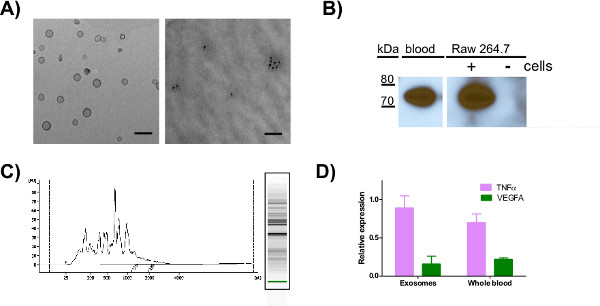
Figure 1. Transmission electron microscopy (TEM), western blot analysis and qRT-PCR for mRNAs using exosomes purified from various sources. A) TEM image of mouse blood exosomes resuspended in 1% glutaraldehyde (left) or RAW cell derived exosomes resuspended in 4% paraformaldehyde, labeled with 10 nm gold and rabbit-anti CD81 (right) and spotted on formvar carbon-coated grids for EM analysis (scale bar = 100 nm). B) Western analysis of HSP70 in exosomes purified from mouse blood or exosome-free media +/- 24 hr incubation with RAW 264.7 murine cells and resuspended in RIPA buffer. C) Bioanalyzer analysis of total RNA purified from exosomes derived from RAW 264.7 cell culture media D) Total RNA purified from whole blood and exosomes obtained from a representative human control was used to compare the expression levels of TNFα and VEGFA. Click here to view larger figure.
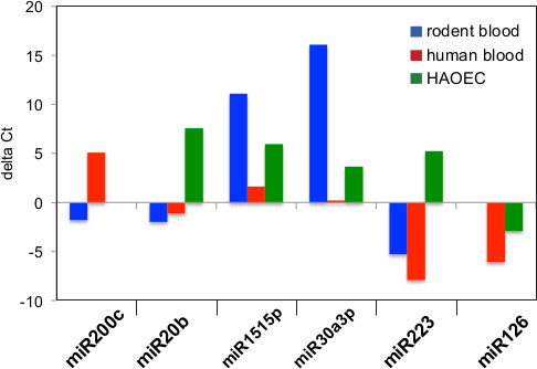
Figure 2. Relative Expression of miRNA fraction found in exosomes. Threshold cycle (CT value) is a relative measure of the concentration of an individual miRNA in the PCR reaction and lower CT value indicates higher expression. qPCR values for six detectable exosomal miRNAs were normalized to U6 RNA and the delta Ct values were graphed for exosomes from three sources. A negative value indicates higher expression in this figure. Exosomes from rodent blood (A) and human blood (B) expressed hsa-miR-223 at higher levels than control, while exosomes from HAOEC cell culture media (C) expressed hsa-miR-223 at lower levels than control. Hsa-miR-226 was absent from rodent blood exosomes and hsa-miR-200c was absent from HAOEC exosomes.
In this protocol, we show the quantification of miRNAs and mRNAs from exosomes purified by differential centrifugation from blood and culture media. Exosomes have diverse components dependent upon their origin and are involved in a number of biological functions, including immune response, antigen presentation, intracellular communication, and the transfer of RNA and proteins 9,11,12,19,20. While size and shape is a determinant of exosome purity, a number of papers showing EM data of exosomes indicate that these vesicles can range in size and morphology based on the source from which they are secreted as well as the method used for fixation and imaging 9,15. The accepted size for exosomes of 30-100 nm is an average range that includes a small population of exosomes with a larger diameter depending on the source 21,22, one reason that multiple methods are used to validate exosomal purity. Secreted miRNAs have many requisite features of good biomarkers 23 in addition to being potential therapeutic targets for various diseases 2,3,24. This purification method provides a noninvasive way to characterize temporal biological changes in exosomal content from a variety of bodily fluids while miRNA profiling by qPCR provides a comprehensive overview of miRNA content 2. Over 4,500 proteins, 1,639 mRNAs, 764 miRNAs and 194 lipids are known to associate with exosomes 25 (www.exocarta.org) and in some cases, changes in the levels of these biomolecules has already been linked to disease states. In our studies, the exosomes purified from human blood had many miRNAs in common with exosomes purified from media from cultured human cell lines but significant differences from exosomal miRNAs found in mouse blood. While there are examples of miRNAs that target only one mRNA, many miRNAs function to partially reduce the levels of multiple targets leading to a strong physiological response. Characterizing both mRNA and miRNA that reside in exosomes provides unique insight into exosome-mediated information transfer and the role of circulating miRNAs in mediating changes in gene expression. Purification and characterization of exosomes from a variety of sources will be beneficial in identifying molecular signatures associated with these secretory vesicles.
The authors have nothing to disclose.
This study was supported by funds from Rita Allen Foundation grant to Seena Ajit. The authors would like to acknowledge Erika Balogh and Dr. Soumitra Ghoshroy from the University of South Carolina Electron Microscopy Center for instrument use, scientific and technical assistance.
Name of Reagent/Material | Company | Catalog Number | Comments |
EDTA coated vacutainer (10 ml tubes) | BD Diagnostics | 366643 | For exosome purification from human blood |
EDTA coated vacutainer (2 ml tubes) | BD Diagnostics | 367841 | For exosome purification from mouse blood |
PAXgene Blood RNA Tube | BD Diagnostics | 762165 | For miRNA isolation from total blood |
miRVana microRNA isolation kit | Ambion | AM1561 | |
Acid-Phenol: CHCl3 | Ambion | 9721G | |
DNase 1 | Qiagen | 79254 | |
TaqMan Universal PCR Master Mix, No AmpErase UNG | Applied Biosystems | 4326614 | For TLDA cards |
Megaplex RT rodent Pool Set v3.0 | Applied Biosystems | 4444746 | |
Megaplex preamp rodent Pool set v3.0 | Applied Biosystems | 4444747 | |
Taqman Array Rodent MicroRNA A+B set V3.0 | Applied Biosystems | 4444909 | |
Megaplex RT Human Pool set V3.0 | Applied Biosystems | 4444745 | |
Megaplex Preamp human pool set v3.0 | Applied Biosystems | 4444748 | |
Taqman Array Human MicroRNA A+B Cards Set v3.0 | Applied Biosystems | 4444913 | |
Taqman MicroRNA RT kit | Applied Biosystems | 4366596 | |
Taqman fast universal PCR master mix | Applied Biosystems | 4366072 | For mRNA qRT-PCR |
Tumor necrosis factor (primer probe) | Applied Biosystems | Hs01113624_g1 | |
Vascular endothelial growth factor A (primer probe) | Applied Biosystems | Hs00900055_m1 | |
Maxima First Strand cDNA Synthesis Kit for RT-qPCR | Thermo Scientific | K1642 | For mRNA |
HSP70 antibody | Abcam | ab94368 | For western blot |
Anti-rabbit IgG-Gold | Sigma | G7402 | For electron microscopy |
Rabbit-anti CD81 | Sigma | SAB3500454 | |
Nickel 300 mesh carbon formvar grids | Electron Microscopy Sciences | FCF300-Ni | |
Copper 300 mesh carbon formvar grids | Electron Microscopy Sciences | FCF300-Cu | |
Table 1. Table of specific reagents. |