Achieving a systems level understanding of cellular processes is a goal of modern-day cell biology. We describe here strategies for multiplexing luciferase reporters of various cellular function end-points to interrogate gene function using genome-scale RNAi libraries.
Genome-scale interrogation of gene function using RNA interference (RNAi) holds tremendous promise for the rapid identification of chemically tractable cancer cell vulnerabilities. Limiting the potential of this technology is the inability to rapidly delineate the mechanistic basis of phenotypic outcomes and thus inform the development of molecularly targeted therapeutic strategies. We outline here methods to deconstruct cellular phenotypes induced by RNAi-mediated gene targeting using multiplexed reporter systems that allow monitoring of key cancer cell-associated processes. This high-content screening methodology is versatile and can be readily adapted for the screening of other types of large molecular libraries.
A variety of luciferase-based reporters for monitoring a diverse array of cell biological processes are commercially available. The majority of these DNA constructs encode luciferase proteins that are induced to express by specific cell stimuli or perturbations. The most robust transcriptionally based reporters place the expression of a luciferase enzyme under the control of synthetic enhancer elements or gene promoter regions well-established for their reliability in reporting a physiologically relevant transcriptional event1,2. There are also trans-reporter luciferase systems that utilize GAL4-UAS to drive luciferase protein expression. Gal4 is a yeast transcriptional activator and the UAS is an enhancer to which Gal4 specifically binds to activate the transcription of gene sequences placed down-stream of it3. These types of luciferase systems are typically supported by two DNA plasmids – one encodes the GAL4 protein fused to the regulatory portion of a protein of interest, and the second encodes a luciferase protein placed under the control of one or more UAS sequences. Thus, the cellular luciferase signal reflects the activity level of the protein of interest. Another common use of luciferase-based reporters is for monitoring protein stability whereby a protein of interest is fused to a luciferase enzyme4. Regardless of the type of reporter, a caveat emptor is noted here as one cannot assume the specificity of any reporter to have been well interrogated. Thus, due diligence is required in the incorporation of new reporter constructs in any research strategy.
The selection of the luciferase enzyme read-out can be as important as the reliability of the DNA elements that control its expression. Whereas firefly luciferase (FL) is the most commonly used enzyme in commercially available constructs, the advent of new luciferase reporter systems that do not require ATP (as in the case of FL-based reactions) or that incorporate more stable enzymes that emit stronger signals promise to improve the efficiency and reliability of this research platform. An important note regarding the selection of luciferase reporters in chemical studies is the susceptibility of FL to chemical inhibition that could give rise to false reports. In our experience, other enzymes such as Renilla or Gaussia luciferase (RL and GL, respectively) that utilize coelenterazine as a substrate are less readily inhibited by small molecules5.
The most common format for multiplexing luciferase read-outs entails the use of FL and RL largely given the availability of easy-to-use kits with the ability to sequentially measure enzymatic activities from a single sample. In most kits, samples are first exposed to luciferin to reveal levels of FL activity followed by a quenching reagent that is simultaneously deposited with coelenterazine to yield a secondary RL signal. With the addition of other luciferases that can be secreted such as GL or that use yet another substrate such as Cypridina luciferase (CL), a greater number of possibilities for generating high content data using luciferases have emerged. An example of a genome-wide RNAi screen using this technique can be found here6. These technological advances have in turn spurred the development of specific mechanisms to quench each type of luciferase enzyme in order to limit cross-talk7. In our hands, we note a greater frequency of “edge effects” (inexplicable trends in cellular activity associated with the edge of high titer culture plates), with secreted luciferases such as GL or CL. On the other hand, such secreted luciferases are useful for kinetic assays as they enable signal sampling without adulterating cell viability.
For our study presented here, we will simultaneously monitor the activity of three cancer-relevant cellular processes: the p53, Kras, and Wnt signal transduction pathways. The reporters incorporated in our study are the pp53-TA-Luc FL reporter from Clontech (henceforth referred to as the p53-FL reporter)8, an Elk1-GAL4/UAS-CL reporter system (henceforth Elk-1 reporter; Agilent), and the 8X TCF RL reporter that incorporates multiple synthetic enhancer elements recognized by the Wnt pathway transcriptional regulators known as T-cell factors (or TCFs)9-11.
Entire protocol takes approximately 4 days.
1. Preparation of Cells for siRNA and Reporter Transfection
We will introduce here a protocol for interrogating siRNA libraries using a small set of siRNAs (384 different siRNA pools array in 96 well format with each pool consisting of 4x siRNAs targeting a single gene) from a genome-wide siRNA library for illustrative purposes. For this particular study, we will exploit HCT116 cells that exhibit deviant Wnt and Kras signaling, and p53 activity. The appropriate cell line in studies aimed at interrogating other cellular processes of interest will have to be identified and evaluated in a similar fashion.
2. Preparing the Reporter Construct Stock Solution
3. Preparing the siRNA/DNA Transfection Mix
In this part of the protocol, we will prepare siRNA/DNA transfection mixes that will be sufficient for transfecting 12 x 96 well plates. For this test library of 4 x 96 well plate of siRNAs, we will transfect each siRNA pool in triplicate. The transfection reagent we will use is called Effectene (Qiagen). In our hands this is the only reagent that works for the simultaneous delivery of siRNA and DNA into cultured cells.
4. Determine Luciferase Activities from Cell Samples
After 36 hr, luciferase activities are ready for measurement in transfected cells. The endpoint should be determined on a per-experiment basis. In our study, a 36 hr incubation time had previously yielded a sufficiently robust signal for meaning outcome determination. As this study incorporates multiple reporters (one secreted into the culture medium, and two others expressed in the cell cytoplasm), we will obtain measurements from both culture medium as well as a cellular lysate.
Despite advances in mapping the mutational landscape of various cancers using massive genome sequencing efforts12-14, we still do not have in place a systematic approach for translating these observations into intervention strategies. In colorectal cancer (CRC), mutations that are predicted to affect components of three cellular processes – Wnt/β-catenin, p53, and Kras signaling – are found in 99% of all tumors suggesting they are key drivers of cellular transformation in the gut13. Thus, cancer genome datasets are likely enriched for genes that support common denominator cancer promoting processes, and that could be exploited for rapid identification of cancer cell vulnerabilities. To investigate this hypothesis, we have devised strategies amenable to high-throughput interrogation of gene function with respect to Wnt/β-catenin, p53, and Kras signaling. We first demonstrate the specificity of each reporter system in single reporter tests (Figure 1) and then provide evidence that these reporters could be used together for simultaneous measurements of gene activity of these three important cellular processes (Figure 2).
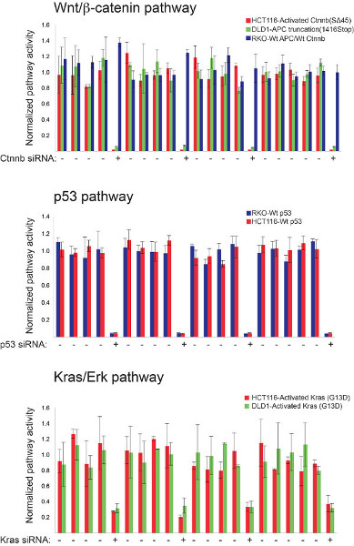
Figure 1. High-throughput luciferase-based assays for monitoring Wnt/β-catenin, p53 and Kras/Erk pathway status in mammalian cells. The robustness of three different screening platforms were individually assessed prior to multiplexing using control siRNAs targeting well-established pathway components. Indicated colorectal cancer cell lines were transfected with the 8X TCF, pp53-TA-Luc, or Elk-1 firefly luciferase-encoding reporter constructs along with pools of siRNAs targeting β-catenin, p53, or Kras, respectively. Pathway-relevant genotypes for each cell line are indicated. The strength of each screening platform was assessed by the reproducibility of these siRNAs-induced effects on the relevant signal transduction pathway. The β-catenin siRNAs have no effect on 8X TCF reporter activity in RKO cells in contrast to DLD-1 and HCT-116 cells that are respectively deficient in the APC pathway suppressor protein and express an activated form of β-catenin. Click here to view larger figure.
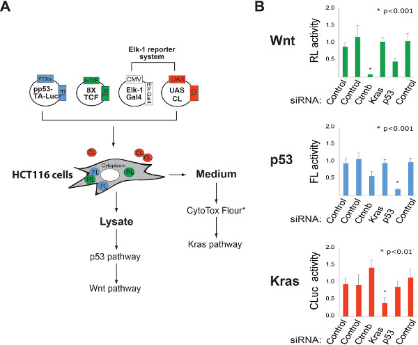
Figure 2. Multiplexing luciferase based reporters for high-content cellular analysis of gene function. A. Schematic representation of a strategy for simultaneously monitoring Wnt/β-catenin, p53, and Kras pathway activities. FL= firefly luciferase, RL= Renilla luciferase, CL= Cypridina luciferase (secreted enzyme). FL and RL activities in lysate are determined using luciferin and coelenterazine substrates. CL activity in the culture medium is measured using oxyluciferin. *Other reporter systems can be incorporated into this protocol to increase the content of each experiment. For example, the CytoTox Flour assay can be used to monitor cellular toxicity by sampling the levels of an intracellular protease released into the culture medium. The addition of the CytoTox assay reagent does not influence the activity of the CL reporter. B. Identification of pathway-specific vulnerabilities using RNAi. The ability of the multiplexed luciferase system described in “A” to quantify gene dedication to cellular function was tested using the indicated siRNA pools. Click here to view larger figure.
There are several purveyors of luciferase-based reporter systems (such as Promega, Targeting Systems, New England BIolabs, and Thermo Fisher) that provide useful on-line resources for those entertaining a high-throughput screen for the first time. In addition, a number of excellent reviews regarding optimizing the use of high-throughput screens for gene discovery and how RNAi-based screening results can be integrated with other -omics-based datasets should be helpful15,16. Methods for the selection of “hits” from high-throughput screens are the subject of another discussion that is beyond the scope of this report. Several reviews on this topic are available17,18. However, an important criterion for validating selected hits from screens based on the use of transcriptionally activated reporters is the ability for the same perturbagen (RNAi- or chemically based) to induce the same effect on validated target genes as measured using RT-PCR, qPCR, or microarray analysis. Moreover, keep in mind that the magnitude of response as measured by the reporter is typically higher than the induction of an endogenous reporter given that a feature of a successfully synthetic reporter is to provide a robust signal. Finally, an advantage of this co-transfection strategy is the ease with which one can rapidly assemble different reporter cocktails for monitoring a desired set of cellular activities. Although more labor intensive, the use of cell lines stably harboring the reporters would likely increase the signal to noise ratio given the variability of response is now primarily limited to siRNA transfection efficiency alone.
The authors have nothing to disclose.
We acknowledge funding support from CPRIT (RP100119) and the Welch Foundation (I-1665).
REAGENTS | |||
PathDetect Elk1 Trans-Reporting System | Agilent Technologies Inc. | 219005 | |
Pathway Profiling Luciferase System 4 pp53-TA-Luc Vector | Clontech Lab Inc. | 631914 | |
Effectene Transfection Reagent | Qiagen Inc. | 301427 | |
Dual-Luciferase Reporter Assay System | Promega Corp. | E1960 | |
Cypridina Luciferase Assay reagent | Targeting Systems | VLAR-1 | |
HCT116 cells | ATCC | CCL-247 | |
CytoTox-Fluo Cytotoxicity Assay | Promega Corp. | G9260 | |
EQUIPMENT | |||
MultiFlo Microplate Dispenser | Labsystems Inc. | Model 832 | |
Biomek FXP Laboratory Automation Workstation | Beckman Coulter Inc. | A31842 | |
PHERAstar FS-multi-mode HTS microplate reader | BMG Labtech Inc. | 0471-101A | |
Bright-Line Reichert Hemacytometers | Hausser Scientific Company | 1490 |