Assaying in vitro β-cell function using isolated mouse islets of Langerhans is an important component in the study of diabetes pathophysiology and therapeutics. While many downstream applications are available, this protocol specifically describes the measurement of intracellular cyclic adenosine monophosphate (cAMP) as an essential parameter determining β-cell function.
Uncontrolled glycemia is a hallmark of diabetes mellitus and promotes morbidities like neuropathy, nephropathy, and retinopathy. With the increasing prevalence of diabetes, both immune-mediated type 1 and obesity-linked type 2, studies aimed at delineating diabetes pathophysiology and therapeutic mechanisms are of critical importance. The β-cells of the pancreatic islets of Langerhans are responsible for appropriately secreting insulin in response to elevated blood glucose concentrations. In addition to glucose and other nutrients, the β-cells are also stimulated by specific hormones, termed incretins, which are secreted from the gut in response to a meal and act on β-cell receptors that increase the production of intracellular cyclic adenosine monophosphate (cAMP). Decreased β-cell function, mass, and incretin responsiveness are well-understood to contribute to the pathophysiology of type 2 diabetes, and are also being increasingly linked with type 1 diabetes. The present mouse islet isolation and cAMP determination protocol can be a tool to help delineate mechanisms promoting disease progression and therapeutic interventions, particularly those that are mediated by the incretin receptors or related receptors that act through modulation of intracellular cAMP production. While only cAMP measurements will be described, the described islet isolation protocol creates a clean preparation that also allows for many other downstream applications, including glucose stimulated insulin secretion, [3H]-thymidine incorporation, protein abundance, and mRNA expression.
The strict maintenance of euglycemia is imperative to prevent morbidities such as neuropathy, nephropathy, and retinopathy, which are all hallmarks of the pathology of uncontrolled type 1 and 2 diabetes1. Reduced β-cell function and mass in both type 1 and 2 diabetes perturb blood glucose concentrations2. Whereas immune-mediated type 1 diabetes results from a devastating loss of insulin-producing β-cells, impaired β-cell insulin secretion and peripheral insulin signaling in type 2 diabetes together promote hyperglycemia, dyslipidemia, and increased hepatic glucose production, which eventually results in both loss of β-cell mass and insulin secretory capacity from individual β-cells3. Understanding the underlying β-cell mechanisms in the progression of type 1 and 2 diabetes will hopefully give rise to novel therapies to prevent and treat these diseases.
In vitro tissue culture models, such as the INS-1 and MIN6 immortalized β-cell lines, can be useful tools for understanding specific β-cell functions. However, the interactions among the different cell types within the islet may themselves regulate β-cell function. For example, the paracrine influence of glucagon (released from α-cells) and somatostatin (released from δ-cells) in increasing and decreasing insulin secretion, respectively, demonstrates the importance of cell cell proximity in the endocrine response4. Moreover, gap junctions between β-cells potentiate the release of insulin5. Furthermore, although strides have been made in generating insulinoma lines that better replicated the physiological response of isolated islets to glucose (e.g., the INS-1- derived 832/13 and 832/3 cell lines), their glucose responsiveness still differs from normal rat islets6,7. Moreover, the response of these clonal insulinoma cell lines to glucagon-like peptide-1 (GLP-1) agonists can differ dramatically from one another, as well as from normal islets6. Therefore, immortalized cell lines may not represent the best model for assaying agents that impact on cAMP production.
In contrast to the insulinoma-derived cell lines, studying β-cell function solely in whole animal models offers its own set of complications. One of the biggest challenges in working with endocrine tissue is measuring the precise concentration of hormone released. Specifically, the liver plays a major role metabolizing insulin, and the pancreas blood flow goes directly to the liver. Thus, a plasma insulin measurement may not accurately portray the amounts of insulin being secreted from the pancreas itself or the impact of different treatments on the rate of insulin secretion8. Furthermore, renal metabolism of glucagon may limit the reliability of glucagon output from islet α-cells9. Therefore, isolating primary mouse islets for in vitro experimentation provides a more precise understanding of how the islet is responding to specific stimuli to complement measurements made in vivo.
The present protocol for the isolation of mouse islets is a well-established protocol used by a number of groups (with slight modifications that may help to increase success)10,11. In addition, the determination of cAMP production allows for a direct read-out of the incretin responsiveness of the β-cells. In conjunction with cAMP measurement, protein content and insulin secretion can also be quantified from the same cAMP sample prep, helping to determine whether a defect in β-cell function lies proximal or distal to cAMP10. The final cAMP content and insulin secretion application in this protocol can be a very powerful tool for understanding the influence of pharmaceutical and dietary constituents, among others, on cAMP and insulin secretion. In addition to stimulation from glucose alone, other compounds can be used to measure changes in cAMP and insulin secretion10,11.
Finally, although insulin is the primary hormone we assay from isolated islets, other hormones, such as glucagon and somatostatin, as well as cytokines, eicosanoids, and cyclic adenosine monophosphate, can also be measured, either by a transient stimulation assay or by quantification of their levels in culture medium12. Finally, although outside of the scope of this manuscript, islet isolation with the described collagenase isolation method allows for islet preservation so that many other downstream applications may be pursued, such as islet transplantation, RNA isolation for quantitative real time PCR or microarray analyses, protein isolation for Western blotting, islet embedding and immunofluorescent imaging, and [3H]-thymidine incorporation as a measure of islet cell replication, some of which have been described in previous JoVE articles13-16. Overall, following the islet isolation procedure described in the protocol may provide a researcher with important and useful information for developing therapies and promoting drug discovery aimed at enhancing β-cell function.
All animal experiments were executed in compliance with all relevant guidelines, regulations and regulatory agencies. The protocol being demonstrated was performed under the guidance and approval of the Institutional Animal Care and Use Committee (IACUC) of the University of Wisconsin-Madison.
1. Preparation of Solutions
2. Preparation of Tools
3. Preparing the Mouse
4. Opening the Thoracic Cavity
5. Inflating the Pancreas
6. Pancreas Removal
7. Washing
8. Picking Islets
9. cAMP Assay
To ensure a high islet yield during isolation, surgical techniques outlined in the protocol should be followed closely. Although the techniques presented here will be tailored to each laboratory, there are a few critical steps that will lead to a successful isolation. In order to make the common bile duct easily accessible, it is recommended that the organs be displaced to the right side of the mouse (Figure 1). Moreover, this will allow the pancreas to inflate with a smaller amount of resistance since there will be less weight restricting expansion. Another critical step to maximize islet yield is the ligation of the common bile duct close to the Sphincter of Oddi (Figures 2A-C). Ligating further away from the Sphincter of Oddi may result in a partial inflation by reducing the amount of collagenase solution entering the major pancreatic duct. Also, a taut knot will prevent collagenase solution from entering the intestine.
A proper incision into the common bile duct should be located far enough away from the bifurcation from the liver but close enough for maximal pancreatic inflation (Figure 3). An incision made too close to the bifurcation may lead to the flow of collagenase solution into the liver. When collagenase solution enters the liver, it will lose its dark-red color and begin to turn whitish. If this occurs, remove the needle and try another cannulation further from the liver. Additionally, the common bile duct incision should only be part way through the duct (about 50%). Completely shearing the duct will make it difficult to seal the duct around the needle tip. A proper incision will result in complete closure of the duct around the needle, producing enough pressure to both fill the common bile and pancreatic ducts (Figure 4A). It is also important to ensure the needle has entered the common bile duct and not the surrounding sheath. Once the needle has been inserted properly, 5 ml of the collagenase solution should fill the pancreas, creating a marbleized tissue.
The pancreas removal process should be performed delicately to promote maximal islet yield (Figure 5). Piercing the pancreas may result in a deflation and loss of collagenase solution, reducing the islet yield. Cutting the pancreas close to the connective tissue and other organs will prevent deflation. Also, harvesting other tissue, such as connective tissue, along with the pancreas is not an issue, as it will be removed in subsequent steps. A properly removed pancreas will remain inflated after excision prior to further collagenase digestion (Figure 6).
Distinct layers in the Ficoll gradient will lead to a clean islet preparation and create an easier environment for picking islets (Figure 7). The gradient ensures the separation of islets from cell debris and connective tissue that remains after digestion and mesh screen filtering. Furthermore, this step is crucial for selecting clean, healthy islets for downstream applications. Specifically, healthy islets will be spherical in shape and have a golden brown to dark brown center (Figure 8). Note: Islets from diabetic mice will be much paler in color, corresponding with decreased insulin content, and can be better distinguished from acinar tissue by their shape, glossiness, and their visible network of capillaries. Any islets connected to acinar tissue should not be used and should be discarded. Moreover, larger islets that develop a dark, necrotic center after an overnight incubation should be discarded as they do not function properly.
As described in the introduction, cAMP production is an important component of β-cell function; in particular, with regards to incretin action. One benefit to the protocol described in this method is that one can simultaneously obtain cAMP production and glucose-stimulated insulin secretion data. Typically, the impact of an agent on cAMP production directly correlates with its impact on insulin secretion17. For simple experiments, this rule holds true fairly well (see Figure 9 for an example). In the present protocol, we use IBMX (a phosphodiesterase inhibitor) in our treatments to prevent the catabolism of cAMP, giving the total production of cAMP.
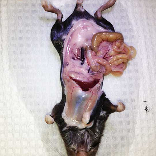
Figure 1. Displacing internal organs. Positioning the internal organs to the right side of the mouse creates an easier work environment and allows the pancreas room to expand during inflation.
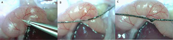
Figure 2. Tying off the Sphincter of Oddi (A-C). Depicted here is the entrance of the common bile duct to the small intestine at the Sphincter of Oddi. Tying off the common bile duct at the Sphincter of Oddi prevents collagenase solution from entering the intestines.
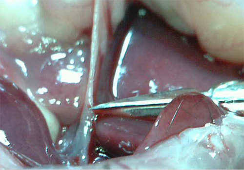
Figure 3. Common bile duct incision. It is best to make a cut in the common bile duct slightly distal to the bifurcation into the liver to prevent flow of the collagenase solution into the liver.
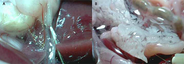
Figure 4. Cannulation of the common bile duct by a blunted 30 G needle and pancreas inflation. A) The 30 G needle is inserted into the common bile duct, creating a seal around the needle tip. It is important to check and to note the needle has been inserted into the common bile duct and not the surrounding sheath. A properly inserted needle will have an opaque appearance. B) A proper pancreas inflation will fill with about 3-5 ml of collagenase solution and have a marbled appearance.
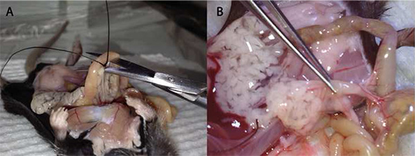
Figure 5. The removal of the pancreas. A) The initial incision made with a curved pair of scissors occurs near the Sphincter of Oddi. The initial removal begins in the direction of the stomach being careful not to puncture the pancreas. B) The final step is removing the pancreas from the connective tissue on the small intestine and the last few connections to the peritoneal cavity.
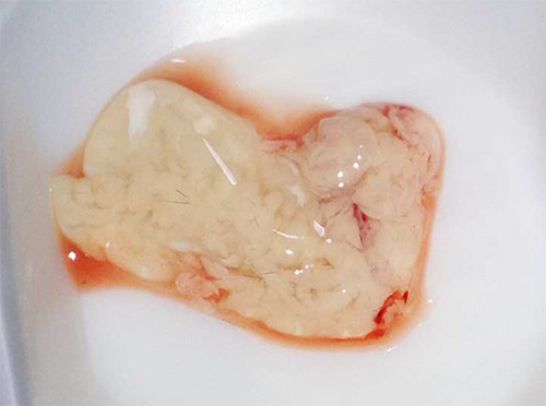
Figure 6. The inflated pancreas. A successful pancreas removal will yield a pancreas perfused with the collagenase solution. Poor excision will leave a smaller, deflated pancreas.
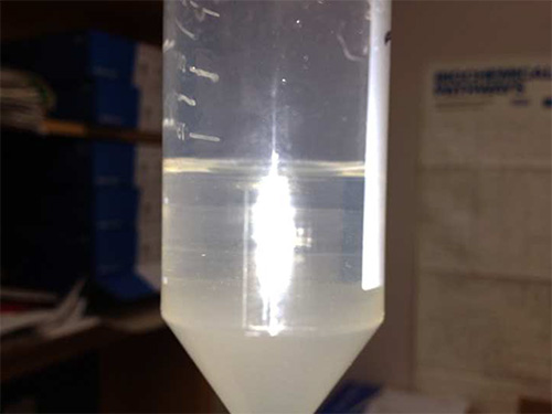
Figure 7. Four layers of the Ficoll gradient. The four distinct layers represent a different Ficoll density from 25% at the bottom to 11% at the top. The distinct layering is imperative to remove debris and exocrine disuse from the islets.
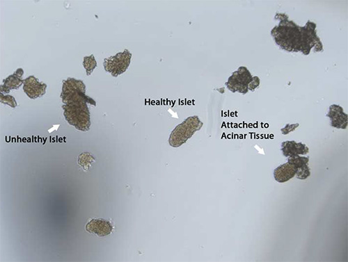
Figure 8. Islet selection. Healthy islets tend to have a golden brown to dark brown color with a round spherical shape and are not connected to acinar tissue. After an overnight incubation (16-20 hr) a necrotic center develops in larger islets, which should be excluded from experiments.
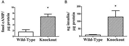
Figure 9. Representative high-quality cAMP production results, demonstrating that secreted insulin can be measured from the cAMP stimulation medium. A) Isolated islets from wild-type or gene knockout mice were stimulated with 11.1 mM glucose and intracellular cAMP production was measured and normalized to the total cellular protein. B) Secreted insulin was measured using a standard insulin ELISA, and also normalized to total cellular protein. In many cases, the change in cAMP production correlates directly with the augmentation in glucose stimulated insulin secretion. n=3 for each group; *, p < 0.05. This figure was adapted from research published in Kimple et al10.
With the prevalence of diabetes projected to affect 7.7% of the world’s population, the requirement of novel research techniques is imperative to both understand and treat diabetes18. The present islet isolation is a well-established protocol used for in vitro experimentation and has been presented previously with slight modifications11,14,16. Although insulin secretion is a common downstream application for isolated islets, focusing on upstream constituents, such as cAMP, may help delineate mechanisms potentiating insulin production and secretion.
The choice of euthanasia must be made thoughtfully to ensure both the humane treatment of laboratory animals as well as the integrity of experimental measurements. For islet isolation, exsanguination under anesthesia is our preferred method for euthanasia as it allows islet perfusion with oxygenated blood for the maximal amount of time prior to pancreas inflation. CO2 inhalation is a poor choice for euthanasia, as if it is performed according to humane protocols, it exposes the islets to poorly-oxygenated blood for at least 5-6 min before the procedure can begin. Cervical dislocation without anesthesia could be an acceptable method with scientific justification, but this would require significant experience with both the method of euthanasia and the mouse dissection and pancreas inflation procedure in order to ensure humane death and preservation of islet function. In choosing exsanguination under anesthesia, Avertin is the preferred anesthetic in this protocol. The particular choice of Avertin is that it is a fast-acting and deep anesthetic with many advantages over other commonly-used anesthetics. First, Avertin does not dilate vasculature and induce nitric oxide release, as does isoflurane19. Pancreatic islets are highly vascularized, and thus their biology may be negatively affected by isoflurane exposure. Second, Avertin does not excessively raise blood glucose levels in the absence of glucose injection, as can ketamine/xylazine, which has been shown to raise blood glucose by 167% up to an hour after injection20. Finally, the therapeutic range for pentobarbital is very narrow in mice21, increasing the chance of hypoxic damage to the islets.
In the present protocol, the use of suture thread to ligate the common bile duct near the Sphincter of Oddi is utilized as an alternative to a hemostat. The thread method allows for greater maneuverability of the common bile duct and helps position the needle for cannulation. However, the use of thread during this step requires holding two objects (thread and syringe) whereas the hemostat allows a free hand to help position the bile duct. Both isolation techniques yield a similar number of and viable islets as shown with previous work in our lab and others10,22. Moreover, cannulation needle size, position, and angle will depend on the user’s preference. Specifically, a 30 G needle is used in this protocol but other sizes may be required based on strain or genetic background. For instance, BTBR mice have a very friable common bile duct and a larger gauge may provide a greater seal around the needle tip (M.E.K., personal observations). Thus, it is recommended to have blunted cannula needles of different sizes on hand during the surgery, ready to switch out if necessary. The position of the needle in the common bile duct will also greatly impact the islet yield. If the needle is placed in front of the bifurcation of the common bile duct, collagenase solution will enter the liver and result in a poor inflation. However, if the needle is placed too close to the Sphincter of Oddi, a partial pancreatic inflation will occur, decreasing the number of isolated islets. Trial pancreatic inflations with various needle sizes and locations on the common bile duct will be required to customize the present procedure and maximize islet yield.
Another important component of this procedure to increase the number of viable islets is to prepare all reagents as per the protocols instructions. Gassing the buffers with 95% O2/5% CO2 to increase oxygenation can a factor in islet survival throughout the protocol. Previous work demonstrates that the oxygen concentration decreases towards the interior of the islet, potentially reducing the energy content of β-cells23. This may be evident after an overnight incubation as a dark, necrotic center will appear in cultured islets, correlating with a decrease in in vitro responsiveness. Thus, providing enough oxygen will help to increase islet number and viability during the isolation and into the overnight, normoxic incubation process. Moreover, proper preparation of the collagenase solution will enhance the digestion of pancreatic tissue and liberate the islets from surrounding tissue. If the collagenase concentration is too low, not only will the islet yield be low, but exocrine acinar tissue may remain attached to the islets, negatively affecting downstream applications. On the other hand, too high of a collagenase concentration may destroy the islets. Therefore, proper quality control measures to ensure collagenase digestion efficiency without the destruction of islets will promote greater yields.
Collagenase isolated from Clostridium histolyticum is the enzyme used for liberating islets from pancreatic tissue in this protocol. The irreproducibility of enzymatic activity between different lots of collagenase has the potential to hinder the quantity and/or quality of isolated islets. In the present protocol, each new lot of collagenase enzyme goes through an internal quality control check to determine both quantity and functionality of islets before they are subject to study. Therefore, downstream applications and lot variation need to be considered when choosing an enzyme for islet isolation. Liberase® (Roche), a purified collagenase blend, is another digestive enzyme alternative that has been successful in many cases; in particular, in obtaining higher quantities of isolated islets24,25. However, the use of Liberase may not be beneficial for isolating functional islets, as it may hinder in vitro function26.
Many limitations exist in the present islet isolation and cAMP determination procedures that may hinder downstream applications. As stated previously, BTBR mice have a very fragile common bile duct, which increases the difficulty of pancreatic inflation and subsequent islet isolation. Therefore, pooling islet preparations from several mice may be required, as many in vitro assays, including the described cAMP assay, require many islets per replicate. Additionally, using IBMX in the cAMP assay blocks phosphodiesterase activity, giving a read-out of cAMP production. Addition of IBMX may not be advisable in cases where the cycling of cAMP production and degradation is important for cellular signaling. However, removing IBMX from this protocol significantly lowers the final cAMP content of the islets, necessitating the addition of many more islets per replicate in order to obtain meaningful cAMP values in the EIA.
The present protocol is an essential tool for an islet biology laboratory or those interested in the endocrine pancreas. The cAMP component to this protocol is just one of many in vitro experiments to understand how islet manipulation through various treatments can enhance or blunt insulin secretion. Although most research utilizing islets primarily focuses on β-cell insulin secretion, other cell types can be assessed for their dynamic relationships within the islet4. In combination with other in vitro and in vivo models, islet experimentation will shed light on mechanisms underlying devastating pancreatic related diseases such as diabetes and insulinomas. Most importantly, understanding how pharmaceutical agents directly impact islets will enhance the efficacy of the treatment and aid in the translation to an in vivo model.
The authors have nothing to disclose.
We would like to thank Renee L. Pasker and Harpreet K. Brar for expert technical assistance on the protocols described in this work. Furthermore, we would like to acknowledge the mentoring of Christopher B. Newgard at Duke University and Alan D. Attie at the University of Wisconsin-Madison, along with the support of their laboratory members, which allowed us the time and support necessary to optimize the described protocols. In particular, we thank Hans Hohmeier, Danhong Lu, and Helena Winfield in the Newgard Laboratory and Mary Rabaglia in the Attie Laboratory for productive discussions and advice. This work was supported by NIH grant DK080845 and Juvenile Diabetes 594
Research Foundation grant 17-2011-608 (to M.E.K.)
Collagenase: Collagenase from Clostridium histolyticum suitable for isolating active islets | Sigma-Aldrich | C7657 |
Ficoll 400 | Sigma-Aldrich | F9378 |
Hanks Balanced Salt Solution 10X | Invitrogen (Gibco) | 14065-056 |
Hepes | Sigma-Aldrich | H3375 |
RPMI 1640 (powder) | Invitrogen (Gibco) | 31800-022 |
Albumin from Bovine Serum (BSA) | Sigma-Aldrich | A7888 |
3/0 Silk Suture Thread | Fine Science Tools | 18020-30 |
Dumont #5 Forceps | Fine Science Tools | 11251-10 |
0.8 mm Forceps | Fine Science Tools | 11050-10 |
Curved Scissors | Fine Science Tools | 14061-10 |
Vannas-Tübingen Spring Scissors – Straight/Sharp/8.5 cm/5 mm Cutting Edge | Fine Science Tools | 15003-08 |
Dissecting Scissors | Fine Science Tools | 14002-14 |
5ml BD Luer-Lok Syringe | BD | 309646 |
1ml BD syringe | BD | 309628 |
30 G BD Needle 1/2" Length | BD | 305106 |
27 G BD Needle 1/2" Length | BD | 305109 |
Sharpening Stone | Fine Science Tools | 29008-01 |
2-2-2 tribromoethanol | Sigma-Aldrich | T48402-25G |
2-methyl-2-butanol | Sigma-Aldrich | 240486-100mL |
Sodium Chloride (NaCl) | Sigma-Aldrich | S9888 |
Potassium Chloride | Sigma-Aldrich | P3911 |
Monopotassium Phosphate (KH2PO4) | Sigma-Aldrich | P0662 |
Sodium Bicarbonate (NaCHO3) | Sigma-Aldrich | S6014 |
CaCl2 *2H2O | Sigma-Aldrich | C3881 |
MgSO4 *7H2O | Sigma-Aldrich | M9397 |
Penicillin-Streptomycin | Invitrogen (Gibco) | 15140-122 |
Heat Inactivated Fetal Bovine Serum (H.I. FBS) | Fisher Scientific | SH30088.03HI |
3-Isobutyl-1-methylxanthine (IBMX) | Sigma-Aldrich | 5879-100MG |