We describe a protocol for the destabilization of the medial meniscus (DMM) model in mice, an effective tool for osteoarthritis (OA) research. In addition, we have demonstrated that deficiency of progranulincan exaggerate OA development and progression by using this model, indicating that progranulin plays a protective role in the pathogenesis of OA.
Destabilization of medial meniscus (DMM) model is an important tool for studying the pathophysiological roles of numerous arthritis associated molecules in the pathogenesis of osteoarthritis (OA) in vivo. However, the detailed, especially the visualized protocol for establishing this complicated model in mice, is not available. Herein we took advantage of wildtype and progranulin (PGRN)-/- mice as examples to introduce a protocol for inducing DMM model in mice, and compared the onset of OA following establishment of this surgically induced model. The operations performed on mice were either sham operation, which just opened joint capsule, or DMM operation, which cut the menisco-tibial ligament and caused destabilization of medial meniscus. Osteoarthritis severity was evaluated using histological assay (e.g. Safranin O staining), expressions of OA-associated genes, degradation of cartilage extracellular matrix molecules, and osteophyte formation. DMM operation successfully induced OA initiation and progression in both wildtype and PGRN-/- mice, and loss of PGNR growth factor led to a more severe OA phenotype in this surgically induced model.
Osteoarthritis (OA), also known as degenerative arthritis, affects 15% of the world's population and over 46 million people within the United States, and is characterized by synovitis, cartilage degeneration, and osteophyte formation1. It can be a result of a complex interplay of genetic, metabolic, biomechanical and biochemical factors. The underlying mechanisms of OA continue to evade the scientific community. There are presently numerous animal models which can mimic the pathogenesis of OA2,3. It is important to establish animal models in mice because of both the availability of various genetically modified mice and the cost effectiveness of experimentation. Among the different kinds of experimental OA models, the surgically induced destabilization of medial meniscus (DMM) model is a well-accepted OA model because of its good reproducibility and a relatively slower progression during OA development. Both of these attributes have been key for the evaluation of OA progression in different treatments or transgenes3-8. However, the consistency of surgical OA model is affected by various factors during the surgery and as a result, the application of surgical mouse model is limited.
Progranulin (PGRN) is a multi-functional growth factor expressed in various cells. It is known that PGRN plays a critical role in various physiological and disease processes such as wound healing 9, tumorigenesis10, and inflammation11-15. Studies also shown that insufficiency of PGRN can cause degenerative diseases of nervous system in both humans and mice16-18. It is known that PGRN is expressed in human articular cartilage, and its level is significantly elevated in cartilage of patients with OA and rheumatoid arthritis19. In addition, PGRN also plays a crucial role in chondrocyte proliferation 20, differentiation and endochondral ossification of growth plate during development 21,22. Recently, we reported that PGRN antagonized TNF-α through binding to TNF receptors and exhibited an anti-inflammatory function in inflammatory arthritis models13,14,23,24. However, the role of PGRN in OA, especially in vivo, remains to be an enigma. Herein, we present the procedure to induce a surgical DMM model, and investigate the role of PGRN in OA development through establishing DMM model in WT and PGRN-/- mice.
All of the surgical procedures relating to the animals should be approved by local Institution's Animal Care and Ethics Committee, with an effort made to minimize pain and discomfort caused by the surgery.
1. Preparation
2. Surgical Process
3. Post-operative Care
4. Histological Scoring of Surgical DMM Model
DMM model was successfully established in mice, and deficiency of PGRN exaggerated surgically-induced OA development.
Sham and DMM operations (Figure 1) were performed in WT and PGRN-/- mice. 8 weeks after operation, the mice were sacrificed, and Safranin O staining was performed on the sections from knee joints, followed by statistical analysis of arthritis score based on histology. As shown in Figure 2A, there was no obvious degeneration of cartilage in both genotypes in sham operation groups based on Safranin O staining, which implied the similar baseline conditions (upper panels). 8 weeks after DMM operation (lower panels), there was loss of proteoglycan and destruction of cartilage structure in both WT and PGRN-/- mice (red arrows). Both the loss of proteoglycan and degradation of cartilage suggest the successful induction of the OA phenotype. The bars of the columns indicated the variation between intra group members following DMM operation. However, the bar is relatively short, and PGRN knockouts displayed a higher loss of proteoglycan and the cartilage structure was more severely destroyed in comparison to the wild type breed. Moreover, the statistical analysis for cartilage score was performed as described previously25, and cartilage score was significantly higher in PGRN knockouts than the WT group. Our results demonstrated that a loss of PGRN resulted in more severe OA phenotype.
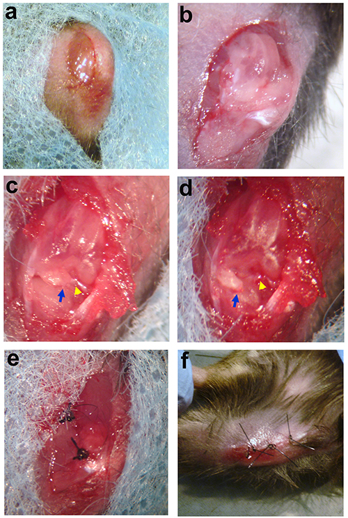
Figure 1. Surgical procedure for DMM induction. a. A medial para-patellar incision is made in skin of knee joint. b. Exposure of knee joint space after lateral dislocation of joint capsule. c. Inner structure of knee joint. Blue arrow indicates medial meniscus. Yellow arrow shows meniscotibial ligament. d. Medial meniscus is destabilized. Blue arrow indicates the medial meniscus which is dislocated from original site. Yellow arrow shows residual part of meniscotibial ligament which has been dissected. d. The joint capsule is closed. f. The skin is closed. Click here to view larger image.
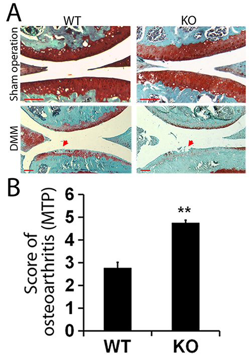
Figure 2. Representative pictures of histology and statistical analysis for score of osteoarthritis. A. Representative histological pictures of WT and PGRN-/- mice 8 weeks following sham operation or DMM, assayed by Safranin O staining. Red arrows indicate cartilage destruction. Loss of red color in cartilage implies loss of proteoglycan. Scale bar, 100 μm. B. Statistical analysis for score of osteoarthritis in both PGRN-/- and WT mice. Click here to view larger image.
It is reported that the strain of mice is very important for DMM model induction, as different strains of mice have varying severity of OA after DMM, with highest level in the 129/SvEv strain, followed by C57BL6, 129/SvInJ and then FVB/n26. A large part of transgenes are established in C57BL6 mice, such as PGRN-/- mice we used in the present study, which are relatively susceptible to DMM. However, if the transgene is based on insensitive strain such as FVB/n mice, it is necessary to backcross these mice with susceptible strains before DMM induction. The gender of mice used for DMM induction is also very important as studies have already shown that female mice are less sensitive than male mice27. Moreover, the age of mice for DMM operation varies from 8 weeks old to 12 weeks old based on literature29, which also plays a critical role in pathogenesis of OA. Therefore, mice of the same age should be operated to eliminate this effect. Post operative pain control is required to enhance the movement of mice after surgery. As DMM model leads to OA through movement mediated wear and tear in cartilage, inadequate movement as a result of post operative pain may cause inconsistency of OA development. Currently there is a discrepancy concerning the definition of DMM regarding whether or not to remove the medial meniscus4,28. In our present study, we merely destabilized medial meniscus but did not remove it. Lesions in the DMM model were mainly located on the central weight-bearing region of medial tibial plateau, and the severity of lesions increased during the time course (unpublished data), and the results were consistent with the previous reports5. Furthermore, the OARSI score following DMM operation was relatively consistent between different members of the same genetic group.
PGRN plays an important role in cartilage development and arthritis14,21.We previously reported that PGRN interplays with metalloproteases ADAMTS-7 and ADAMTS-12, and protects cartilage matrix protein COMP from degradation19. Moreover, it is well-established that TNF-α plays a critical role in cartilage destruction24. Recently, we found that PGRN antagonized TNF-α and protected against cartilage destruction in inflammatory arthritis models14. In this study, we induced DMM model in WT and PGRN-/- mice, and as expected, PGRN-/- mice exhibited exaggerated progression of OA following induction of DMM, which was indicated by more severe loss of proteoglycan in Safranin O staining and significantly higher arthritis score based on histology (Figure 2). Moreover, the expressions of catabolic markers were dramatically elevated in PGRN-/- group (unpublished data), suggesting accelerated OA progression in deficiency of PGRN.
In conclusion, our protocol for establishment of DMM model successfully induced OA phenotype in WT and PGRN-/- mice, and the induction was consistent and reproductive. The successful generation of DMM model in C57/B6 mice provides a useful tool for evaluating therapeutic agents and OA related genes for OA in future. In the current study, the result of DMM model documents a protective role of PGRN in OA development.
The authors have nothing to disclose.
This work was supported partly by NIH research grants R01AR062207, R01AR061484, R56AI100901, K01AR053210, and a Disease Targeted Research Grant from Rheumatology Research Foundation (to C. J. Liu).
No. 10 Surgical blades | FEATHER | 25-2976#10 | |
6-0 suture | APPLIED DENTAL | WG-N53133 |