Establishment of human models of the blood-brain barrier (BBB) can benefit research into brain conditions associated with BBB failure. We describe here an improved technique for preparation of a contact BBB model, which permits coculturing of human astrocytes and brain endothelial cells on the opposite sides of a porous membrane.
The blood-brain barrier (BBB) comprises impermeable but adaptable brain capillaries which tightly control the brain environment. Failure of the BBB has been implied in the etiology of many brain pathologies, creating a need for development of human in vitro BBB models to assist in clinically-relevant research. Among the numerous BBB models thus far described, a static (without flow), contact BBB model, where astrocytes and brain endothelial cells (BECs) are cocultured on the opposite sides of a porous membrane, emerged as a simplified yet authentic system to simulate the BBB with high throughput screening capacity. Nevertheless the generation of such model presents few technical challenges. Here, we describe a protocol for preparation of a contact human BBB model utilizing a novel combination of primary human BECs and immortalized human astrocytes. Specifically, we detail an innovative method for cell-seeding on inverted inserts as well as specify insert staining techniques and exemplify how we use our model for BBB-related research.
The BBB is a specialized interface between the peripheral blood circulation and the central nervous system, crucially responsible for the maintenance of brain hemostasis. It comprises distinct brain microvascular endothelial cells (BECs) which are functionally influenced by few cellular and acellular components (below) to form a tight and dynamic gateway into the brain. Under physiological conditions the BBB restricts the passage of blood cells, plasma components and harmful substances, all potentially neurotoxic, into the brain. In parallel, the BBB selectively exchanges key ions and nutrients (glucose and amino-acids) and metabolic waste products between the brain and the circulation to precisely maintain the brain environment1,2. In recent years it is becoming evident that failure of the BBB occurs in a variety of chronic brain pathologies, such as neurodegenerative or inflammatory-related diseases (e.g. Alzheimer's disease and multiple sclerosis, respectively)3, as well as in acute conditions like ischemic stroke4.
The unique BBB properties of brain endothelial cells (BECs) are largely induced by their cerebral environment5, and in particular by astrocytes6,7. There is a growing understanding that other cell-types, such as pericytes8, neurons and microglia1,3, as well as the basement membrane9, support BECs and form together a functional unit termed the "neurovascular unit" (NVU) which simultaneously couples neuronal metabolic demands to their supplying capillaries10.
The involvement of the BBB in pathological situations underlies numerous attempts to develop in vitro BBB models to assist in BBB-related research11,12. These models aim to mimic as close as possible in vivo BBB characteristics according to the NVU principle. In vitro BBB models generally rely on a monolayer of tight-junction-forming BECs (mainly from bovine13, human14, rat15, mouse16, and porcine17,18 origins), cultured on a porous membrane together with supporting astrocytes (extensively reviewed by Deli et al. 200511).
Astrocytes can be grown in non-contact conditions on the bottom of a tissue culture well, separated from BECs (cultivated on the upper surface of the membrane) by the culture medium yet communicating with BECs via soluble factors16. In more advanced models which better resemble the anatomical structure of the BBB in vivo, astrocytes are maintained in contact conditions and cultured directly on the opposite side of the membrane in close proximity to BECs13,15,17 (Figure 1). This configuration enables physical contact between BECs and astrocytes, established when astrocytes project their processes through the porous membrane. Importantly, for a true contact to occur the pores should be ≥1µm in diameter, since astrocytic end-feet cannot pass through smaller pore sizes (i.e. 0.4 µm)14,15. Notably, contact BBB systems are demonstrated in some studies to be superior to their non-contact counterparts regarding their trans-endothelial electrical resistance (TEER) and endothelial permeability values of various tracers13,17,18. An additional dimension of media flow was recently added in a number of in vitro BBB models to apply shear forces to the endothelium for closer simulation of the brain vasculature12,19.
One technical obstacle to overcome when generating a contact BBB model is the seeding of astrocytes against gravity on the abluminal surface of the porous membrane. Previous protocols13,20, where astrocytes were simply seeded in a drop of media on top of an inverted insert, allowed only short seeding times (i.e. 10 min13 or 2 hr20) which were found in our hands to be insufficient for proper cell attachment. Using this basic method, a longer astrocyte attachment period requires constant monitoring of the inserts by frequent opening of the incubator (causing fluctuations in temperature, pH and humidity) and is also prone to uneven cell seeding due to leakage of media through the pores, especially if pores larger than 1 µm are employed.
Here, we describe a general protocol for preparation of a contact BBB model. Our procedure includes an alternative method for cell-seeding on inverted inserts, which addresses the above mentioned limitations. The method permits undisturbed adherence of astrocytes onto the abluminal membrane surface, in an equilibrated incubator, for an extended period of time. As a result, a uniform seeding of astrocytes is achieved which increases barrier quality and minimizes basal permeability variations between inserts.
As the use of human cells is important for human-relevant research21, we additionally demonstrate in this article the specific utilization of a novel combination of primary human BECs and immortalized human astrocytes for establishment of a contact human BBB model with a high throughput screening capacity. Since viewing of cells on porous membranes can be difficult, we also detail staining techniques which can assist in determination of confluence and cell morphology on the porous membranes. Finally, we exemplify how our human BBB model can be utilized to examine the effect of tissue-type plasminogen activator (t-PA) – a clot busting enzyme which serves as a sole treatment option for acute ischemic stroke – on the BBB.
1. Cell Culture (3-7 Days Prior to BBB Assembly)
1.1. Primary Human Brain Microvascular Endothelial Cells (BECs)
BECs were commercially obtained. The cells were produced by dispase dissociation of normal human brain cortex tissue and provided frozen at passage 3 (<12 population doublings).
1.2. SVG Human Fetal Astroglial Cell-line
SVG human fetal astroglial cells22 were originally derived from primary cultures of human fetal brain transformed with replication-deficient simian virus 40.
2. Assembly and Coculturing of the Human In vitro BBB Model (Day 0)
Comment: The human in vitro contact BBB model is prepared according to published protocols 13,23, with modifications.
2.1. Coating of Inserts
2.2. Seeding SVGs on the Underside (Abluminal) Membrane Surface
2.3. Seeding of BECs into the Luminal Chamber above the Astrocytes
Seed 2×10⁴ BECs in 200 μl onto the collagen-coated luminal surface and return the inserts into the incubator.
2.4. Coculturing
Coculture the SVG/BEC-seeded inserts for 3 days in BEC medium without media change before experimentation.
3. Experimentation with the Human BBB Model (Day 3)
To account for fluctuations in baseline permeability between experiments, analyze changes in permeability relative to a coated insert without cells (which serves as a reference for maximal permeability) and to the vehicle-treated group using the formula: permeability (% of max) = (permeability value of experimental insert – average permeability value of the vehicle group) / (permeability value of the blank insert – average permeability value of the vehicle group) X 100.
4. Visualization of Cells on Porous Membranes
Comment: Unstained cells on insert membranes are difficult to observe by phase contrast or differential interference contrast (DIC) microscopy since the membrane is quite opaque and interferes with light transmission. To deal with this issue we developed various staining procedures of cells on inserts, which greatly enhance cell appearance on the membrane. Generally, stain the insert membranes while still attached to the insert, in the wells. The membranes should be removed only at the end of the staining procedure for mounting purposes.
4.1. Hematoxylin Staining
4.2. Scanning Electron Microscope (SEM)
4.3. Immunofluorescence
In order to establish a human, contact BBB model we had to cultivate SVGs and BECs on porous membranes with a 3 µm pore-size, shown to permit passage of astrocyte end-feet for contact with endothelial cells14,15,27,28. A schematic representation of the complete contact model is illustrated in Figure 1 (left illustration). The main technical challenge presented by a contact system is the need to seed astrocytes against gravity on the abluminal surface of the membrane. We have succeeded in this task by principally creating a removable silicone well on top of the abluminal membrane surface while preventing leakage of media with an additional silicone plug inserted into the luminal cavity (Figure 2). The method allowed an optimal, uninterrupted seeding period of SVGs (at least 4 hr), which in turn became strongly attached to the porous surface with minimal cell loss. Indeed, 3 days post-seeding both SVGs and BECs appeared confluent and covered the membrane uniformly, as judged from images of hematoxylin-stained inserts (see section 4.1; Figure 3A, left panels). Additionally, both cell types maintained their cell-specific markers on porous membranes (a diffuse staining pattern of glial fibrillary acidic protein in SVGs22 and expression of von Willebrand factor in Wiebel-Palade bodies and cytoplasmic vesicles in BECs; Figure 3A, right panels), observed by immunofluorescence (section 4.3). Furthermore, from SEM imaging (section 4.2) it became apparent that SVGs and BECs were capable of growing directly over the membrane pores (Figure 3A, middle panels, arrow heads), a phenomenon which artificially contributes to the physical barrier function of in vitro BBB contact models13,28. Functionally, in the presence of SVGs the endothelial permeability value (Pe)16,24 of fluorescent albumin28 improved ~4-fold relative to BECs alone (P<0.01; n=4), from 0.574±0.199 x10⁻³ to 0.126±0.028 x10⁻³ cm/min without or with astrocytes (SVGs), respectively. These albumin Pe results are comparable to another study using mouse BECs with rat C6 glioma cells29. Our seeding protocol was successfully applied also to freshly-isolated primary mouse BECs and primary mouse astrocytes grown on 1 µm pores. In this mouse contact model we obtained a reasonable physical barrier for the small hydrophilic tracer sodium fluorescein (Pe=0.83±0.22 x10⁻³ cm/min; n=3). Hence, our seeding method can be extended for preparation of BBB models from other species.
The most fundamental feature of a contact BBB model is the ability of astrocytes to project their end-feet through the membrane pores to physically contact endothelial cells. A few studies made use of scanning electron microscopy to identify astrocytes processes which pass from the abluminal membrane surface (where astrocytes are seeded) to the luminal surface, as a proof of principle that contact between astrocytes and EC is plausible14,15,27. Following these studies we imaged by SEM the luminal membrane after seeding SVGs on the abluminal surface. As shown in Figure 3B, astrocytic (SVG) end-feet could be observed passing through 3 µm pores into the luminal compartment, confirming that contact can potentially exist between SVGs and BECs seeded on pores of this dimension. Collectively, our seeding method was simple, efficient, and yielded an authentic contact BBB model.
We have utilized our human contact BBB model to explore the effect of t-PA, a fibrinolytic agent, on the BBB. A number of articles have suggested that t-PA may be able to induce opening of the BBB during its intravenous administration for the treatment of acute ischemic stroke30-33. This interaction between t-PA and the BBB may contribute to bleeding complications associated with t-PA-induced thrombolysis4,34,35 and is thus a subject for current research efforts. Our studies in vitro confirmed that t-PA indeed increased the permeability of the intact BBB in a concentration- (Figure 4A) and time-dependent manner (Figure 4B), which were both within a pharmacologically-relevant range36,37. Furthermore, dramatic morphological changes of SVG astrocytes could be observed on the porous membranes post exposure to t-PA (Figure 4C), suggesting that astrocytic responses to t-PA underlie t-PA-induced BBB opening. Subsequent investigation identified that t-PA stimulates astrocytes via activation of plasminogen – its natural substrate – into the potent and broad-spectrum protease plasmin. Furthermore, t-PA and plasmin were found to activate Rho-kinase (ROCK) signaling in astrocytes, and blockade of the ROCK pathway prevented t-PA-induced BBB opening28. This is a good example of how utilization of BBB models can lead to identification of novel biological processes and therapeutic opportunities.
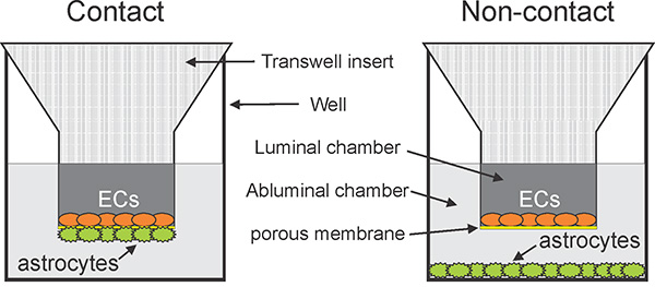
Figure 1. Insert-based configurations of in vitro BBB models. Schematic representations of static (without flow) in vitro BBB models. In basic models brain endothelial cells (ECs; orange) are cultured alone on a porous membrane (yellow). In more advanced approaches ECs are cocultured with astrocytes (green). Astrocytes can be grown either in non-contact conditions, on the bottom of the well (right), or on the abluminal surface of the porous membrane in contact conditions (left). Click here to view larger image.
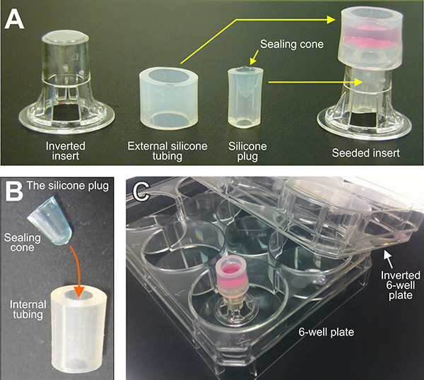
Figure 2. An improved method for astrocyte seeding on the abluminal membrane surface for preparation of a contact BBB model. (A) The insert is inverted, a short piece of silicone tubing ("external tubing") is assembled around its perimeter and a silicone plug fitted into the luminal cavity. This assembly creates a well on top of the abluminal surface and prevents media from leaking through the pores. Cells can be seeded in the well and allowed to adhere undisturbed for extended periods of time (right) (B) The silicone plug is prepared from another piece of silicone tubing ("internal tubing"), sealed at one end with a sealing cone which is inserted into its cavity. (C) Once seeded, the assembled inserts are transported in between two 6-well plates (one plate inverted over the other) to minimize the risk of contamination. Click here to view larger image.
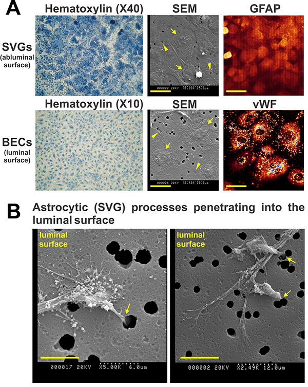
Figure 3. Visualisation of brain endothelial cells (BECs) and astrocytes on a 3 µm porous membrane. A) Hematoxylin (left panels), scanning electron microscope (SEM; middle panels) and immunofluorescence images (right panels) of SVGs (on the abluminal surface, 40,000 per 6.5 mm insert, top panels) and BECs (20,000 per 6.5 mm insert on the collagen I-coated, luminal surface, bottom panels) grown on a 3 µm porous membrane for 3 days (without coculturing). Both cell types reach confluence within this time frame and maintain their cell-specific markers (glial fibrillary acidic protein (GFAP) for SVG and von Willebrand factor (vWF) in Wiebel-Palade bodies for BECs, right panels) on the membrane. Arrow heads in the SEM panels further show the ability of both cell types to directly grow over and cover the pores, while arrows represent cell nuclei. Scale bars on SEM images represent 25 µm (top) or 20 µm (bottom). Scale bars on immunofluorescence images represent 40 µm. (B) SEM images of SVG end-feet passing through 3 µm pores into the luminal (BEC) surface 3d post seeding of SVGs (alone) on the abluminal membrane surface. These images validate the potential for development of true contact between SVGs and BECs in our human BBB model. Scale bars represent 6 µm on the left panel and 12 µm on the right panel. Click here to view larger image.
* For further clarification of the cells position please refer to Figure 1, left panel.
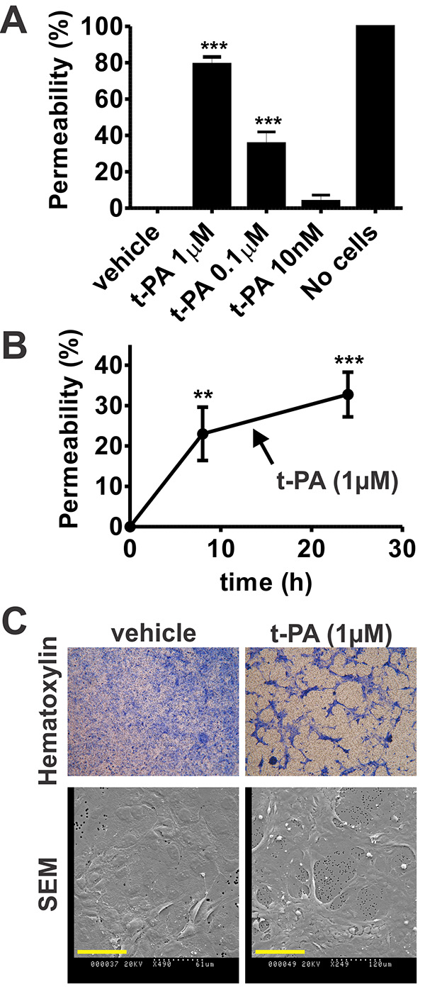
Figure 4. tissue-type plasminogen activator (t-PA)-mediated increase in BBB permeability is driven by morphological changes of astrocytes. (A) Increasing t-PA concentrations were added to the luminal chamber of the human in vitro BBB and permeability was assessed 24 hr later by passage of fluorescent albumin into the abluminal chamber. t-PA increased BBB permeability in a concentration-dependent manner. n=4-6 for all groups but t-PA 10nM, where n=2. ***P<0.001 compared to all other groups by one way ANOVA with Newman-Keuls post hoc. (B) t-PA (1 µM) was added to the luminal chamber of the human in vitro BBB for 8 hr and 24 hr before permeability assessment. As shown, t-PA-mediated BBB opening was already substantial 8 hr post stimulation (70.26% of the maximum at 24 hr) but a further increase was observed at 24 hr. n=6, ***P<0.001, **P<0.01compared to zero by one way ANOVA with Newman-Keuls post hoc. In both (A) and (B), bars/data points represent mean±SEM. (C) Representative bright-field images of Haematoxylin-stained SVGs (top panels) or scanning electron micrographs of SVGs (bottom panels) on polyester insert membranes (3 µm pore size) 24 hr post treatment with vehicle or t-PA (1 µM). Pronounced t-PA-induced morphological changes and disruption of the SVG monolayer integrity can be observed, suggesting that astrocytic responses to t-PA underlie the effect of t-PA on BBB permeability. Images are representative of two independent experiments. Scale bars on SEM images represent 61 µm (left) or 120 µm (right). Click here to view larger image.
Medical research into brain pathologies suffers much translational difficulty. In the area of acute ischemic stroke, for example, many drugs which showed great promise in animal models failed at the clinic38,39. The reasons for these disappointing results are diverse and include infidelity of the preclinical test systems to the human stroke scenario and overstatement of results obtained from animal studies21. One strategy to improve the predictive value of preclinical findings to the clinical phase is additional development of human cell-based in vitro models; these may provide potent and cost-effective tools for screening novel drugs and mechanisms and allow better focusing on clinically-relevant avenues 21. Within this context, we described here in detail the process undertaken in our laboratory for the establishment of a human contact BBB model utilizing a novel combination of human immortalized astrocytes (SVGs) and human primary BECs for examination of stroke-related phenomena.
Under static conditions (without flow), contact BBB models (Figure 1) resemble best the in vivo anatomical structure and are shown to be superior in their barrier properties13,17,18. Importantly, true contact between BECs and astrocytes occurs only on membranes with larger pore diameter (≥1 µm), which permit passage of astrocytic end-feet and subsequent induction of tighter (less permeable) BEC monolayers14,15,27. However, the seeding of astrocytes on inverted inserts with higher pore size – the core technical element in the preparation of a contact BBB model – presents a double technical challenge, since in addition to the limited medium volume which can be added to the inverted 6.5 mm inserts (~50 ul), the membranes are also prone to leakage of medium due to their large pore diameter. These limitations can make the astrocytic seeding awkward and inconsistent.
Our innovative technique for seeding astrocytes on inverted inserts with 3 µm pores offers a simple yet effective solution for this procedure. By creating a removable, impermeable silicone well on top of the abluminal surface we greatly improved the astrocytes' ability to successfully adhere to the porous membrane. This enables uniform growth of astrocytes on the abluminal surface and allows reproducible preparation of a large number of inserts for simultaneous screening of many experimental parameters (for example, our recently performed screening of the effect of five different plasminogen-activators on the BBB28) . Hence, despite its simplicity, we found our method to dramatically improve the throughput and quality of our research using this contact BBB model. One limitation of our astrocyte seeding method is the requirement for extra manual handling of the inserts, which harbor thin (10 µm thick) membranes. Excessive or rough handling can cause micro-tears and damage to the membrane. Hence, the use of soft and flexible silicone tubing is essential as the assembly/disassembly has to be performed with minimal force.
Regarding its overall concept, our human BBB model was established based on a previously-described protocol14,23 which originally utilized primary human cells. Despite their superiority for BBB modeling, one drawback in employment of primary human brain cells is the inability to routinely access live human brain tissue for regular production of primary BECs and astrocytes and/or the cost involved in commercially obtaining them. Since many BBB models successfully substitute primary astrocytes with immortalized astrocytes (for example with a rat C6 glioma cells29), it was reasonable to employ SVG astrocytes22 as a novel, abundant and cost-effective option for human BBB modeling. The ability of SVGs to positively influence the BEC barrier function validated in principle their suitability for BBB studies. In contrast, we chose to keep primary BECs in our BBB model for their critical role in the BBB in vivo1,2,9. Importantly, prolonged culturing of primary BECs can result in de-differentiation and deterioration of their BBB-specific phenotype. This indeed may be evident in our system by a rather high endothelial permeability values for albumin. For this reason, we have used our model mainly in a relative fashion28 (Figure 4), taking into account its elevated basal permeability. If a very tight human BEC monolayer is desired the barrier function can be further improved by addition of chemicals such as the glucocorticoid hydrocortisone17 or cyclic AMP with phosphodiesterase inhibitors13. Immortalized human BECs, such as the hCMEC/D3 cell line40, could also be employed for their ability to better maintain tight junctions over time. Yet, BEC cell-lines often display reduced barrier qualities compared to low-passage primary BECs41,42 and needs to be carefully considered because of other potential changes (i.e. altered expression of cell-surface receptors).
Collectively, our modification of the astrocytic cell type (i.e. the employment of SVGs) and the substantial improvement to the astrocyte seeding protocol resulted in generation of a reproducible, easy to assemble and authentic human contact BBB model. Bearing in mind its limitations, this system can serve as a useful tool for in vitro investigation not only of acute ischemic stroke, as we exemplified here28, but also of several other brain pathologies involving modulation of the BBB.
The authors have nothing to disclose.
This study was funded by grants awarded to R.L.M. from the National Health and Medical Research Council of Australia (grant # 606658).
Attachment Factor | Cell-Systems Corporation | 4Z0-210 | |
Brain microvascular endothelial cells (BECs), primary, human | Cell-Systems Corporation | ACBRI 376 | |
Complete medium | Cell-Systems Corporation | 4Z0-500 | Supplemented with CSC JetFuel |
Complete serum-free medium | Cell-Systems Corporation | SF-4Z0-500 | Supplemented with CSC RocketFue |
DMEM/F-12 with 15 mM HEPES | Life Technologies | 11330-032 | |
Endothelial cells growth supplement (from bovine origin) | Sigma-Aldrich | E2759-15MG | |
External silicone tubing | Watson-Marlow | 913.A080.016 | Pumpsil brand, 8 mm internal diameter, 1.6 mm wall |
Foetal calf serum | Lonza | 14-501F | |
Gentamycin sulfate | Life Technologies | 15750-060 | |
Heparin sodium | Pfizer | 1,000 U/ml | |
Hexamethyldisilazane (HMDS) | Sigma-Aldrich | H4875 | |
Human brain microvascular endothelial cells | Cell-Systems Corporation | ACBRI 376 | |
Internal silicone tubing | Watson-Marlow | 913.A032.016 | Pumpsil brand, 3.2 mm internal diameter, 1.6 mm wall |
L-Glutamine | Life Technologies | 25030 | |
Mayer’s hematoxylin solution | Amber Scientific | MH | |
Minimum essential medium with Earle’s balanced salt solution | HyClone Laboratories | SH30244.01 | |
Penicillin/Streptomycin | Life Technologies | 15140 | |
Rat collagen I | Trevigen | 3440-100-01 | Cultrex brand |
Tissue culture inserts | Corning Life Sciences | 3472 | Transwell brand, 6.5 mm in diameter, with polyester porous membrane, 3 µm pore-size |