1. Priming the SynVivo-SMN Microfluidic Device
- Each port (inlet/outlet) of the device is comprised of two parallel ports – one for flowing in surface coating moieties (adhesion molecules, growth matrices, etc.) and/or cells for seeding and the other for running the assay (Figure 1A).
- Completely submerge the SynVivo-SMN microfluidic device (Figure 1B) in a Petri dish containing sterile deionized (DI) water and place the dish into a vacuum desiccator. Allow the desiccator to run until all of the air is removed from the channels of the device. This should take approximately 15 min.
- Before removing the device from the water, place Tygon tubing (O.D. of 0.06″ and I.D. of 0.02″) primed with water into each port of the device with fine-point forceps. The tubing should be approximately 1 inch in length. The device can now be removed from the water. Figure 1C shows image of the device with the tubing.
2. Coating the Microfluidic Device with Desired Protein (e.g., Avidin)
- Using a pipette, place a drop of water (approximately 100 µl) around the base of the one inlet port tubing. Carefully remove the tubing used to prime the device. The drop of water will prevent air from entering the device.
- Prepare a 1 ml syringe loaded with avidin at a concentration of 20 µg/ml. Connect the syringe to a 24 G stainless steel needle and tubing. Insert the tubing to one of the inlet ports of the device. Clamp the inlet port not being utilized with a jaw clamp.
- Inject avidin at a flow rate of 1 µl/min for 10 min to allow complete perfusion of the device. At the end of the flow time, clamp the tubing with the jaw clamp and place the device at 4 °C overnight.
3. Flowing the Biotinylated Particles for Adhesion Experiments
- Allow the device to come to room temperature. Place the device on an inverted fluorescence microscope equipped with a motorized stage and a high performance camera.
- Prepare a solution of 2 µm biotinylated particles at a concentration of 5 x 10⁶ particles/ml in Phosphate Buffer Saline (PBS). Load the particles into a 1 ml syringe. Prepare a second 1 ml syringe of PBS only. Load each syringe on a syringe pump and connect to needle and tubing.
- Using a pipette, place a drop of water (approximately 100 µl) around the base of the inlet port tubing. Carefully remove the tubing used to coat the device. The drop of water will prevent air from entering the device.
- Carefully insert the tubing’s for biotinylated particles and PBS from step 3.2 into each of the inlet ports. Figure 2A shows image of the set-up.
- Start injecting biotinylated particles at a flow rate of 2.5 µl/min. Monitor the inlet port on the microscope. At the first sign of particles, begin the timer and continue flow for 3 min.
- At the end of the 3 min, stop the flow of biotinylated particles while simultaneously staring the flow of PBS at a flow rate of 2.5 µl/min. Allow PBS to flow in the device for 3 min to wash off unbound particles.
4. Acquiring Images and Making Area of Interest (AOI) Measurements Using Imaging Software (NIKON Elements)
- Use the “scan large image” function in the imaging software to acquire the image of the entire device.
- Sequentially number the bifurcations in the device and create a circular AOI with twice the diameter of the channels. In this case, set the AOI diameter to 200 µm since the channel diameter is 100 µm.
- Use the automated count function in the imaging software to export the number of particles in each AOI to an MS Excel sheet.
- Likewise, use the automated count feature to export the number of particles in the entire device.
5. Particle Flux Analysis Using Computational Fluid Dynamics (CFD) Models
- CFD simulations are run using commercially available software (CFD-ACE+, ESI Inc.) for the SynVivo-SMN device topology. The results are stored in a database for analyzing experimental observations. The simulation results store information on wall shear rates, velocity, particle flux, and adhesion in the device.
- The simulation results are used to determine the number of particles entering each AOI based on a given inlet particle concentration.
6. Generating Shear Adhesion Map
- Calculate % of adhesion by dividing the adhered particles in the bifurcation by the particles flowing in the bifurcation as shown in equation 6.1.

where the number of particles adhered and the particles flowing are obtained from protocol steps 4.3 and 5.2, respectively. - Plot the shear adhesion map using the shear rate at each bifurcation of the networks obtained from the database in step (5.1) and the % adhesion values obtained from equation 6.1.
Figure 1A shows a schematic and a bright field image of SynVivo-SMN device. Figure 1B shows the SynVivo-SMN device mounted on a glass slide. Figure 1C shows the device with tubing following priming with water in a vacuum desiccator.
Figure 2A shows an image of the experimental-set up. Figure 2B shows a typical avidin-coated SynVivo-SMN device following binding of 2 µm biotinylated particles. Note that particles preferentially adhere near the bifurcations in the network.
Figure 3A shows the numbered bifurcations in the SynVivo-SMN network. Figure 3B shows sample wall shear map generated by the CFD model of the device. The shear rate varies in the device ranging from 250 sec-1 to 15 sec-1 as observed in the microvasculature in vivo. Note that these varying shear rates cannot be obtained simultaneously in linear microfluidic channels as they provide a constant shear rate for each flow rate. Figure 3C shows a plot of the values of the shear rate at each of the bifurcations of the network. The data shows that the shear rate patterns are complex and cannot be obtained with a simple analytical relationship unlike linear flow channels. Furthermore, multiple bifurcations (AOIs) are present in the network that fall in the same shear bin, adding to the statistical confidence of the data.
Figure 4A shows sample results of the CFD analysis for particle fluxes in the network which are used to compute the particle fluxes in the branches and bifurcations of the experimental network. Figure 4B shows the shear adhesion map computed from the single SynVivo-SMN experiment. The shear adhesion maps follows the inverse relationship as observed from shear adhesion map obtained from linear channel experiments. However, in contrast using a SynVivo assay, a single experiment allows generation of this shear map unlike multiple experimental runs required from the linear channels resulting in significant savings of time and resources. Note that experiments are run in triplicates for maximal statistical analysis.
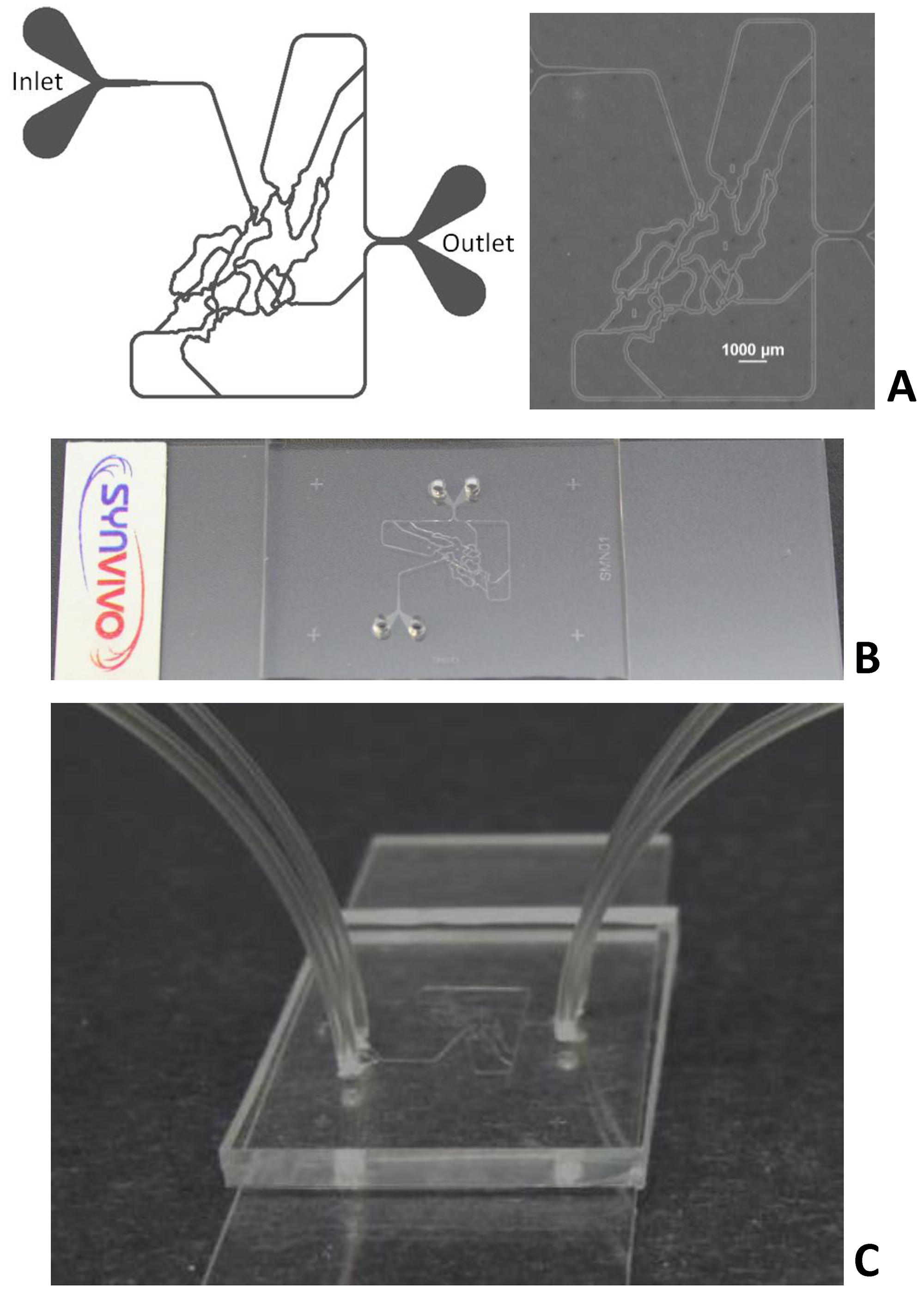
Figure 1. SynVivo-SMN devices. Left panel (Figure 1A) shows a schematic of the device. Right panel (Figure 1A) shows bright field image of the device. Figure 1B shows the SynVivo-SMN device mounted on microscope glass slide. Figure 1C shows the device with Tygon™ tubing attached to the inlet/outlet ports following priming with water.
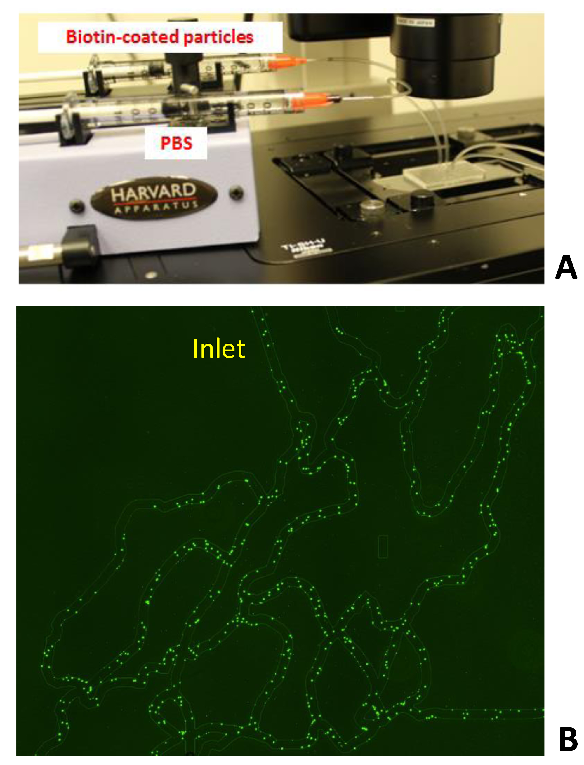
Figure 2. Typical adhesion assay in SynVivo-SMN. Figure 2A shows an image of the experimental-set up. Figure 2B shows biotinylated particles following binding in the device. Note that particle adhesion is found to be localized near bifurcation compared to the branches of the network.
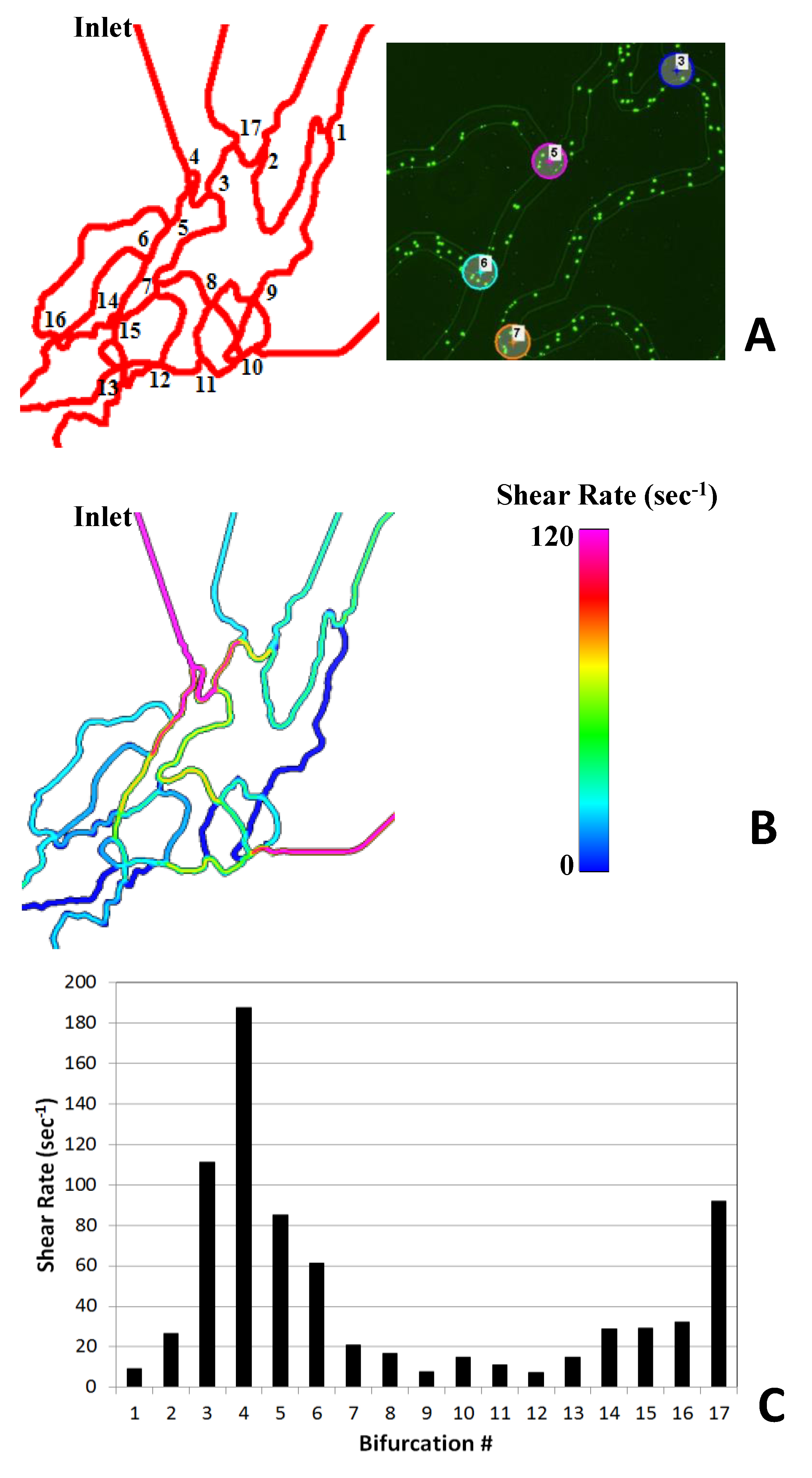
Figure 3. Shear analysis in SynVivo-SMN. Figure 3A shows all the bifurcations numbered in the network and a magnified view of bifurcations from the experiments. Figure 3B shows the qualitative wall shear map in the bifurcations in the network. Figure 3C shows the quantitative information on shear in the branches in the network. Note that the shear patterns in the network are complex and span the physiological ranges of shear rates (0-240 sec-1) found in vivo.
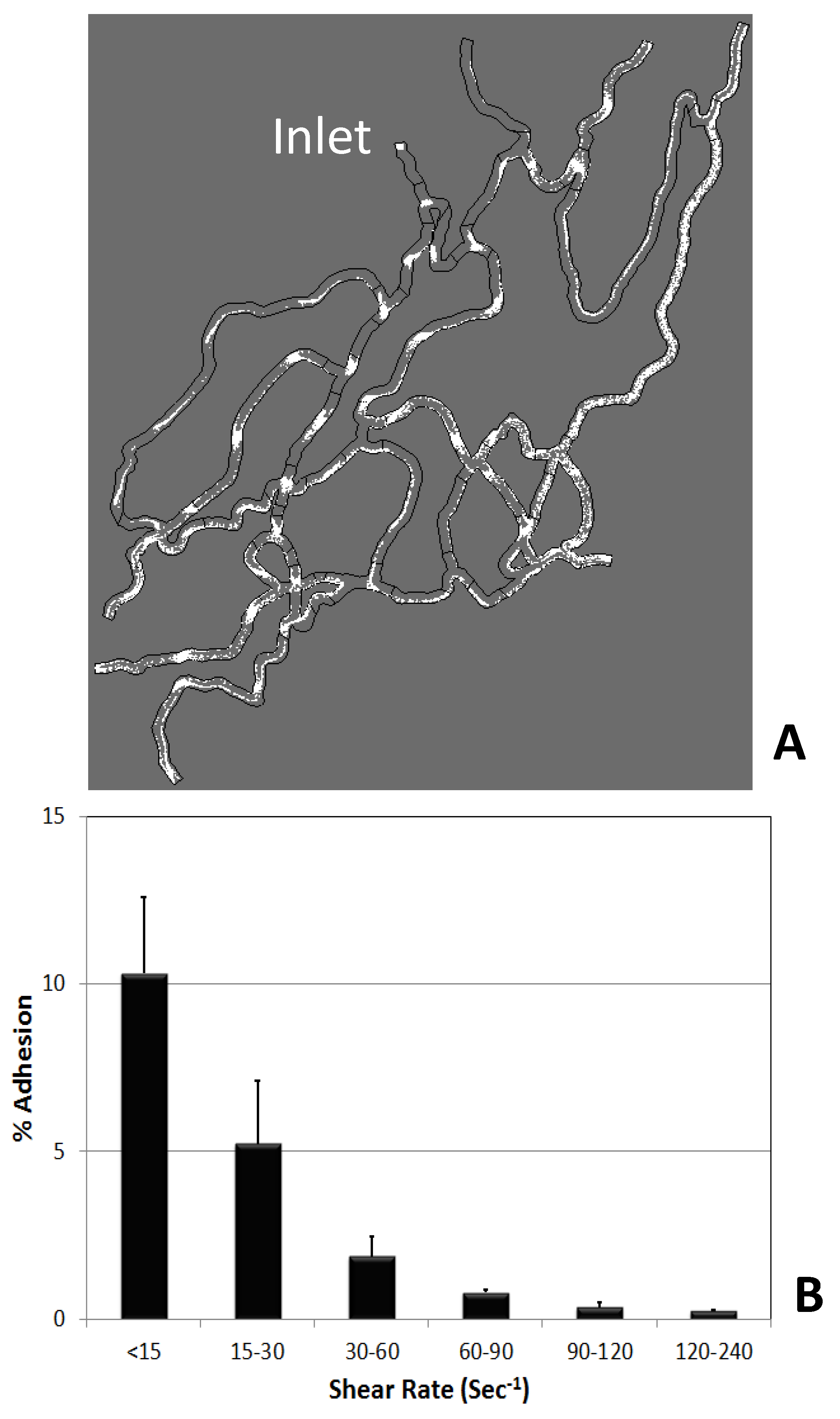
Figure 4. Generation of shear adhesion map. Figure 4A highlights the particle trajectories (shown in white) in the networks obtained from the CFD simulation. These trajectories are post-processed to obtain particle fluxes in the different branches and the bifurcations. Figure 4B shows the shear adhesion map obtained from a single SynVivo-SMN experiment.
