A highly sensitive ribozyme-based assay, applicable to high-throughput screening of chemicals targeting the unique process of RNA editing in trypanosomatid pathogens, is described in this paper. Inhibitors can be used as tools for hypothesis-driven analysis of the RNA editing process and ultimately as therapeutics.
Substantial progress has been made in determining the mechanism of mitochondrial RNA editing in trypanosomes. Similarly, considerable progress has been made in identifying the components of the editosome complex that catalyze RNA editing. However, it is still not clear how those proteins work together. Chemical compounds obtained from a high-throughput screen against the editosome may block or affect one or more steps in the editing cycle. Therefore, the identification of new chemical compounds will generate valuable molecular probes for dissecting the editosome function and assembly. In previous studies, in vitro editing assays were carried out using radio-labeled RNA. These assays are time consuming, inefficient and unsuitable for high-throughput purposes. Here, a homogenous fluorescence-based “mix and measure” hammerhead ribozyme in vitro reporter assay to monitor RNA editing, is presented. Only as a consequence of RNA editing of the hammerhead ribozyme a fluorescence resonance energy transfer (FRET) oligoribonucleotide substrate undergoes cleavage. This in turn results in separation of the fluorophore from the quencher thereby producing a signal. In contrast, when the editosome function is inhibited, the fluorescence signal will be quenched. This is a highly sensitive and simple assay that should be generally applicable to monitor in vitro RNA editing or high throughput screening of chemicals that can inhibit the editosome function.
The process of RNA editing, a post-transcriptional mRNA modification, was first discovered in trypanosomatids1. Since then, substantial work has been conducted in studying the mechanism behind RNA editing in Trypanosoma brucei2,3. In a series of enzymatic reactions, the editosome, a core complex of about 20 proteins, creates mature mitochondrial mRNAs for multiple components of the energy generating oxidative phosphorylation system. The order of catalytic events is endonucleolytic cleavage, uridylate (U) addition or deletion, and ligation, as dictated by guide RNAs (gRNAs)4.
In addition to the core editosome complex proteins, a number of accessory factors have also been identified5-7. These proteins are mostly seen grouped in independent complexes. However, the order of protein assembly in the core editosome complex and the interaction patterns of the core complex with the accessory complexes are yet to be determined. Targeting the RNA editing process in trypanosomatids may provide chemical dissectors that aid in studying the assembly and function of the editosome complex. Furthermore, functional studies on several editosome proteins have shown essentiality across different life stages, indicating their potential as drug targets8-12. Therefore, the found inhibitors of the editosome may also act as lead compounds against trypanosomatids. This is timely, as drugs currently available against diseases caused by trypanosomatid are toxic, inefficient and expensive13,14.
An efficient and convenient in vitro assay is necessary to explore the chemical universe for specific inhibitors that block RNA editing. Three assays have been developed and used to monitor editosome activities: (a) full round in vitro RNA editing assay15, (b) pre-cleaved in vitro RNA editing assay16,17, and (c) hammerhead ribozyme (HHR)-based assay18. The first two assays rely on direct visualization of the edited product (ATPase 6 mRNA) with the help of radioactivity. The HHR-based assay uses a modified version of the ATPase 6 mRNA that is modeled to behave as a ribozyme upon editing. The functional ribozyme then specifically cleaves a radiolabeled RNA substrate, serving as a reporter. Recently, Moshiri et al. developed a ‘mix and measure’ HHR-based in vitro reporter assay to monitor RNA editing where the radiolabeled RNA substrate is replaced with a fluorescence resonance energy transfer (FRET) substrate19. The principle advantages of this assay are: (a) it is a rapid and convenient mix and measure type of assay, as the production of active ribozyme and substrate cleavage occur simultaneously in the same tube in low volume (i.e. 20 μl), (b) it avoids the use of radioactively labeled materials, (c) sensitivity that is afforded by fluorescence instrumentation in a micro-titer plate format, and (d) a high signal to noise ratio. Using this assay, the effect of known RNA editing ligase inhibitors against purified editosome was confirmed19. This experiment validated the assay for rapid identification of RNA editing inhibitors, primarily against whole editosomes from T. brucei.
Figure 1 is a detailed step-by-step schematic of the fluorescence-based in vitro RNA editing assay. This protocol can either be used for monitoring RNA editing in vitro or easily be adapted for screening compound libraries of various scales.
The protocol below describes the procedure for performing the fluorescence-based RNA editing assay. The assay can be performed in a single PCR tube, 96-well, or 384-well plates depending on the scope of the experiment. Subsequently the fluorescence signal can be read on a suitable real time PCR detection system. The assay here is described in the context of 384-well plates.
1. Culturing T. brucei Cells
2. Isolation of Crude Mitochondria
NOTE: All the steps should be performed on ice or at 4 °C to preserve editosome activity.
3. Editosome Purification
4. RNA Preparation
5. Fluorescence-based RNA Editing Assay
To demonstrate the necessary steps required for setting up a large-scale screen, Figures 2-5 are representative control experiments related to the quality of the assay. These are essential control experiments for a consistent assay over several days of screening or for comparison of different screens.
Assessing the Fluorescence Signal-to-noise Ratio
To ensure the stability and quality of the fluorescein-labeled oligoribonucleotide substrate in a large-scale setup, Z’-factor, defined as the difference between the assay background and the maximum signal was calculated using the active ribozyme molecule (A6Rbz). Assays with Z’-factor >0.5 are considered acceptable for high-throughput screen. Figure 2 shows representative data using the FRET substrate labeled with 5’ fluorescein (FAM; emitter) and 3’ N,N’-tetramethylrhodamine (TAMRA; quencher) (A6Rbz_F/T). This substrate produced a Z’-factor of 0.64 when calculated using 72 replicates in the presence and absence of A6Rbz.
In this assay an alternative substrate using Iowa Black dark quencher (A6Rbz_F/Ib) can yield a better signal-to-noise ratio compared to TAMRA. This is because Iowa Black, unlike TAMRA, emits absorbed energy as heat and not light. The Z’-factor obtained using the FAM/Iowa Black (F/Ib)-labeled substrate was 0.68. The improvement in the Z’-factor for the F/Ib-labeled substrate over TAMRA-labeled substrate is because of its relatively lower background. These representative results show that both substrates are viable options for use in a high-throughput screen.
Determining the Editing Activities of Glycerol Gradient Fractions
To determine and select the most active editosome fractions for experiments, the glycerol gradient fractions were tested for in vitro editing using the fluorescence-based assay (Step V). These data show (Figure 3) fractions 7-12 as the most active fractions (≥50% editing activity; with the most active fraction as 100%). These fractions can be combined when more editosome is required.
Calculating the Z’-factor when the Editosome Fractions were Combined
To determine the effect of combinig the editosome fraction, the Z’-factor value of 0.6 was calculated when the most active fractions (F8+F9+F10) were used as the source of editosome. To calculate the Z’-factor 72 replicates were tested in the presence and absence of the editosome (Figure 4). The F/Ib substrate was used in this experiment. These data show that based on the Z’-factor, combining the glycerol gradient fractions have minimal effect on the quality of the assay.
Representative Control Experiment for the Assay
Representative data comparing different control samples were used to analyze the variables in the assay. As shown in Figure 5, reactions missing any RNA editing components (reactions 1-4) do not cleave the substrate. Cleavage of the substrate as measured by the fluorescence and relative editing activity is only observed in the presence of all components of the editing reactions in the absence of an inhibitor (reaction 5) or in the presence of all editing components and an inactive compound (reaction 6). In contrast, an inhibitory compound can block RNA editing and is used as a positive control (reaction 7). Here, Mordant black (MrB) and C53 compounds were used as the positive and negative controls, respectively.
Figure 1. Hammerhead ribozyme-based in vitro RNA editing assay. A) Step-by-step schematic representation of the fluorescence-based in vitro editing assay: (a) Hybridization of the pre-edited hammerhead ribozyme (pre-edited A6Rbz) with its guide RNA (gA6Rbz). (b) Recognition and interaction of the RNA duplex by the purified editosome complex. (c) Deletion RNA editing catalyzed by the editosome. (d) Dissociation of the edited A6Rbz from the editosome and gA6Rbz by heating at 85 °C and addition of guide RNA competitor (gA6Rbz_comp). (e) Hybridization of the FRET substrate with the active A6 hammerhead ribozyme (active A6Rbz). (f) Detection of FAM signals following cleavage of the FRET substrate by the active ribozyme. B) The pre-edited hammerhead ribozyme (pre-edited A6Rbz) is shown in association with gA6Rbz that specifies the deletion of three Us (double-headed arrow) from the editing site (ES). As a result of RNA editing in the presence of functional editosome the inactive ribozyme is edited into its active form (edited A6Rbz) that can now cleave the FRET substrate (cleavage site indicated by an arrow). The conserved 5′-CUGA-3′ of the edited A6Rbz in the catalytic core essential for ribozyme activity (edited site) is highlighted (This figure has been modified from Moshiri19). Please click here to view a larger version of this figure.
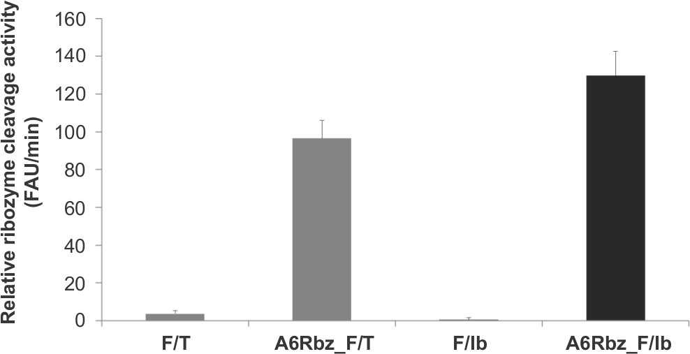
Figure 2. Signal-to-noise ratio comparison between FRET substrates harboring different quenchers. Ribozyme (A6Rbz) activity using FAM/TAMRA (F/T) and FAM/Iowa Black FQ (F/Ib) substrates. The y-axis represents fluorescence arbitrary unit (FAU) per minute.
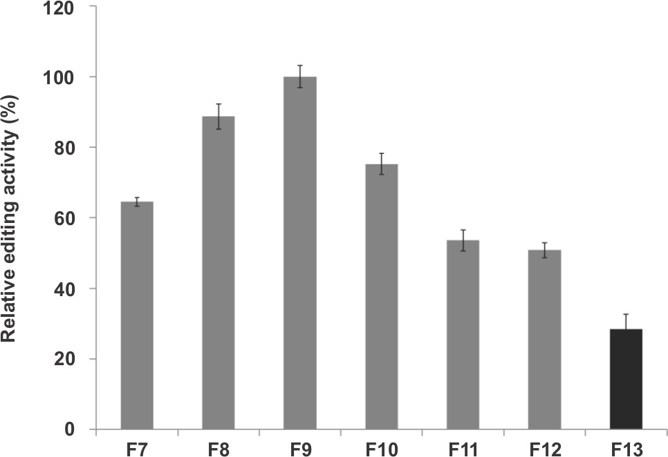
Figure 3. RNA editing activity of select fractions obtained from glycerol gradient centrifugation of the mitochondrial lysate. Fraction #9 (F9) has the highest activity. The y-axis represents relative editing activity in percentage, considering F9 as 100%.
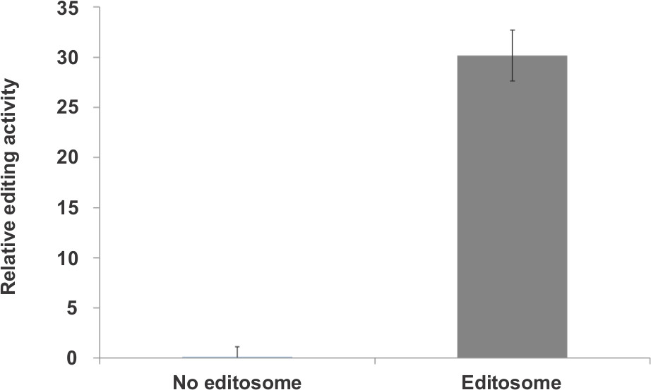
Figure 4. Z’-factor calculation using the most active fractions (F8+F9+F10) as the editosome source. Experimental variation was obtained from 72 replicates of each type of reaction. Z’-factor was calculated as 0.6. The y-axis represents relative editing activity.
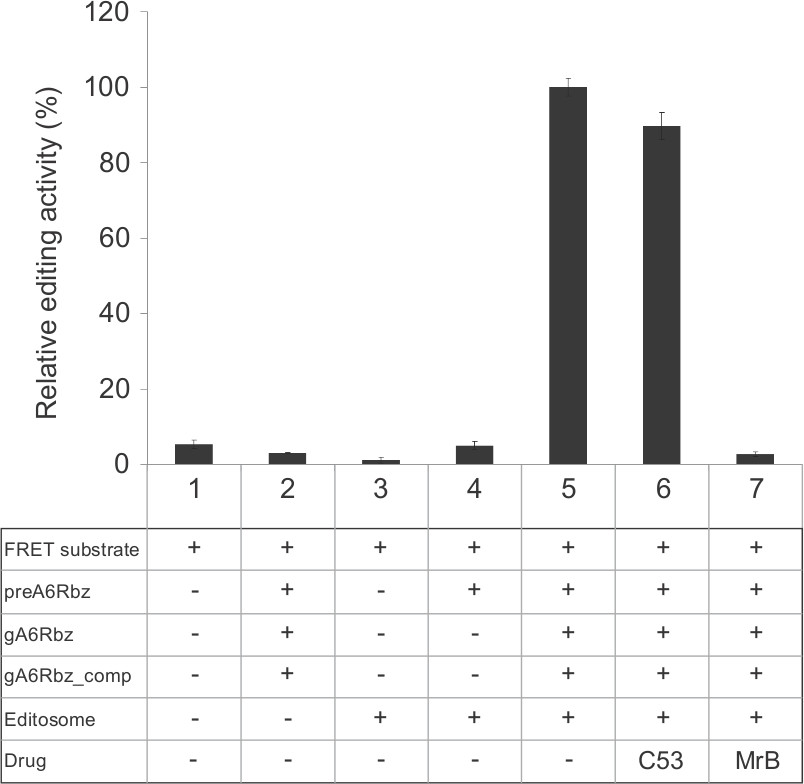
Figure 5. Representative experiment for the fluorescence-based RNA editing assay. Reactions were performed in a final volume of 20 µl per well and CFX384 TouchTM real-time PCR detection system was used for measuring the fluorescence signal. The graph presents various controls in addition to a complete editing reaction that contains all components for the assay (#5). The fluorescence was measured in the presence of FRET substrate alone (#1) to assess the integrity of the substrate. Sample #2 was performed in the absence of the editosome to monitor any ribozyme activity prior to editing. To assess the effect of denatured editosome on the substrate, the fluorescence was measured in the presence of the editosome and FRET substrate only (#3). To test the guide-directed editing specificity, a sample in the absence of gA6Rbz was used (#4). A non-inhibitory (C53, #6) and an inhibitory compound (MrB, #7) were used to test the effect on the editing assay. While RNA editing is inhibited significantly by MrB, C53 has negligible effect. The error bars correspond to an experimental variation (standard deviation) between 10 replicates. The y-axis represents relative editing activity in percentage, considering the complete reaction (#5) as 100%.
10% | 30% | |
2x HHE gradient buffer | 5 ml | 5 ml |
Glycerol | 1 ml | 3 ml |
DEPC H2O | 4 ml | 2 ml |
1 M DTT | 10 µl | 10 µl |
Table 1. 10% & 30% Glycerol Solution (10ml each).
Pre-edited A6 Ribozyme (preA6Rbz) | |
preA6Rbz DNA template | 5’-ACATTTGATCTATTGTTTCGTCCTCACGGACTCA TCAAAAGTCACAACTTTCCCTTTCTCTCCTCCCCCTAACCTTT CCCCCTATAGTGAGTCGTATTA-3’ |
preA6Rbz RNA | 5’-GGGAAAGGUUAGGGGGAGGAGAGAAAGGGAAA GUUGUGACUUUUGAUGAGUCCGUGAGGACGAAACAAUAGA UCAAAUGU-3’ |
RNA stock conc. | 1 μM |
Guide A6 Ribozyme (gA6Rbz) | |
gA6RBz DNA template | 5′-AAAAAAAAAAAAAAAAATAATTATCATATCACTGT CAAGGGAAAGTTGTGAGGGTGATGAGTCCGTGTATATCCCC CTATAGTGAGTCGTATTA-3′ |
gA6Rbz RNA | 5′-GGGAUAUACACGGACUCAUCACCCUCACAACUU UCCCUUGACAGUGAUAUGAUAAUUAUUUUUUUUUUUUUUUUU-3’ |
RNA stock conc. | 2.5 μM |
Guide A6 Ribozyme Competitor (gA6RBz_comp) | |
gA6Rbz_comp DNA template | 5′-GGATATACACGGACTCATCACCCTCACAACTTTC CCTTGACAGTGATATGATAATTATTTTTTTTTTTTTTTTTCCCTAT AGTGAGTCGTATTA-3′ |
gA6Rbz_comp RNA | 5’-AAAAAAAAAAAAAAAAAUAAUUAUCAUAUCACUG UCAAGGGAAGUUGUGAGGGUGAUGAGUCCGUGUAUAUCC-3’ |
RNA stock conc. | 12.5 μM |
Active A6 Ribozyme (A6RBz) | |
A6Rbz DNA template | 5′-ACATTTGATCTATTGTTTCGTCCTCACGGACTCAT CAGTCACAACTTTCCCTTTCTCTCCTCCCCCTAACCTTTCCCC CTATAGTGAGTCGTATTA-3′ |
A6Rbz RNA | 5’-GGGAAAGGUUAGGGGGAGGAGAGAAAGGGAAA GUUGUGACUGAUGAGUCCGUGAGGACGAAACAAUAGAUCA AAUGU-3’ |
RNA stock conc. | 1 μM |
FRET substrate | |
TAMRA quencher | 5’-FAM-GAUCUAUUGUCUCACA-TAMRA-3' (Eurogentec) |
Iowa Black quencher | 5’-FAM- GAUCUAUUGUCUCACA-Iowa Black-3' (IDT) |
RNA stock conc. | 15 μM (for both) |
Table 2. DNA Templates and RNA Substrates.
Composition | Editing reaction (μl) |
10x HHE | 1.5 |
0.1 M CaCl2 | 1 |
100 mM ATP | 0.2 |
10% Tritonx-100 | 0.2 |
500 ng/μl Torula Yeast RNA | 1 |
Editosome | 5 |
RNase free H2O | 7.1 |
1 μM preA6Rbz | 1 |
2.5 μM gA6Rbz | 1 |
Total | 18 |
Table 3. Reaction Composition and Master Mix.
A novel high-throughput screening method to identify inhibitors against the RNA editing complex of Trypanosomes was presented, providing a new tool for drug discovery to counter diseases caused by trypanosomatids. FRET-based ribozyme assay has been extensively used for different purposes20-22; however, we have utilized the capacity of FRET-based ribozyme assay for in vitro monitoring of RNA editing activity19. This assay could potentially be adapted to other types of RNA editing in eukaryotes, such as nucleotide substitution editing of nucleus-encoded RNAs of mammals23.
The novelty of this assay is not in establishing FRET-based ribozyme assays per se, but in developing a method that incorporates this technique into an RNA editing assay that is amenable to high-throughput screening of chemicals against editosome. The ribozyme assay-based method has several advantages over other screens and assays currently utilized for this purpose. Specifically, the technique offers a sensitive and reproducible “mix-and-measure” assay relying on a fluorescent substrate as opposed to a radiolabeled one, thereby making it fit for high-throughput screening19. Real-time monitoring of a fluorescence signal after addition of the substrate instead of a simple end-point signal makes it possible to more accurately determine IC50 values of assayed compounds19. Furthermore, it permits testing of the inhibitory effects of compounds on the whole-editosome complex as opposed to individual recombinant proteins. Given the dynamic nature of interactions within protein complexes, it can be suggested that this method allows identification of inhibitors against editosome proteins in a more biologically representative setting24. In addition, targeting the whole-editosome complex provides an opportunity to potentially arrest the progression of RNA editing at various transient steps that have not been previously identified. Thus, the technique will allow gaining a better perspective as to interactions and activities that are crucial to the process of RNA editing. This approach has been found to be successful in the past as exemplified by the elucidation of prokaryotic ribosome complex assembly and function utilizing antibiotics, such as viomycin and erythromycin, acting as inhibitors of the complex24,25.
To ensure the successful completion of the method, certain adjustments in protocols may be necessary. First, selection of the proper glycerol gradient mitochondrial extract fraction is crucial. Here, fractions 7 through 11, corresponding to the ~20S region of the gradient, showed the highest editing activity, with fraction 9 exhibiting maximum activity. Although this fraction was selected for all of the tests presented, it is essential to test all fractions beforehand in order to assess in which fraction the editing activity peaks. Second, to validate fraction selection, it is critical to perform preliminary tests to assess the signal-to-noise ratio by calculating the Z’-factor value. If proper mitochondrial extract glycerol gradient fractionation has been completed, a Z’-value of 0.6 can be achieved. Next, it is recommended to re-evaluate the reaction volume of mitochondrial extract necessary to achieve maximal editing activity via titration19.
An important limitation of the technique presented in this paper concerns the laborious and time-consuming process of mitochondrial extract glycerol gradient fractionation by which the editosome complex is purified19. Despite these disadvantages, this technique is preferentially suggested to obtain crude editosome fractions given its low cost versus yield potential, thereby qualifying it for application to high-throughput screening. In contrast, other methods such as TAP-tag purification, which are able to give more purified editosome fractions, are advisable for low to medium throughput assays such as in the case of secondary assays. Moreover, in order to ensure specific inhibitory effect of compounds, it is necessary to include controls to test them against the activity of ribozyme (A6Rbz) in the absence of the editosome. It should be noted that previous studies have highlighted that this method may be ineffective at calculating IC50 values for dye-like compounds due to the possible interference at high compound concentrations19.
To further increase the sensitivity of the HTS technique presented, developing a similar fluorescence-based assay involving pre-cleaved pre-edited ribozyme is beneficial. This would allow bypassing the rate-limiting endonucleolytic cleavage step catalyzed by endonucleases in the editosome complex. Another advantage of this modified assay would be the application as a secondary screening tool to monitor the effect of the inhibitors identified through the primary screen on ExoUase, TUTase and ligation catalytic activities. The currently proposed assay is limited to the detection of inhibitors against the deletion type of RNA editing. Therefore, another avenue to explore would be the modification of the presented assay to enable monitoring of compound effects on the insertion type of RNA editing.
The authors have nothing to disclose.
Najmeh Nikpour and Fiona Alum provided suggestions and edited this manuscript. Department of Biochemistry at McGill University supported HM and VM with the CIHR Training Initiative in Chemical Biology. This work was supported by Canadian Institute of Health Research (CIHR) grant 119464 (to R. S.).
Name of Material/ Equipment | Company | Catalog Number | Comments/Description |
SDM-79 Medium | Gibco by life technologies | ||
Fetal Bovine Serum | life technologies | 12483-020 | heat inactivation at 550C for 1 h |
Hemin, minimum 80% | Sigma | H5533-10G | |
Penicillin-Streptomycin Sollution | Fisher Scientific | MT-30-002-CI | |
Dnase 1 recombinant, Rnase Free | Roche | 4716728001 | |
T7 RiboMax Express Large scale RNA production system | Promega | P1320 | |
Kimble Kontes Dounce Tissue Grinders | Fisher Scientific | K885300-0040 | |
Gradient Master, ver 5.25 | Biocomp | 107-201M | |
Ultra Clear Tube, 13.2ML | Beckman Coulter | 344059 | |
Optima L-100XP Ultracentrifuge | Beckman Coulter | 392052 | |
SW 41 Ti ROTOR | Beckman Coulter | 331336 | |
MicroSeal 'B' Seal, Seals | Biorad | MSB1001 | |
CFX 384 Touch Real-Time PCR Detection System | Biorad | 185-5484 | |
Acryl/Bis solution (19: 1), 40% (w/v) | Bio Basic | A0006-500ML | |
Urea, Molecular biology grade, 1Kg | life technologies | AM9902 |