Here we present a protocol to synthesize novel, high-aspect ratio biocomposites under biological conditions and in liquid media. The biocomposites scale from nanometers to micrometers in diameter and length, respectively. Copper nanoparticles (CNPs) and copper sulfate combined with cystine are the key components for the synthesis.
The goal of this protocol is to describe the synthesis of two novel biocomposites with high-aspect ratio structures. The biocomposites consist of copper and cystine, with either copper nanoparticles (CNPs) or copper sulfate contributing the metallic component. Synthesis is carried out in liquid under biological conditions (37 °C) and the self-assembled composites form after 24 hr. Once formed, these composites are highly stable in both liquid media and in a dried form. The composites scale from the nano- to micro- range in length, and from a few microns to 25 nm in diameter. Field emission scanning electron microscopy with energy dispersive X-ray spectroscopy (EDX) demonstrated that sulfur was present in the NP-derived linear structures, while it was absent from the starting CNP material, thus confirming cystine as the source of sulfur in the final nanocomposites. During synthesis of these linear nano- and micro-composites, a diverse range of lengths of structures is formed in the synthesis vessel. Sonication of the liquid mixture after synthesis was demonstrated to assist in controlling average size of the structures by diminishing the average length with increased time of sonication. Since the formed structures are highly stable, do not agglomerate, and are formed in liquid phase, centrifugation may also be used to assist in concentrating and segregating formed composites.
Copper is a highly reactive metal that in the biological world is essential in some enzyme functions 1,2, but in higher concentrations is potently toxic including in the nanoparticulate form 3,4. Concern over copper toxicity has become more relevant as CNPs and other copper-based nanomaterials are utilized, due to the increased surface area/mass for nanostructures. Thus, even a small mass of copper, in nanoparticle form, could cause local toxicity due to its ability to penetrate the cell and be broken down into reactive forms. Some biological species can complex with and chelate metal ions, and even incorporate them into biological structures as has been described in marine mussels 5. In studying the potential toxic effects of nanomaterials 4, it was discovered that over time, and under biological conditions used for typical cell culturing (37 °C and 5% CO2), stable copper biocomposites could be formed with a high-aspect ratio (linear) structure.
By a process of elimination, the initial discovery of these linear biocomposites, which occurred in complete cell culture media, was simplified to a defined protocol of essential elements needed for the biocomposites to self-assemble. Self-assembly of two types of highly linear biocomposites was discovered to be possible with two starting metal components: 1) CNPs and 2) copper sulfate, with the common biological component being cystine. Although more complex, so called “urchin” and “nanoflower” type copper-containing structures with nanoscale and microscale features have been previously reported, these were produced under non-biological conditions, such as temperatures of 100 °C or greater 6-8. To our knowledge, synthesis of individual, linear copper-containing nanostructures that are scalable in liquid phase under biological conditions has not been previously described.
One of the starting materials utilized for synthesis of nanocomposites, namely CNPs, has been reported previously to be very toxic to cells 4. It has recently been reported that after the nanocomposites are formed, these structures are less toxic on a per mass basis than the starting NPs 9. Thus, the synthesis described here may be derived from a biological and biochemical reaction that has utility in stabilizing reactive copper species, both in the sense of transforming the NP form into larger structures and in producing composites less toxic to cells.
In contrast to many other nanomaterial forms which are known to aggregate or clump upon interaction with biological liquid media 10,11, once formed, the highly linear composites described here avoid aggregation, possibly due to a redistribution of charge which has been previously reported 9. As detailed in the current work, this avoidance of aggregation is convenient for the purposes of working with the structures once formed for at least 3 reasons: 1) composite structures once formed may be concentrated using centrifugation and then easily dispersed again using vortex mixing; 2) formed structures can be decreased in average size by sonication for different periods of time; and 3) the formed linear structures may provide an additional tool for avoiding the recently described “coffee ring effect” 12 and thus provide a dopant for creating more evenly distributed coatings of materials, especially those containing spherical particulates.
1. Planning of Experiments
2. Preparation of Materials
3. Synthesis Using Copper Sulfate
4. Characterization and Handling of Biocomposites Post-synthesis
Figure 1 shows a flow-chart schematic of the synthesis steps to form the linear biocomposites described in this work. CNPs or copper sulfate as starting materials are combined with sterile water to form a 2 mg/ml solution, this solution is mixed and sonicated to provide an even mixture, and this copper solution is then mixed in the following ratio for synthesis: 949 parts sterile water: 50 parts copper mixture: 1 part cystine stock solution. The actual volumes may be increased or decreased according to these ratios to scale up or scale down the final synthesis yield. After incubation for at least 2 hr as indicated, linear biocomposite structures are formed which over time can be observed under normal white light microscopy or by eye as the liquid solution changes in appearance.
Figure 2 shows a representative result from the initial discovery of these linear structures in complete cell culture media over a period of 82 hr. Our laboratory was carrying out evaluation of the potential toxicity of different nanomaterials, including CNPs on normal cells and cancer cells, and the cells shown in Figure 2 are a fast-growing brain tumor cell line from ATCC (CRL-2020). A key supplement to the complete media used for these cells is cystine, which turned out to be the essential component to the discovery of why these linear structures were forming in the cultures (see below and discussion section). From an initial even dispersion of CNPs which quickly aggregate into microstructures (Figures 2A and 2B), over time the smaller particles are cleared and larger aggregates are formed (Figures 2C and 2D). Finally, larger aggregates with fine, linear structures appear in the same wells (Figures 2E-H), forming the “urchin” type structures previously reported in the literature using non-biological methods 6. Comparison of the final two time points, at 69 and 82 hr (Figures 2G and 2H, respectively), shows that development of the large urchin type structures remains quite stable, as indicated by imaging the same exact field.
To explain why these urchin-like structures under these conditions in the cell cultures were observed, we started eliminating media components to determine if the essential elements could be isolated. We discovered that a key component and supplement to the cell culture media was cystine, which by the process of elimination was ultimately identified as an essential component for the self-assembly process. By simplifying the synthesis components (see Figure 1), we could ultimately form high-aspect ratio (linear) structures in liquid, that could be shown over time to transform from nanoparticle form to linear form, without the need of cells, or any of the other cell culture components (Figures 3A-C).
Figure 4 shows characterization of the discovered novel structures using electron microscopy, including a transmission electron microscopy (TEM) image capturing nanoparticle starting material and forming linear nanostructures (Figure 4A). Representative scanning electron micrograph (SEM) images are shown for starting material CNPs, linear structures formed from the NPs, and linear structures formed from copper sulfate starting material (Figures 4B-F, respectively).
To verify that the biocomposites contained cystine or cystine-derived material as an essential biological component of the composites, the formed structures were analyzed using EDX with SEM microscopy. Representative screen shots from analyzed materials are shown in Figure 5. Importantly, when comparing CNPs and biocomposites from the CNPs, a prominent sulfur peak appears (Figure 5B), which is not present in the CNP starting material (Figure 5A). For the biocomposites using copper sulfate as starting material (Figure 5C), carbon and nitrogen peaks appear (Figure 5D), which is consistent with the presence of cystine for this biocomposite.
At this time, a method for controlling length and size of composites during synthesis has not been identified. However, to investigate if average size of structures could be controlled after synthesis, linear biocomposites were sonicated for different periods of time as shown in Figure 6. With increased time of sonication, it was shown that the average size of the linear biocomposites decreased, as shown by bright field microscopy (Figures 6A-D). As a method for concentrating and segregating formed composites, centrifugation can be used, and as shown in Figures 6E-G, a visible pellet can be developed depending upon the volumes and centrifugation forces used.
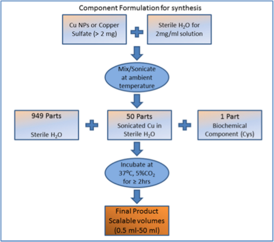
Figure 1. Representative flow chart of synthesis design. Cu NPs = copper nanoparticles; Cys = cystine. Please click here to view a larger version of this figure.
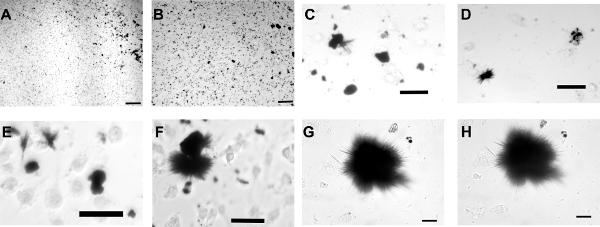
Figure 2. Representative formation of copper biocomposite structures in brain tumor cell cultures with complete cell culture media. A representative experiment tracked over a total of 82 hr is shown in cell culture containing brain tumor cells and CNPs (50 µg/ml). Panels A and B show the culture wells at time 0, after CNPs have settled to the bottom of the well. Panels C-H show subsequent time points at 17, 24, 36, 49, 69, and 82 hr, respectively. Panels G and H represent the same field at 69 and 82 hr. All images were obtained using brightfield microscopy to enhance contrast of copper material. Scale bars = 100 microns for A+B, 50 microns for C-E, and 25 microns for F+G. Please click here to view a larger version of this figure.
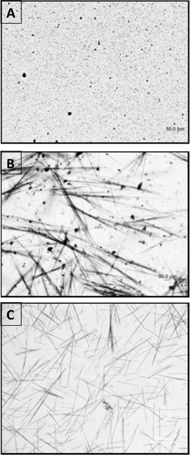
Figure 3. Transformation of CNPs to linear biocomposites. CNPs were combined with cystine and water as indicated in Figure 1 and in protocol section with a total volume of 7 ml. Panel A shows the synthesis vessel at time 0, panel B shows 3 hr, and panel C shows 6 hr. Images were obtained using brightfield microscopy with scale bar indicated (50 microns). Please click here to view a larger version of this figure.
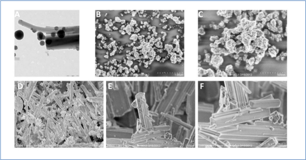
Figure 4. Electron microscopy characterization of synthesized biocomposites. Panel (A): TEM of CNP starting material (round) with the forming linear composites. Panels (B) and (C) show characterization of the starting CNPs using SEM. Panel (C) is a zoomed image of (B). Panel (D) shows SEM of composites formed from CNPs and cystine. Panels (E) and (F) show SEMs of the copper sulfate biocomposites. Panel (F) is a zoomed image of (E). Scale bars are indicated in all images and = 200 nm in (A), 1 micron in (B), 500 nm in (C), 5 microns in (D), 2 microns in (E), and 1 micron in (F). Please click here to view a larger version of this figure.
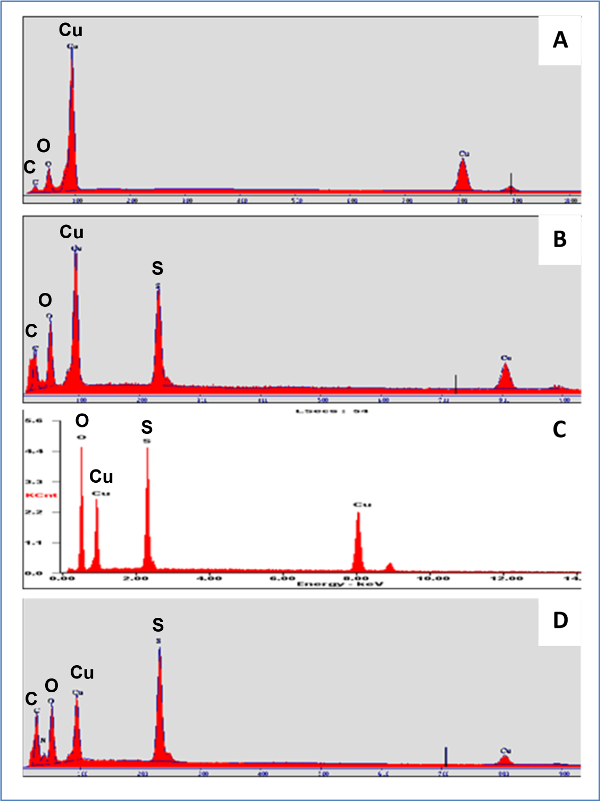
Figure 5. EDX (SEM) analysis of starting materials and synthesized linear composites. Screen snapshots of SEM scanned samples using EDX analysis for elemental content. Panel (A) = starting CNPs; Panel (B) = biocomposites from CNPs and cystine; Panel (C) = copper sulfate starting material, and Panel (D) = composites from copper sulfate and cystine. For peak labeling, C = carbon, O = oxygen, Cu = copper, S = sulfur, and N = nitrogen. Larger labels for elemental identity have been placed above key peaks for ease of viewing. Please click here to view a larger version of this figure.
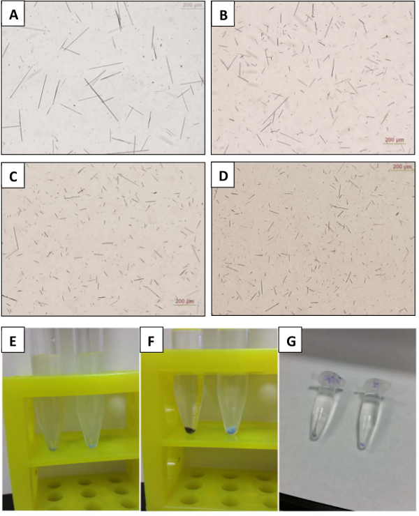
Figure 6. Modification of linear composite size and concentration post synthesis. Linear structures synthesized from copper sulfate were sonicated for 0, 15, 30, or 60 min, respectively, as shown in panels (A-D). Images were obtained using brightfield microscopy with scale bar of 200 microns indicated in all images. Panels (E-G), concentration of biocomposites using centrifugation: 6 ml of linear structures derived from CNPs (E, left) and copper sulfate (E, right) are shown settling under gravity after 10 min. With 10 min of centrifugation at 500 x g, a compacted pellet is formed (panel F). A smaller volume (500 µl) of the same material was concentrated as shown in (panel G) (CNP-derived structures are shown in the left tube and copper sulfate-derived structures in the right tube). Please click here to view a larger version of this figure.
While evaluating potential toxic effects of nanomaterials including CNPs, it was observed that over the long-term, CNPs were transformed from an initially more dispersed particulate distribution to a larger, aggregated form (Figure 2). In some cases, these highly aggregated formations which were produced in the cell culture dish, under biological conditions, formed highly linear projections from the central aggregate, reminiscent of previously described copper- containing “urchins” 6. It should be pointed out that under the conditions shown here, the concentration of CNPs added to the cells was sub-maximal, thus not killing all of the cells in culture (see Figure 2). From these initial observations, experiments were then continued by eliminating successively more components of the cell culture conditions (including ultimately the cells themselves) to find the remaining key components needed for synthesis of linear copper biocomposites.
We discovered that cystine, a supplemented component of the original cell cultures, when combined with CNPs under the correct biological conditions, could result in transformation of the nanoparticulate form into a highly linear biocomposite that contained both the metal and biochemical components as indicated by EDX analysis by scanning electron microscopy of the prepared samples (Figure 5). Thus, sulfur, which is not present in the starting CNP material, shows a prominent peak in the synthesized biocomposites, indicating that both copper and components of the biochemical material cystine are essential for the formed linear structures.
It was further shown that by using similar synthesis conditions, but replacing CNPs with copper sulfate, highly linear biocomposites could also be formed. In fact, synthesis using copper sulfate and cystine tended to result in “cleaner” end products, in that no unreacted CNPs were ever present, since copper sulfate is fully soluble in water, which was used as a solvent in all of the syntheses reported here. Further, copper-sulfate biocomposites distinguished themselves from CNP-derived composites in retaining a blue color which was apparent upon centrifugation of the material.
Other groups have previously studied copper-cystine complexes and defined some key chemical properties. For example, Kahler et al. demonstrated the formation of copper-cystine complexes in the form of fine fibers, which were evident upon drying but unstable in solution 13. In other examples, complexation studies with L-cystine and different metal cations confirms copper and cystine complex formation in an aqueous medium 14 and formed complexes may be mononuclear or polynuclear, with the sulfur from cystine contributing to the complex formation 15. A number of studies have reported interactions between cystine and copper in biological systems. For example, at physiological pH, copper (II) ions coordinate with histidine and cystine in simulated plasma to form complexes 16, and sulfur containing amino acids were shown to be protective against copper toxicity in chicks 17. These findings thus support the essential role of cystine in complexing with copper starting material in our synthesis methods for forming linear biocomposites.
At this time a synthesis strategy has not yet been identified that can directly control the length of individual linear biocomposites shown here. However, the critical steps identified in the described synthesis include: 1) good dispersion by sonication of starting materials in the case of synthesis incorporating CNPs; 2) use of freshly prepared CNPs, copper sulfate, and cystine for effective synthesis of composites; 3) allowing synthesis in the flask to remain undisturbed in the incubator for at least 6 hours; and 4) avoiding “over-reaction” conditions in which branched “urchin”-type, aggregated composites form.
After synthesis is completed, it was shown that sonication of the structures can be utilized effectively to decrease the average size (length) of the structures. Sonicating to make smaller structures may aid in applications such as cell uptake or other biocomposite-cellular interactions. As has been previously reported, charge stabilization of the CNPs during this synthesis by combining with cystine altered the measured zeta potential from positive to a less charged (negative) form 9. This change in charge may help explain why the formed composites show very little aggregation in the dried form or liquid media, which makes handling of the structures much more convenient.
Since the reported synthesis of these CNP-derived and copper sulfate-derived structures is carried out in liquid media, it is anticipated that the process may be highly scalable, meaning that with the correct ratio of components and synthesis conditions, the milliliter synthesis recipe used here could be scaled up or down to include many hundreds of milliliters or more, and would thus be expected to yield more final product, as well. Due to the metal (copper) component of these biocomposites, it is quite straightforward to concentrate synthesized product by centrifugation (Figure 6). Biocomposites formed from CNP starting material retain a darker color once concentrated into a pellet, possibly due to unreacted copper oxide nanoparticles (Figure 6). In comparison, copper sulfate biocomposites when concentrated into a pellet have a blue color, consistent with properties of copper sulfate with various levels of hydration 18.
The novel synthesis reported here is also scalable in the sense that the self-assembled linear structures scale from the nano-scale to the micro-scale as shown using electron microscopy (Figure 4) and traditional white light microscopy (Figures 3 and 6). It is interesting to note that recently, engineered coiled-coil protein microfibers were reported that could incorporate curcumin which allowed for the formed fibers to be observed under fluorescence illumination 19. Similar strategies may be possible to incorporate spacers or tagging agents into the linear composites reported here to provide production of larger structures and/or enhancements for imaging.
The authors have nothing to disclose.
The authors would like to acknowledge the technical assistance of Alfred Gunasekaran in electron microscopy studies at the Institute of Micromanufacturing at Louisiana Tech University, and Dr. Jim McNamara for assistance with additional microscopy studies. The work described was supported in part by Louisiana board of Regents PKSFI Contract No. LEQSF (2007-12)-ENH-PKSFI-PRS-04 and the James E. Wyche III Endowed Professorship from Louisiana Tech University (to M.D.).
Mini Vortexer | VWR (https://us.vwr.com) | 58816-121 | |
CO2 Incubator Model # 2425-2 | VWR (https://us.vwr.com) | Contact vendor | Current model calalog # 98000-360 |
Eppendorf Centrifuge (Refrigerated Microcentrifuge) | Labnet (http://labnetinternational.com/) | C2500-R | Model Prism R |
Cell Culture Centrifuge Model Z323K | Labnet (http://labnetinternational.com/) | Contact vendor | Current model Z206A catalog # C0206-A |
Sonicator (Ultrasonic Cleaner) | Branson Ultrasonics Corporation (http://www.bransonic.com/) | 1510R-MTH | |
Balance | Sartorius (http://dataweigh.com) | Model CP225D similar model CPA225D | |
Olympus IX51 Inverted Light Microscope | Olympus (http://olympusamerica.com | Contact vendor | |
Olympus DP71 microscope digital camera | Olympus (http://olympusamerica.com | Contact vendor | |
external power supply unit- white light for Olympus microscope | Olympus (http://olympusamerica.com | TH4-100 | |
10x, 20, and 40x microscope objectives | Olympus (http://olympusamerica.com | Contact vendor | |
Scanning Electron Microscope | Hitachi (http://hitachi-hitec.com/global/em/sem/sem_index.html) | model S-4800 | |
Transmission Electron Microscope | Zeiss (http://zeiss.com/microscopy/en_de/products.html) | model Libra 120 | |
Table Top Work Station Unidirectional Flow Clean Bench | Envirco (http://envirco-hvac.com) | model PNG62675 | Used for sterile cell culture technique |