Here, we selectively target antibodies against a specific member of a highly conserved family of proteins by immunizing animals with their most divergent regions followed by removing cross reactive antibodies by pre-adsorption.
The nuclear receptor subfamily 4 (NR4A) is composed of 3 related proteins sharing a DNA binding domain (DBD) and a ligand-binding domain (LBD). The nuclear receptor related 1 protein (Nurr1 or NR4A2) plays a key role in the maintenance of the dopaminergic system. Dopamine dysfunctions associated with the Nurr1 gene include Parkinson’s disease, schizophrenia and manic depression among others. Furthermore, recent evidence indicates that Nurr1 is also expressed in other brain areas such as the hippocampus and plays critical roles for learning and memory. The other members of the family are nerve growth factor IB (Nur77 or NR4A1) and neuron-derived orphan receptor 1 (NOR1 or NR4A3). To help investigate the precise functional roles of Nurr1 in dopaminergic and other brain region-related neuronal dysfunctions antibodies devoid of cross-reactivities against Nur77 and NOR1 were needed. Since the proteins are more divergent in their LBDs than in their DNA binding domains immunization with purified LBDs should yield antibodies specific for Nurr1 with minimal reactivities against Nur77 and/or NOR1. Although anti-Nurr1 antibodies were successfully generated these showed significant immunoreactivity against the other members of the family. Affinity chromatography over immobilized Protein A followed by pre-adsorption against immobilized Nur77 and NOR1 LBDs yielded Nurr1 specific antibodies free of cross-reactivity. Here, we selectively target antibodies against a specific member of a highly conserved family of proteins by immunizing animals with their most divergent regions followed by removing cross reactive antibodies by pre-adsorption. The goal of the protocol is to increase polyclonal antibodies specificity through pre-adsorption against cross-reactive antigens.
The transcription factor Nurr1 and its homologs (Nur77 and Nor1) belong to the nuclear receptor subfamily 4A (NR4A)1. They are also orphan receptors because their endogenous ligands are not identified yet. Nurr1 was first cloned in 1992 and although known to be expressed in the brain2, its essential role for development and maintenance of midbrain dopamine neurons was revealed by knockout mouse studies3. In addition, its important role for maintenance of midbrain dopamine neurons was recently demonstrated by a conditional knockout study4. Due to these elegant studies showing Nurr1’s critical roles for midbrain dopamine neurons, many subsequent studies have largely focused on the midbrain dopamine neurons and dopamine-related neurodegenerative disorder, Parkinson’s disease (PD)5.
Notably, Nurr1 is expressed not only in the midbrain dopamine (mDA) neurons, but also in diverse brain areas2, suggesting that it may have functional roles in many non-DA areas, which is strongly supported by more recent studies showing that Nurr1 plays important roles in various brain functions. For examples, it was shown that memory-inducing activities such as learning, or other hippocampus-dependent tasks result in up-regulation of Nurr1 expression in the hippocampus6,7. In addition, knock down of Nurr1 expression in the hippocampus was sufficient to impair long-term memory and/or synaptic plasticity8-11, strongly suggesting that Nurr1 plays diverse roles in many brain areas. Thus, to further understand the cell-type-specific and subcellular expression of Nurr1, it is desirable to use Nurr1-specific antibodies, which do not exhibit any cross-reactivity to its homologs Nur77 or Nor1. This paper describes a pre-adsorption protocol to generate Nurr1-specific antibodies and present additional data showing its specificity.
Note: The composition of all solutions cited below can be found in the Materials/Equipment Table.
1. Protein A Column Antibody Purification
2. Coupling of LBD Proteins to AminoLink Coupling Resin
3. Blocking Remaining Sites
4. Purification of Nurr1 Specific Antibodies
5. ELISA-based Analysis of Purified Anti-Nurr1 Antibody
A comparison of Protein A column purified Nurr1-specific antibodies with Protein A purified anti-Nurr1 antibodies followed by passage through Nur77 LBD and Nor1 LBD columns is shown in Figure 1. As can be seen, Protein A-purified Nurr1 antibody exhibited a strong binding to Nurr1 LBD. However, it also showed significant binding to Nur77 LBD and to Nor1 LBD. When Protein A-purified Nurr1 antibodies were further purified against Nur77 LBD and Nor1 LBD, the final affinity purified Nurr1 antibodies exhibited specific binding to Nurr1 LBD with undetectable binding to Nur77 LBD or Nor1 LBD, demonstrating that its cross-reactivity to Nur77 and Nor1 was completely removed.
This specificity was further demonstrated by Western blot analysis of extracts from CHO cell transfected with expression vectors carrying full length Nurr1, Nor1, or Nur77 fused to Myc tagging protein (Figure 2). As expected, anti-myc antibodies revealed that each full-length protein was expressed at its expected molecular weight. In agreement with the ELISA results, Protein A-purified Nurr1 specific antibody robustly detected full-length Nurr1 but also detected Nor1 and Nur77 although less efficiently. In contrast, the fully purified Nurr1 antibody did not exhibit any detectable cross-reactivity to Nor1 and Nur77 by ELISA or Western blot analyses.
Finally, the fully purified Nurr1 antibody was used for immunohistochemical analysis of mDA neurons. It is well documented that Nurr1, but not its close homologs Nor1 and Nur77, is prominently expressed in rodent and human mDA neurons at both mRNA and protein levels14,15. The use of the fully purified Nurr1 antibody confirmed that Nurr1 is almost exclusively expressed in the nucleus of mDA neurons in the substantia nigra area (Figure 3).
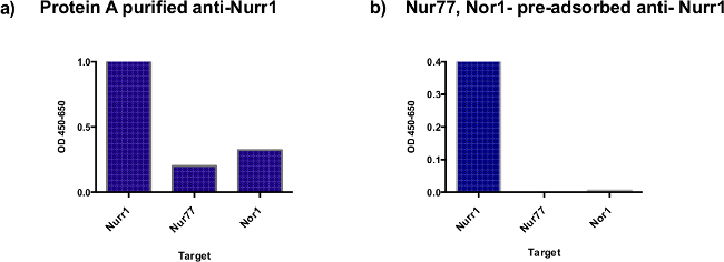
Figure 1: ELISA comparison of cross-reactivities of anti-Nurr1 antibodies before and after purification by passage through Nur77 and Nor1 LBDs coupled columns. Protein A purified anti-Nurr1 antibodies (0.156 μg/ml) (a) or protein A purified followed by chromatography onto Nor1 and Nur77 LBD-coupled columns anti-Nurr1 antibodies (0.156 μg/ml) (b) were added to wells of 96 well plate coated with 100 μl of 2.5 μg/ml of either Nurr1 LBD, Nor1 LBD or Nur77 LBD. The ELISA was carried out as described in the method section.
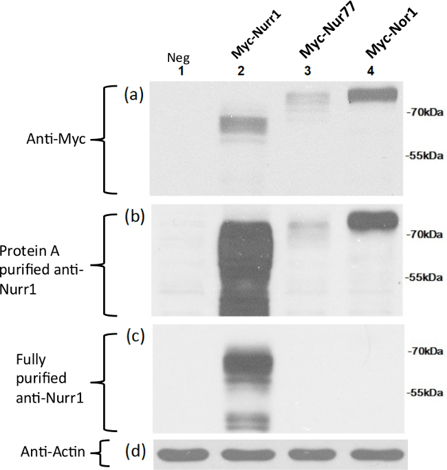
Figure 2: Specific detection of full-length Nurr1 expressed in CHO cells using the fully purified Nurr1-specific antibody. Full-length Nurr1, Nor1, and Nur77 proteins were expressed as myc tagged fusion proteins in CHO cells and detected by Myc specific antibodies (a), Protein A-purified Nurr1 antibody (b), and fully purified Nurr1 antibody (c). Anti-actin antibodies used for protein loading control (d). Each Myc-tagged full length DNA (molecular weight of 65, 66, and 69 kDa for Nurr1, Nur77, Nor1, respectively) was transiently transfected into CHO cells and the same amount of cell extracts were loaded in each lane. Lane 1: negative control consisting of CHO cells extracts transfected with an empty vector; lane 2: Myc-Nurr1; lane 3: Myc-Nur77; lane 4: Myc-Nor1.
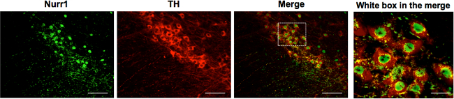
Figure 3: The fully purified Nurr1-specific antibody specifically detects tyrosine-hydroxylase-positive dopamine neurons. Midbrain dopamine neurons in the substantia nigra are positive for Nurr1, as examined by immunohistochemistry using the fully purified Nurr1-specific antibodies. Notably, Nurr1 was localized in the nucleus of mDA neurons. TH (tyrosine hydroxylase). Scale bar = 100 μm. Scale bar in white box merge is 10 μm. Please click here to view a larger version of this figure.
The success of this protocol relies on the availability of pure proteins for characterization of the antibodies raised against a specific member of the protein family of interest. There is no need to purify the antibodies using protein A/G until it is clearly established that there are significant cross-reactivities by ELISA and Western blotting.
As stated in the text, it is important to avoid suspending the protein(s) to be cross-linked to the column in an amine-containing buffer such as Tris or glycine. Furthermore the optimal protein concentration used for cross-linking must be determined empirically. Too much protein on the column could result in steric hindrance while too little would reduce the efficiency of adsorption. Therefore, it is a good idea to test protein concentrations ranging from 2 to 10 mg/ml and to assess the quality of the resulting antibodies.
Several factors must be considered when this technique fails to yield highly specific antibodies. These include: cross-linking efficiency, loss of immunoreactive domain, and cross-linked protein denaturation. As stated in the protocol, monitoring cross-linking efficiency allows the determination that too much or too little protein is being immobilized on the column. If cross-linking using reactive amines is suspected of destroying immunoreactive domain(s), immobilization through the carboxylic group or through carbohydrates should be considered.
It is important to monitor the efficiency of the procedure to consistently yield highly specific antibodies. For regeneration purposes, the columns must be stripped of the cross-reactive antibodies bound to them using harsh conditions (high or low pH). This causes an increasingly high percentage of cross-linked proteins to be effectively denatured, reducing the column performance.
The addition of blocking agents in ELISA and Western blotting is commonly used with a fair level of success. However, the approach described in this video increases the probability of success and reduces the need for maintaining large amounts of the cross-reactive proteins on hand. As the reagents and methods to immobilize proteins to chromatography media keep improving, this approach will contribute to the identification of circulating transformed cells specific antibodies.
This approach is limited by the availability of pure proteins. Another limitation of this protocol is the dependence on proper folding when using protein domains. If the protein domains do not fold as in the whole protein, the pre-adsorption strategy may not yield antibodies that can readily identify the protein member of interest when analyzing cell or tissue extracts.
Many proteins of interests belong to (sub)families of proteins with high amino acid sequence homologies. Functional characterization and identification of the distribution of proteins often rely on the availability of specific antibodies against each family member. The high amino acid homologies between protein members in the same family contribute to the difficulties in generating highly specific antibodies. Using these cross-reactive antibodies often lead to conflicting results.
One such example is the orphan nuclear receptor Nurr1 that belongs to the NR4A subfamily of nuclear receptors, together with Nur77 and Nor1. Since these three factors share a high degree of amino acid homology, antibodies against each factor show significant cross reactivities, which hamper precise analysis of each factor.
Using these NR4A members as an example, it was possible to generate highly specific Nurr1 antibodies free of reactivities to Nor1 or Nur77. Since the LBDs share less homology than the DNA binding domains, the LBD of each protein was expressed and purified and then used to improve the specificity of the anti-Nurr1 antibodies. Initially, antibodies against Nurr1’s LBD exhibited modest but significant levels of cross reactivity to the other proteins, as examined by ELISA and Western blot analyses. However, when Protein A-purified Nurr1 antibodies were further purified through pre-adsorption against cross-reactive LBD domains of Nur77 and Nor1, the resulting Nurr1 antibody did not show any cross reactivity, as examined by Western blot and ELISA analyses of prominently detected Nurr1-expressing DA neurons in the midbrain areas. The Western blot data and ELISA analyses closely agree with each other, indicating that these analyses can be complementary and support each other.
Taken together, this antibody purification strategy will be extremely useful in generating a specific antibody by eliminating the cross reactivity to homologous protein members.
The authors have nothing to disclose.
This work was supported by NIH grants (NS070577 and NS084869).
Purified Ligand Binding Domain from Nurr1, Nor 1 and Nur77 | Column Storage solution | ||
AminoLink Coupling Resin and Kit | Thermo Scientific | 44890 | |
Protein A spin column | Thermo Scientific | 89978 | |
Dulbecco's Phosphate-Buffered Saline | Corning | 21-031-CV | |
96 well Flate-bottom plates, High Flange, 330μL | Thermo Scientific | 3455 | |
10% Normal Goat Serum | KPL | 50-675-69 | |
Milk Diluent/ Blocking Concentrate | KPL | 50-82-01 | |
IgG Elution Buffer | Thermo Scientific | 21004 | |
UltraPure 1 M Tris-HCI Buffer, pH 7.5 | Life Technologies | 15567-‐027 | |
Micro BCA Protein Assay Kit | Thermo Scientific | 23235 | |
Horse Radish Peroxidase conjugated Goat anti-Rabbit IgG (H+L) | Thermo Scientific | 31460 | |
Ni-NTA Resin | Thermo Scientific | 88221 | |
3, 3', 5, 5' Tetramethylbenzydine | Thermo Scientific | 34028 | |
2N H2SO4 | Macron Fine Chemicals | H381 05 | |
Protein A IgG Binding Buffer | Thermo Scientific | 21001 | |
IgG Elution Buffer | Thermo Scientific | 21004 | |
Neutralization Buffer | 1M Tris-‐HCl pH 8.5 | ||
Column Storage solution | Phosphate Buffered Saline containing 0.02% sodium azide | ||
Spectra/Por 7 pre-treated dialysis tubing | Spectrum Labs | 132128 | |
10 ml Disposable columns | Thermo Scientific | 29924 | |
AminoLink Coupling Buffer | 0.1M sodium phosphate, 0.05% NaN3, pH 7.0 | ||
Quenching buffer | 1M Tris. HCI, 0.05% NaN3, pH 7.4 |
||
Wash Solution | 1M NaCl, 0.05% NaN3 | ||
ELISA Blocking buffer | 1% Normal Horse serum (NHS), 1% Normal Goat Serum (NGS) in 1X KPL milk diluent | ||
Storage Solution | PBS pH 7.4 containing 0.02% sodium azide | ||
Micropipettes: 10 μl; 20 μl; 200 μl; 1000 μl |