In this protocol, we describe a novel BrdU-ChIP-Slot-Western technique to examine proteins and histone modifications associated with newly synthesized or nascent DNA.
Histone deacetylases 1 and 2 (HDAC1,2) localize to the sites of DNA replication. In the previous study, using a selective inhibitor and a genetic knockdown system, we showed novel functions for HDAC1,2 in replication fork progression and nascent chromatin maintenance in mammalian cells. Additionally, we used a BrdU-ChIP-Slot-Western technique that combines chromatin immunoprecipitation (ChIP) of bromo-deoxyuridine (BrdU)-labeled DNA with slot blot and Western analyses to quantitatively measure proteins or histone modification associated with nascent DNA.
Actively dividing cells were treated with HDAC1,2 selective inhibitor or transfected with siRNAs against Hdac1 and Hdac2 and then newly synthesized DNA was labeled with the thymidine analog bromodeoxyuridine (BrdU). The BrdU labeling was done at a time point when there was no significant cell cycle arrest or apoptosis due to the loss of HDAC1,2 functions. Following labeling of cells with BrdU, chromatin immunoprecipitation (ChIP) of histone acetylation marks or the chromatin-remodeler was performed with specific antibodies. BrdU-labeled input DNA and the immunoprecipitated (or ChIPed) DNA was then spotted onto a membrane using the slot blot technique and immobilized using UV. The amount of nascent DNA in each slot was then quantitatively assessed using Western analysis with an anti-BrdU antibody. The effect of loss of HDAC1,2 functions on the levels of newly synthesized DNA-associated histone acetylation marks and chromatin remodeler was then determined by normalizing the BrdU-ChIP signal obtained from the treated samples to the control samples.
Defective DNA repair and/or DNA replication are a major cause of genome instability, which can trigger cell death. A single unrepaired double strand break is sufficient to cause cell death1. Chromatin organization is transiently altered during both replication and repair2,3, and failure to maintain epigenetic information during these processes will result in a threat to genome integrity. Loss of HDAC3 or HDAC1,2 function impedes S-phase progression, DNA replication and repair leading to genotoxic stress (DNA damage) and cell death4-9. It is therefore a practical strategy to use selective HDAC inhibitors to disrupt replication, repair and chromatin in cancer cells and cause DNA damage, which in turn can stop growth and induce cell death selectively in rapidly growing cancer cells.
During DNA replication, chromatin is rapidly disassembled and then reassembled following DNA duplication. Newly synthesized histone H4 is acetylated at K5 and K12 residues (H4K5K12ac) and following deposition onto chromatin, they are rapidly deacetylated by histone deacetylases (HDACs)10,11. Failure of cells to maintain nascent chromatin integrity by histone deacetylation can lead to fork collapse, which in turn can result in DNA damage and cell death. We recently showed that selective inhibition of HDAC1,2 increases histone acetylation (H4K5ac, H4K12ac and H4K16ac) and inhibits SMARCA5 chromatin remodeler activity on nascent chromatin, which correlates with reduced replication fork progression, increased fork collapse and increased replication stress-induced DNA damage6. Thus, HDAC inhibitor treatment can alter nascent chromatin structure to trigger DNA damage and death very quickly in cancer cells, as these cells cycle rapidly and pass many times through the S-phase. It is therefore important to understand how HDACs function to regulate histone acetylation and protein binding to maintain DNA replication in mammalian cells.
To quantitatively measure the amount of histone acetylation and SMARCA5 chromatin remodeler associated with nascent DNA during DNA replication, we devised a modified ChIP assay called the BrdU-ChIP-Slot-Western technique. Following chromatin immunoprecipitation (ChIP) of a desired protein or histone modification, the amount of nascent DNA (labeled using thymidine analog, BrdU) in the ChIP sample can be detected using western analysis of BrdU-labeled ChIP DNA transferred onto a membrane using a Slot Blot apparatus. Using this technique, we showed that H4K16ac (a mark involved in chromatin packaging) and the ISWI family member SMARCA5 (SWI/SNF-related matrix-associated actin-dependent regulator of chromatin) chromatin remodeler are associated with nascent DNA in S-phase cells6. We also found that the nascent H4K16ac mark is deacetylated by HDAC1,2 during DNA replication6. H4K16ac inhibits chromatin remodeler activity of the ISWI family member SMARCA512. Hence, using the BrdU-CHIP-Slot-Western technique, we could connect the functions for HDAC1,2 in regulating chromatin remodeling during DNA replication. Hence, the BrdU-ChIP-Slot-Western Blot technique is a powerful approach to quantitatively measure the association and dissociation dynamics of proteins or their post-translational modifications that are bound to nascent DNA.
1. Chromatin immunoprecipitation (ChIP) following BrdU Labeling
2. Slot Blot Analysis of ChIP DNA
To determine the specificity of HDAC1,2-selective inhibitors, Hdac1,2FL/FL and Hdac3FL/FL fibrosarcoma cells were used. Adenovirus-containing Cre recombinase (Ad-Cre) was used to delete Hdac1,2 and Hdac3 in these cells. Following Ad-Cre infection of Hdac1,2FL/FL cells, a robust increase in H4K5ac was observed. Treatment of Hdac1,2 knockout cells with 233 or 898 did not result in any further increase in H4K5ac confirming that 233 and 898 inhibit Hdac1,2 and not Hdac3 in these cells. In contrast, addition of 233 or 898 to Hdac3 knockout cells resulted in a significant increase in H4K5ac compared to the increase seen in Hdac3 knockout cells due to an additive effect on H4K5ac levels as a result of inhibition of Hdac1,2 and 3. Hence, 233 and 898 are Hdac1, 2-selective inhibitors. These results are shown in Figure 1.
To validate the BrdU-ChIP-Slot technique, BrdU-pulse chase analysis was performed to look at the kinetics of PCNA loading on to the nascent DNA in HeLa cells. Our results showed that PCNA association with nascent DNA occurs rapidly within 15 min and disappears after a 30 min chase in agreement with previously published results13. These results are shown in Figure 2.
BrdU-H4K16ac ChIP-Slot-Western technique was used to determine the amount of H4K16ac associated with nascent DNA in the absence of HDAC1,2 function. A robust enrichment in H4K16ac associated with nascent DNA was observed when compared to the rabbit IgG control (Figures 3A and 3B). The increase in nascent DNA-associated H4K16ac following inhibition of HDAC1,2 activities or knockdown of Hdac1,2 is shown in Figure 3C, 3D, and 3E.
The level of SMARCA5 chromatin remodeler on nascent DNA was determined using BrdU-SMARCA5 ChIP-Slot-Western technique. Our results showed that SMARCA5 associates with nascent DNA in mammalian cells. Furthermore, HDAC1,2 inhibition or knockdown of Hdac1,2 did not change the amount of nascent DNA-associated SMARCA5 chromatin remodeler as shown in Figure 4.
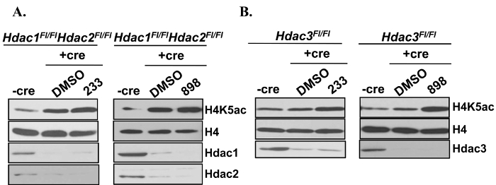
Figure 1. Confirmation of the Specificity of HDAC1,2-selective Inhibitors. Western analysis of whole cell lysates prepared from Hdac1FL/FL Hdac2FL/FL or Hdac3FL/FL fibrosarcoma cells following Ad-Cre infection and treatment with 898 or 233. Cells were treated with 3 µM 898 or 233 for 24 hr following a 48 hr Ad-Cre infection. This figure is derived from our previous published work6. Please click here to view a larger version of this figure.
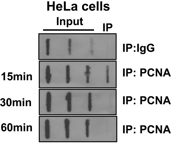
Figure 2. Dynamics of PCNA Association with the Nascent DNA in HeLa Cells. HeLa cells were labeled with bromodeoxyuridine (BrdU) for 30 min. Cells were then washed to remove unincorporated BrdU and cultured in media without BrdU for indicated periods of time (chase). Chromatin immunoprecipitation (ChIP) was performed with anti-proliferating cell nuclear antigen (PCNA) antibody. BrdU labeled DNA present in input DNA and those associated with PCNA were assessed in slot blot analysis using an anti-BrdU antibody as shown in Figure 2. This figure is derived from our previous published work6. Please click here to view a larger version of this figure.
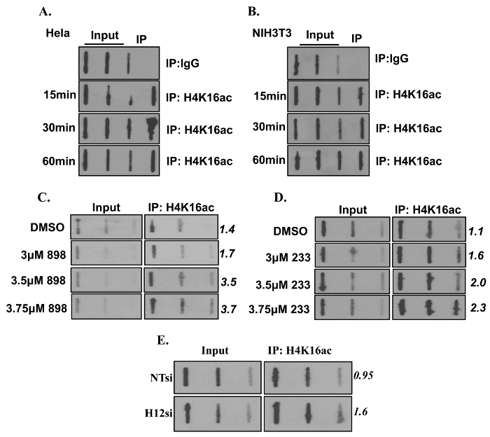
Figure 3. Loss of Histone Deacetylase 1 and 2 Increases H4K16ac on Nascent DNA. (A-B) Bromodeoxyuridine (BrdU) pulse chase was performed in HeLa and NIH3T3 cells to determine association of H4K16ac with nascent DNA at the indicated time points. (C-D) NIH3T3 cells were treated with either DMSO or a HDAC1,2-selective inhibitor (898 or 233) for 24 hr. (E) NIH3T3 cells were either transfected with non-targeting (NT) or Hdac1,2 (H12) siRNA. Cells from (C – E) were labeled with BrdU and used for ChIP with anti-H4K16ac followed by Slot blotting. The membrane was probed with anti-BrdU antibody. Numbers indicate to Image J quantitation of the average BrdU signal of high and medium volume of ChIP DNA spotted. This figure is derived from our previous published work6. Please click here to view a larger version of this figure.
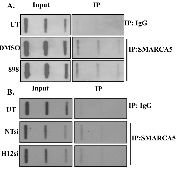
Figure 4. SMARCA5 Associates with Nascent DNA. The amount of SMARCA5 on nascent chromatin following loss of HDAC1,2 function is shown. NIH3T3 cells were either treated with an HDAC1,2-selective inhibitor (898) or transfected with non-targeting (NT) or Hdac1,2 (H12) siRNA. Cells were labeled with BrdU following the above-mentioned treatments and ChIP with anti-SMARCA5 or rabbit IgG (negative control) was performed. Increasing volumes of ChIP DNA was spotted on the slot blot and the membrane was probed with anti-BrdU antibody. This figure is derived from our previous published work6. Please click here to view a larger version of this figure.
The protocol described in this manuscript is a relatively quick method to demonstrate the presence of proteins or their post-translationally modified forms on newly replicated or nascent DNA. Additionally, this technique permits one to measure the association-dissociation kinetics of a protein or its modified form with nascent DNA. This technique is complementary to the elegant iPOND technology13. In the iPOND technology, newly synthesized DNA is labeled with ethyl deoxyuridine (EdU). A biotin conjugate is then added onto EdU using the click chemistry. Biotin-tagged nascent DNA is then immunoprecipitated using streptavidin beads and co-purifying proteins are detected by western blotting. On the other hand, in our BrdU-ChIP-Slot-Western technique, a protein or its modified form associated with nascent DNA is immunoprecipitated using protein-specific or modified form-specific antibody and the amount of BrdU-labeled nascent DNA bound to the protein is then determined quantitatively by Slot-Western blotting using an anti-BrdU antibody.
There are critical steps in this protocol that needs special attention. It is critical to ensure that the antibody of interest works well in standard ChIP assays with a high efficiency. It is important to test the efficiency of an antibody in ChIP assays by quantitative-PCR before performing the BrdU-CHIP-Slot-Western assay. In order for the quantitation of the BrdU-CHIP-Slot-Western technique to be accurate, a serial dilution of the BrdU-labeled DNA should be applied to slot blot and western analysis using the anti-BrdU antibody should be initially performed in order to determine the linear range of detection. Determining the DNA concentration of ChIP and Input DNA enables one to ensure that the amount of BrdU-labeled DNA is within the linear range of detection in Western blotting. For example, we determined 50 ng to 12.5 ng of input DNA to be in the linear range in our pilot experiment. Also, if the concentration of the ChIP samples is very high, it is imperative to dilute the ChIP DNA before performing slot. The limitation of this technique is its resolution, which is dependent on the efficiency of chromatin shearing. The shearing of chromatin by sonication determines the proximity of a given protein or its modified form to the newly synthesized DNA.
In the past, the study of changes in the chromatin and chromatin-modifying enzymes during DNA replication in mammalian cells remained a difficult question to address due to the unavailability of appropriate techniques. Moreover, since Hdac1,2-null cells arrest in G1 phase, it was difficult to examine functions for Hdac1,2 within S-phase. In our study, we showed functions for these two Hdacs during DNA replication using a combination of first-in-class selective inhibitors to inhibit their activities within S-phase and the novel BrdU-ChIP-Slot-Western technique. In future, this technique could also be used to study the dynamics of DNA damage response and DNA repair proteins at the stalled fork in addition to studying the histone modifications and chromatin-modifying enzyme at the fork. Moreover, this technique is not limited to mouse NIH3T3 cells. This technique can be successfully used in other cell lines in order to investigate how other inhibitors of enzymes or proteins affect DNA replication and replication stress-induced chromatin changes at the replication fork.
The authors have nothing to disclose.
The work in this manuscript was supported by funds from Dept. of Radiation Oncology and Huntsman Cancer Institute and the National Institute of Health grant (R01-CA188520) to SB. I thank Danielle Johnson and Steven Bennett in my lab for demonstrating the protocol and explaining its benefits. I am grateful to Dr. Mahesh Chandrasekharan (Huntsman Cancer Institute) for critical comments on the manuscript.
Anti-BrdU (westerns) | BD Biosciences | B555627 | |
Anti-SMARCA5 | Abcam | ab3749 | |
Anti-H4K16ac | Active Motif | 39167 | |
Zeta-Probe GT Membrane | Bio-Rad | 162-0197 | |
Formaldehyde | Fisher | BP531-500 | |
Protein A agarose | Millipore | 16-156 | |
Rnase A | Qiagen | 19101 | |
Proteinase K | Sigma | P4850 | |
PCR Purification Kit | Qiagen | 28106 | |
Glycine | Sigma | G7403 | |
Protease inhibitor cocktail | Roche | 11836170001 | |
BrdU | Sigma | B9285 | |
Rabbit IgG | Millipore | 12-370 | |
HEPES | Sigma | H3375 | |
NaCl | Sigma | S-3014 | |
Triton-X-100 | Sigma | 93443 | |
NP40 | USB Corporation | 19628 | |
Sodium deoxycholate | Sigma | D6750 | |
Sodium bicarbonate | Sigma | S-4019 | |
Ethanol | Decon Laboratories | 04-355-222 | |
DEPC-treated water | Sigma | 95284 | |
Tris | Fisher | BP-152-5 | |
EDTA | Invitrogen | 15575-020 | |
SDS | Ambion | AM9820 | |
Sodium hydroxide | Mallinckrodt GenAR | MAL7772-06 | |
20X SSC | Life Technologies | 15557-036 | |
Non-fat dry milk | Lab Scientific Inc | M0841 | |
ECL | Thermo Fisher | 80196 | |
X-ray film | Genesee Sci | 30-101 | |
Developer | Konica Minolta | SRX-101A | |
UV Crosslinker | Stratagene | XLE-1000 | |
Sonicator | Branson Sonifier Digital Ultrasonic Cell Disruptor | Model: 450 | |
Centrifuge | Eppendorf | Model: 5810R | |
Water bath | Fisher Scientific Isotemp | Model: 2322 | |
NanoDrop 1000 spectrophotometer | ThermoScientific | Model: 1000 | |
Slot Blot apparatus | Schleicher and Schuell Minifold II | 44-27570 | |
Tissue Culture Incubator | Thermo Scientific | Series II 3110 Water-Jacketed CO2 Incubators |