We describe a method to produce an animal model of liver fibrosis in the rat, and assess the degree of fibrosis by histological examination of the liver. The model can be used to study the development of liver disease as well as to test the efficacy of potential anti-fibrotic agents.
Four to six week old, male Wistar rats were used to produce animal models of liver fibrosis. The process requires four weeks of administration of 10 mg/kg dimethylnitrosamine (DMN), given intraperitoneally for three consecutive days per week. Intraperitoneal injections were performed in the fume hood as DMN is a known hepatoxin and carcinogen. The model has several advantages. Firstly, liver changes can be studied sequentially or at particular stages of interest. Secondly, the stage of liver disease can be monitored by measurement of serum alanine aminotransferase (ALT) and aspartate aminotransferase (AST) enzymes. Thirdly, the severity of liver damage at different stages can be confirmed by sacrifice of animals at designated time points, followed by histological examination of Masson's Trichome stained liver tissues. After four weeks of DMN dosing, the typical fibrosis score is 5 to 6 on the Ishak scale. The model can be reproduced consistently and has been widely used to assess the efficacy of potential anti-fibrotic agents.
DMN is a potent liver specific toxin. Its metabolism, tissue distribution, and ability to cause injury to livers of rats was reported by Magee1, and the mechanism of hepatocyte damage and cell death by apoptosis was described by Pritchard and Butler2. Intermittent administration of this compound was reported to induce liver fibrosis in dogs and rats3,4.
The mechanisms and morphologic changes of liver fibrosis have been extensively investigated using this model. In early studies using the rat, 3-week treatment with DMN produced centrilobular hemorrhagic necrosis followed by micronodular cirrhosis without steatosis5. It was shown that in early fibrosis, the collagen formed was more cross linked with type III being more prominent than type I6. In common with other causes, DMN-induced fibrotic changes were associated with an increase in Kupffer cells; hepatic macrophages that reside in the sinusoids. These cells morph into myofibroblasts and produce excessive amounts of extracellular matrix which is the primary problem in fibrosis7.
In terms of cellular signaling, Nakamura et al. demonstrated that TGF-β plays a critical role in the progression of liver fibrosis8. They used an adenovirus expressing a truncated type II TGF-β receptor, to specifically inhibit TGF-β signaling. Liver fibrosis in these rats was spectacularly halted during DMN treatment when compared to control groups. Other studies have confirmed that suppression of TGF-β leads to alleviation of liver fibrosis development9,10. The model was also used in a global gene profiling study to identify other fibrosis markers and proteins which could be used as drug targets for anti-fibrotic therapy11.
Other chemical agents used to induce liver fibrosis include thioacetamide (TAA) and carbon tetrachloride (CCl4). TAA was used first in rats and later in mice12. The advantages of this model include: ease of chemical administration in drinking water, the reproducibility of the model with characteristic micronodular cirrhosis and biochemical changes. The disadvantages include: the long time period of 3 months for liver fibrosis to develop and the lack of understanding of the molecular mechanism for induction of liver fibrosis. As for CCl4, its use has declined for the following reasons: it does not mimic human liver disease, has harmful effects on the ozone layer, causes pain and distress to animals, is very toxic to humans and requires extra precautions in its handling and disposal13,14.
Prolonged bile duct obstruction (by surgical intervention) as an experimental model for liver cirrhosis was first reported by Kountouras et al.15. This method is nontoxic to humans and animals. However, the time required for liver fibrosis to develop varies. A review of 30 reports by Marques et al.16, found that it took from seven days to four weeks after surgery for liver fibrosis to develop. The pathological changes described mimic those of human chronic biliary fibrosis and the model would be more suited for researchers interested in this area.
In summary, the intermittent administration of a constant dose of DMN in the rat over 4 weeks produces liver fibrosis that mimics the human disease. Dosed rats show progressive development of liver damage and parenchymal fibrosis4,17. Disease progression and severity can be monitored via blood samples or sacrifice of animals at specific time points and the effect is highly reproducible18. Thus the model has been widely used to study the mechanisms of liver fibrosis and cirrhosis as well as to screen for potential anti-fibrotic agents10,19,20.
All animal experiments were approved by the animal care and use committee of the School of Applied Science, Temasek Polytechnic.
1. Preparation of DMN
2. Intraperitoneal Injection of DMN
3. Gross Examination and Harvest of Liver
4. Processing, Embedding and Sectioning of the Liver
5. Masson Trichrome Staining
6. Fibrosis Scoring on Masson's Trichome Stained Sections of Liver
Score | Description |
0 | No fibrosis |
1 | Fibrous expansion of some portal areas, with or without short fibrous septa |
2 | Fibrous expansion of most portal areas, with or without short fibrous septa |
3 | Fibrous expansion of most portal areas, occasional portal to portal (P-P) bridging |
4 | Fibrous expansion of portal areas with marked bridging (portal to portal (P-P) as well as portal to central (P-C) |
5 | Marked bridging (P-P and/or P-C) with occasional nodules (incomplete cirrhosis) |
6 | Cirrhosis, probable or definite |
Table 1: Fibrosis Score Used for Reading the Masson's Trichome Stained Liver Sections25.
DMN treated rats lose weight and become less vigorous with ruffled hair coat. There is significant loss in average body weights of DMN treated rats; first detectable after 2 weeks of DMN treatment, and this difference remains through weeks 3 and 4 after DMN treatment (Figure 1a). As the rats receive DMN over successive weeks, damage to the liver causes it to become smaller. The liver index; which is the percent of liver weight at final body weight was significantly lower for the DMN treated rats (Figure 1b).
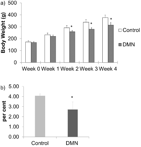
Figure 1: a) Body weights of DMN treated rats. The data are represented as the means ± SD (n = 6 – 8). *P < 0.05 compared with normal control group. b) Liver index; percent liver weight at final body weight of DMN treated rats after 4 weeks of DMN treatment. The data are represented as the means ± SD (n = 6 – 8). *P < 0.05 compared with normal control group. Please click here to view a larger version of this figure.
At sacrifice, after 4 weeks of DMN treatment, the liver is smaller and harder (Figure 2a) compared to those from aged matched control animals (Figure 2b). Fibrin may be present on the liver surface and adjacent liver lobes are adhered. About 20% of rats have ascites.
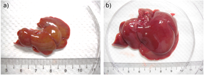
Figure 2: a) Liver from a rat after 4 weeks of DMN treatment. b) Liver from aged matched control rat. In (a) the liver is shrunken, firm and pale with a yellowish tinge when compared with the liver from its aged matched control. Please click here to view a larger version of this figure.
Injury to the liver causes increased permeability of the hepatocyte cell membrane. Increased serum ALT and AST are indicators of hepatocyte damage. Serum ALT (Figure 3a) and AST (Figure 3b) of the DMN treated group are significantly higher than the control group after weeks 2 and 4 of DMN injection. Serum ALT and AST levels typically increase after each week of DMN treatment.
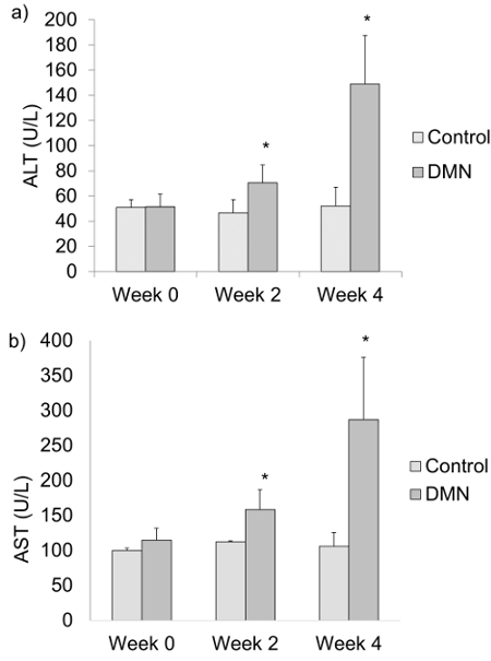
Figure 3: a) Serum alanine aminotransferase (ALT) and b) serum aspartate aminotransferase (AST) levels of DMN treated rats at weeks 0, 2 and 4 after the last DMN injection. The data are represented as the means ± SD (n = 6 – 8). *P < 0.05 compared with normal control group. Please click here to view a larger version of this figure.
Histological examination of livers from DMN treated rats show that there is progressive increase and expansion of fibrous septa, with loss of hepatocytes, over time compared with control rats. Masson's Trichome stain is commonly used to highlight collagen deposits in liver tissue: these are stained blue.
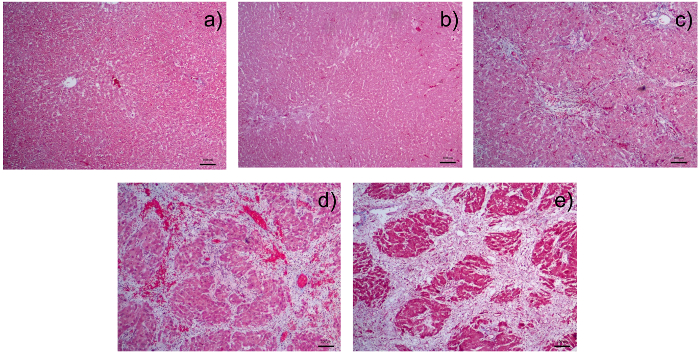
Figure 4: Photomicrographs of Liver Sections Stained with Masson's Trichome: a) liver section from a normal control rat; b) liver section from a rat after receiving 1 week of dimethylnitrosamine (DMN); c) liver section from a rat after receiving 2 weeks of DMN. There is fibrous expansion of most portal areas with occasional portal to portal bridging. d) Liver section from a rat after receiving 3 weeks of DMN. Note the fibrous expansion of portal areas with marked portal to portal as well as portal to central bridging. e) Liver section from a rat after receiving 4 weeks of DMN. There is cirrhosis with nodule formation. The control liver is from the group sacrificed together with rats after 4 weeks of DMN injection. There is a pattern of progressive increase of fibrosis score from 0 in (a); 2 in (b); 3 in (c) 4 in (d) to 5/6 in (e). All photomicrographs were taken at a magnification of 40X with 1 unit length of scale bar equivalent to 100 µm. Please click here to view a larger version of this figure.
We have described a method to make an animal model of liver fibrosis and to assess the severity of liver fibrosis. It is important to deliver the correct dose of DMN and adhere to the schedule of weekly intraperitoneal injections. As the experiment progresses, it is crucial to weigh the rats and re-calculate the dose at the start of each week of DMN injections. Keep in mind that DMN is toxic and needs to be handled in the fume cabinet. We perform the intraperitoneal injections in the fume cabinet as well. With the need for repeated injections, the correct restraint and technique of injection is important to avoid introduction of contaminants and damage to internal organs. We rarely have mortality from the intraperitoneal injections.
Rats should be monitored 2x a day throughout the experimental period. During the last week of DMN injection, animals should be observed even more frequently and closely for moribund signs. This is the period when liver damage is most severe and affected rats will be inappetent, lose body weight and condition. 25 – 40% of rats can become severely sick during this period and die if there is no veterinary intervention. Hence close monitoring allows the researcher to assess the condition of the animal and plan appropriate termination of the experiment in order that organs can be freshly harvested from a euthanized rat instead of being lost to decay from unexpected death.
We have found weekly measurement of ALT and AST levels to be useful indicators of the progression of liver damage. We have also observed that AST is less specific than ALT and attribute this to its release from damaged erythrocytes and muscle tissue in addition to liver.
Ensure that sufficient volume of blood is collected to yield the required volume of serum for analysis. We typically collect 200 – 300 µl of blood per rat.
During the collection of liver tissue for histological examination, we recommend the sample be obtained from the same lobe. This is done after checking that the changes in the liver are uniform. DMN causes generalised changes in the liver. In the early stages of the study, we sampled from the left lateral and medial lobes as well as the right medial lobe. We did not observe any differences in the microscopic changes between these lobes.
Liver sections should be fixed in 10% buffered formalin for at least 24 hr and processed soon after. Prolonged immersion beyond a week can result in the tissue becoming brittle and difficult to section after paraffin embedding. Cutting suitable 5 µm thin sections requires practice and patience. Sections that are assessed to be suitable with the naked eye (Step 4.11) can be examined with the microscope to check that they do not have artefacts like folds or tears within the section. The staining procedure is best completed according to the time periods for slide immersion at each step without rest breaks. During the staining process, ensure that the sections are kept moist at all times and transferred from one staining solution to the next as soon as possible.
Evaluation of the Masson's Trichome stained sections for the degree of fibrosis requires the input of a veterinary pathologist. He/She is a crucial team member who should be part of the study from the beginning, as he/she would be able to advise and participate in the correct harvesting of organs at all stages of the experiment. Thus the current gold standard for liver fibrosis scoring is the assessment of appropriately stained tissue sections by a pathologist. Though it would be optimum to have the input of more than one pathologist, this may not be possible in smaller organizations. In such cases, the objectivity can be improved by the use of imaging technology26. We have found that results from imaging technology are comparable to pathological scoring (unpublished data), and recommend that it be used as a supplementary method of evaluation.
In summary, the DMN induced model of liver fibrosis has many advantages over other animal models. It is a comparatively easy model to make as it does not require surgical skills. DMN is environmentally safer and less toxic to humans and animals than CCl4 and takes a shorter time to induce disease than TAA. Compared to liver disease produced by CCl4, TAA, and bile duct ligation, DMN produces a closer representation of human liver fibrosis. For these reasons, we foresee that the model will continue to be used widely to study the mechanisms of liver fibrosis as well as to screen for potential anti-fibrotic agents.
The authors have nothing to disclose.
The authors acknowledge the funding support from the Ministry of Education, Singapore, grant number MOE2010-IF-1-025.
Dimethylnitrosamine | Wako | 147-03781 | |
Formalin | Sinopharm chemicals | F63257009 | |
Ethanol | Sigma | 64-17-5 | |
Xylene | Fisher | 1330-20-7 | |
Masson trichome stains | |||
Aniline Blue | Electron Microscopy Sciences | #42755 | |
Acid Fuschin | Electron Microscopy Sciences | RT42685 | |
Scarlet Red | Electron Microscopy Sciences | #26905 | |
Phosphotungstic Acid Hydrate | ALFA AESAR | ALFA40116.4 | |
Phosphomolybdic Acid Hydrate | Sigma SG | 221856-25G | |
Weigert's Iron Hematoxilyn | Merck | 1.15973.0002 | |
DPX Mounting Medium | Merck | HX066873 | |
Tissue processor | Leica | Leica TP 1020 | |
Embedding machine | Sakura | Sakura Tissue Tek TEC5 Embedding System | |
Microtome | Leica | Leica RM 2235 | |
Vet Test Analyzer | Idexx | Vet Test 8008 |