A method to establish an in vitro model of blood-brain barrier based on a co-culture of rat brain microvascular endothelial cells and astrocytes is described and validated. This system proved to be a valid tool to study the effect of nanoformulation on the trans-barrier permeation of fluorescent molecules.
Brain microvascular endothelial cells, supported by pericytes and astrocytes endfeet, are responsible for the low permeation of large hydrosoluble drugs through the blood-brain barrier (BBB), causing difficulties for effective pharmacological therapies. In recent years, different strategies for promoting brain targeting have aimed to improve drug delivery and activity at this site, including innovative nanosystems for drug delivery across the BBB. In this context, an in vitro approach based on a simplified cellular model of the BBB provides a useful tool to investigate the effect of nanoformulations on the trans-BBB permeation of molecules. This study describes the development of a double-layer BBB, consisting of co-cultured commercially available primary rat brain microvascular endothelial cells and astrocytes. A multiparametric approach for the validation of the model, based on the measurement of the transendothelial electrical resistance and the apparent permeability of a high molecular weight dextran, is also described. As proof of concept for the employment of this BBB model to study the effect of different nanoformulations on the translocation of fluorescent molecules across the barrier, we describe the use of fluorescein isothiocyanate (FITC), loaded into ferritin nanoparticles. The ability of ferritins to improve the trans-BBB permeation of FITC was demonstrated by flux measurements and confocal microscopy analyses. The results suggest this is a useful system for validating nanosystems for delivery of drugs across the BBB.
The resistance of central nervous system (CNS) diseases (i.e. cancer, epilepsy, depression, schizophrenia and HIV-associated neurological disorder) to pharmacological therapies is due to various different mechanisms, including arduous drug permeation across the blood-brain barrier (BBB). The BBB is the boundary that isolates brain tissues from the substances circulating in the blood. Within this barrier, a layer of brain microvascular endothelial cells (BMECs), supported by pericytes and astrocytes endfeet, is responsible for the high selectivity of the BBB to those hydrosoluble drugs with a molecular weight higher than 400 Da1. Another drug-related resistance mechanism is linked to the presence on BMECs of drug efflux transporters (P-glycoprotein and multidrug resistance proteins), which co-operate to reduce drug penetration into the CNS and facilitate their extrusion from the brain2.
In the last decade, a large number of nanotechnological approaches have been developed to meet the clinical and biological challenge of delivering drugs across the BBB3-6. In this context, ferritin nanospheres (FnN) represent a completely innovative and promising solution. FnN are 12 nm spheres of 24 self-assembling ferritin (Fn) monomers, which are arranged in a hollow spherical structure of 8 nm inner diameter. Ferritin subunits can be disassembled at acidic pH and reassembled in a shape-memory fashion by bringing the pH to neutrality, allowing various organic molecules to be encapsulated. Therefore, FnN represent an interesting model for the development of multifunctional drug delivery systems7,8. Moreover, FnN may interact with BMECs thanks to the specific recognition of Transferrin Receptor (TfR) 1, which is expressed on the luminal membrane of these cells9.
So far, different in vitro models of the BBB have been developed in order to elucidate trans-BBB permeability to various drugs, toxicity toward the BBB, or the interaction of molecules with efflux transporters. Indeed, these models are considered valid in vitro approaches for a rapid screening of active molecules before proceeding with in vivo studies. These models consist of a single endothelial layer of BMECs or co-cultured BMECs and astrocytes (more rarely pericytes), obtained from animal (rat, mouse, pig and bovine) or human cell lines10,11,12. The TransEndothelial Electrical Resistance (TEER) and the apparent permeability (Papp) of tracers with a defined molecular weight represent two critical parameters that are used to determine the quality of the in vitro model. Here we describe the employment of a BBB in vitro model, based on a co-culture of rat BMECs (RBMECs) and rat cortical astrocytes (RCAs) to study the trans-BBB permeation of ferritin nanocages encapsulating fluorescein isothiocyanate (FITC).
1. Establishing the BBB Model
Note: For establishing the BBB model we suggest using commercially available primary RBMECs and RCAs. All steps must be performed with sterile reagents and disposables, handled in a laminar flow hood.
2. BBB Validation
3. Trans-BBB Permeation of FITC-loaded Ferritins (FnN)
Note: A recombinant variant of human ferritin (Fn), produced in Escherichia coli and assembled in nanocages (FnN) for the encapsulation of different fluorescent molecules, is available from the NanoBioLab of Prof. Prosperi (University of Milan-Bicocca, Italy). FnN are loaded with FITC, according to a previously described protocol13 and the concentrations of both Fn and the loaded molecules are accurately determined.
During the establishment of the BBB model, cell attachment and growth on the inserts can be monitored using a light microscope thanks to the transparent nature of the PET membranes. RCAs, seeded at a density of 35,000 cells/cm2, attach efficiently to the bottom side of the insert after 4 hr of incubation at RT (Figure 2A) and grow to cover the membrane surface in 3 days, taking a spindle-shaped morphology (Figure 2B). RBMECs, seeded at a density of 60,000 cells/cm2, are visibly attached to the upper face of the PET membrane after about 3 hr of incubation at 37 °C (Figure 2C). The developing RBMEC layer can be difficult to visualize over the following days with an optical microscope, because of the overlap with the underlying RCA layer (Figure 2D).
A correct validation of the BBB model always requires TEER measurement and this can be confirmed by evaluating the trans-BBB Papp of a low permeability tracer, such as FD40.
The TEER values, recorded over the co-culture period, represent the first clear indication of the correct formation of the endothelial barrier. Three days after RBMEC seeding, the recorded TEER, subtracted from the TEER of the astrocytes-bearing inserts, is mainly due to the contribution of the non-electrogenic layer of RCAs and the developing layer of RBMECs. At this time point, our BBB gives values ranging between 20 and 40 Ω x cm2. During the following days, usually between the 4th and the 5th day of co-culture, the TEER values increase because of the formation of tight junctions between adjacent endothelial cells14, reaching values usually between 55 and 110 Ω x cm2, and in exceptional cases higher values14. The values measured at the 4th/5th day of co-culture remain stable at least until the 7th – 8th day before they start to decrease; therefore, there is a very narrow time window available for undertaking the trans-BBB flux experiments.
Between the 5th and the 7th day of co-culture, the integrity of the experimental models can be confirmed by evaluating the FD40 trans-BBB permeability. Figure 3 shows an example of the trans-BBB flux of FD40 (1 mg/ml), compared to the flux across empty inserts, over 3 hr of incubation; the BBBs are at the 6th day of co-culture and the recorded TEER is 55.6 ± 15.8 Ω cm2 (mean ± SE, n = 3). The flux is linear between 1 and 3 hr of incubation and the mean Papp calculated between 1 and 2 hr, and between 2 and 3 hr of incubation is 0.12 ± 0.01 x 10⁻⁶ cm sec–1 (± SE, n = 6).
Once at least three consecutive successful BBBs, in terms of TEER and FD40 Papp, have been obtained in independent experiments the measurement of tracer permeability can be avoided; however, the TEER always needs to be recorded for each experiment.
The trans-BBB permeability of fluorescent molecules and the effect of the nanocomplex on their delivery can be investigated using the rat BBB models described above. Figure 4 shows the permeation of the model dye FITC upon encapsulation in FnN across BBBs with TEER of 100.4 ± 3.5 Ω cm2 (n = 16) at the 7th day of co-culture. The histograms, representing FITC concentration in the lower chamber after 7 and 24 hr from the addition of free or nanoformulated dye in the upper compartment, indicate that FnN is able to significantly increase the delivery of FITC across the BBB.
Confocal microscope images of the upper side of the insert after 7 and 24 hr of incubation with FITC-FnN (Figure 5A, C) or FITC (Figure 5B, D) show that while free FITC is not internalized by the RBMECs, its loading into the FnN allows it to enter the cells.
Before processing the inserts for confocal microscopy, a further check of the TEER is necessary to ensure there are no FnN-mediated effects upon BBB integrity.
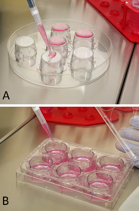
Figure 1: Cell Seeding onto Inserts. RCA (A) and RBMEC (B) seeding procedure onto the two opposite sides of the multi-well plate inserts. Please click here to view a larger version of this figure.
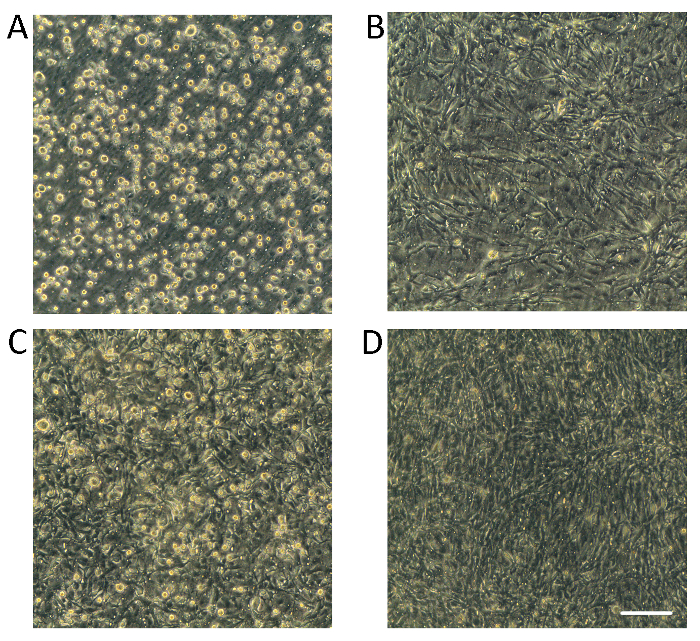
Figure 2: Optical Microscope Images of RCAs and RBMVECs on Inserts. Inserts seeded with RCAs and RBMECs are observed with a light microscope (20X optical zoom). (A) Four hr after seeding, round and translucent RCAs are visibly attached to the bottom surface of the insert; (B) Spindle-shaped RCAs are visible on the 3rd day of culture; (C) Round and translucent RBMECs are attached on the upper side of the insert after 3 hr of incubation; (D) On the 4th day of co-culture both the surfaces of the insert are completely covered with cells, and the two layers are not easily distinguishable. Scale bar = 100 µm. Please click here to view a larger version of this figure.
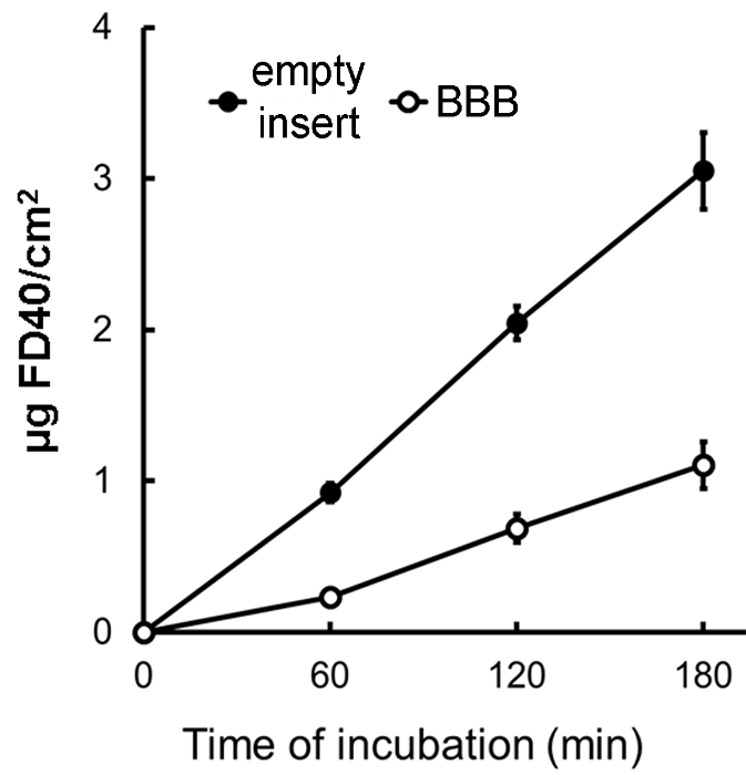
Figure 3: FD40 Flux across the BBB. Flux of FD40 (1 mg/ml) from the upper to the lower side of the BBB in vitro system, compared to that across the empty insert. The amount of FD40 in lower chamber has been measured at 60, 120 and 180 min post the addition of the dye to the upper chamber. Means ± SE; n° inserts = 3. Please click here to view a larger version of this figure.
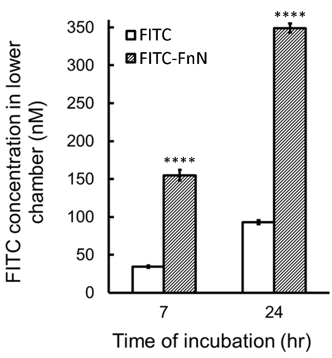
Figure 4: Effect of FnN Encapsulation on FITC Permeation Across the BBB. Concentration of FITC in the lower chamber of the BBB in vitro system calculated at 7 and 24 hr after the addition of FITC or FITC-FnN into the upper chamber. Mean ± SE of 4-5 replicates; ****P <0.0005, FITC-FnN vs. FITC (Student's t-test). Please click here to view a larger version of this figure.
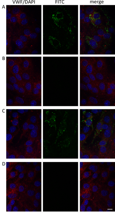
Figure 5: Confocal Microscopy of RBMECs on Inserts. Confocal laser-scanning micrographs (single optical sections) of RBMECs after 7 hr (A, B) or 24 hr (C, D) of incubation with free FITC (B, C) or FITC-FnN (A, C). FITC is green; endothelial cells are immunodecorated with anti-VWF (red) and DAPI (blue). Panels represent, from left to right, merged images of blue and red channels, green channel images, and merged images of all channels. Scale bar = 10 µm. Please click here to view a larger version of this figure.
The in vitro method described here represents a useful validated approach to study the trans-BBB delivery of fluorescent molecules upon nanoformulation with nanoparticles. Here we use FnN, which represents a good candidate to study the translocation of cargo molecules across the BBB. FnN is considered the gold nanovector for trans-BBB delivery of drug/agents since it is specifically recognized by the TfR1 receptor, which is expressed on the luminal membrane of BMECs and mediates the nanoparticle uptake using a receptor-mediated internalization pathway. Moreover, FnN is a natural nanoparticle and it has therefore a good biocompatibility profile. Finally, FnN is able to encapsulate low dimension hydrosoluble molecules by a simple and efficient mechanism. The trans-BBB permeation of other different types of organic or inorganic nanoparticles could be also assessed with this protocol, according to few considerations regarding nanoparticles features. First prerequisite to study the permeation of nanoformulated drugs/agents is the safety of the void nanoparticle. Another crucial issue is the interaction of the investigated nanoparticle with the PET filter. This aspect, has to be evaluated preliminarily in order to exclude a significant retention into the membrane pores and avoid alterations of their permeation across the BBB. Our group has recently employed the proposed strategy to study a polymer-coated iron oxide nanoparticle as a trans-BBB delivery system for fluorescence-labeled antiretroviral drugs14. In that case, we associated electron microscopy localization of nanoformulations in RBMECs to the fluorescence detection. Moreover, other highly sensitive diagnostic methods, such as Inductively Coupled Plasma (ICP)-MS could be employed to evaluate the trans-BBB delivery of inorganic nanosystems.
The BBB in vitro model used in this protocol is based on co-culture of rat BMECs and astrocytes. In the wide scenario of the BBB models10,11,12, some of which have been accurately described in literature with a well-detailed protocol15, our model represents a good quality alternative. Indeed, even though the recorded TEER was below the best standards suggested in the literature (150-200 Ω cm2)10, the trans-BBB permeability of the high dimension tracer Dextran 40 was in total agreement with the formation of a tight barrier.
This in vitro model, obtained with commercially available rat BMECs and astrocytes, has a few advantages: (1) optimal growth efficiency of the endothelial cells, (2) a suitable period of time for the production of the final BBB model (no longer than 13 days), (3) the compliance with ethical issues, avoiding the use of cells from human brain tissue (autopsy materials, surgical specimens, and fetal tissue) and 4) saving of time and costs related to animal housing, and primary cells extraction. Nevertheless, a limitation of the present model is related to the high costs of non-immortalized primary RBMECs, which must be purchased (frozen at passage one) for each experimental setting. Indeed, our preliminary studies of BBB production have demonstrated that the ability of endothelial cells to produce a tight BBB is strictly associated with the number of passages in culture of the RBMECs after the first post-purchase thawing. Further implementation of the BBB model described here with endothelial supplements, such as cAMP and hydrocortisone, could be considered in order to improve endothelial tightness and increase TEER values.10,11,16
The present technique is related to the possibility to take advantage of a single BBB model for different assays, thus providing several data sets from each experimental setting. With a single BBB system exposed to free or nanocomplexed fluorescent molecules, it is possible: (1) to measure the trans-barrier flux of the molecule by analyzing the fluorescence intensity of sECM aliquots collected from the lower chamber at different time points of incubation; (2) to investigate the nano-mediated internalization of the molecules in RBMECs and their intracellular trafficking by confocal microscopy analysis of the cells on inserts; (3) to get an indication of the status of the BBB cells upon exposure to the nanoformulations, by measuring TEER at the end of the experiment or by analyzing endothelium integrity by electron microscopy.
In conclusion, here we described a protocol for the study of the permeation of nanocomplexed fluorescent molecules across a high-quality BBB in vitro model. We consider this methodology a useful tool for investigating the effect of nanoformulation on drug delivery across the BBB.
The authors have nothing to disclose.
The authors acknowledge Assessorato alla Sanità, Regione Lombardia and Sacco Hospital (NanoMeDia Project) for research funding.
Rat Brain Microvascular Endothelial Cells | Innoprot | P10308 | isolated from Sprague Dawley rat brain tissue, cryopreserved at passage one and delivered frozen |
Cortical Astrocytes | Innoprot | P10202 | isolated from 2 days rat brain tissue, cryopreserved at passage one and delivered frozen. |
Endothelial Cell Medium kit | Innoprot | P60104 | ECM (500 ml) and fetal bovin serum (25 ml), endothelial cell growth supplement (5 ml) and penicillin/streptomycin (5 ml). Warm in 37 °C water bath before use and protect from light |
Trypsin-EDTA without Phenol Red | EuroClone | ECM0920D | Warm in 37 °C water bath before use |
Fluorescein isothiocyanate-dextran 40000 | Sigma | FD40S | protect from light |
paraformaldehyde | Sigma | 158127 | diluition in chemical hood |
Dulbecco's phosphate buffer saline w/o Ca and Mg | EuroClone | ECB4004L | |
Triton X-100 | Sigma | T8787 | |
bovine serum albumin | Sigma | A7906 | |
goat serum | EuroClone | ECS0200D | |
mouse monoclonal anti-Von Willebrand Factor | Dako | M0616 | |
AlexaFluor 546-conjugated antibody against mouse IgGs | ThermoFischer Scientific | A-11003 | protect from light |
DAPI (4’ ,6-diamidino-2-phenylindole) | ThermoFischer Scientific | D1306 | protect from light |
ProLong Gold Antifade Mountant | ThermoFischer Scientific | P36934 | |
Poly-L-lysine Hydrobromide | Sigma | P1274 | the same solution can be used several times |
fibronectin from bovine plasma | Sigma | F1141 | the same solution can be used several times |
Polyethylene terephthalate (PET) inserts | Falcon | F3090 | Transparent Polyethylene terephthalate (PET) membranes; surface area: 4.2 cm2; pore size 0.4 µm/surface area |
T75 Primo TC flask | EuroClone | ET7076 | |
T175 Primo TC flask | EuroClone | ET7181 | |
EVOM2 Epithelial Tissue Volt/Ohmmeter | World Precision Instruments Germany | EVOM2 | |
Endohm- 24SNAP cup | World Precision Instruments Germany | ENDOHM-24SNAP | |
Light/fluorescence microscope with camera | Leica Microsystems | DM IL LED Fluo/ ICC50 W Camera Module | inverted microscope for live cells with camera |
Confocal Microscope | Leica Microsystems | TCS SPE |