Here, we describe the preparation of viable ventricular slices from adult mice and their use for sharp electrode action potential recordings. These multicellular preparations provide a preserved in vivo like tissue structure, which makes them a valuable model for electrophysiological and pharmacological studies in vitro.
Murine cardiomyocytes have been extensively used for in vitro studies of cardiac physiology and new therapeutic strategies. However, multicellular preparations of dissociated cardiomyocytes are not representative of the complex in vivo structure of cardiomyocytes, non-myocytes and extracellular matrix, which influences both mechanical and electrophysiological properties of the heart. Here we describe a technique to prepare viable ventricular slices of adult mouse hearts with a preserved in vivo like tissue structure, and demonstrate their suitability for electrophysiological recordings. After excision of the heart, ventricles are separated from the atria, perfused with Ca2+-free solution containing 2,3-butanedione monoxime and embedded in a 4% low-melt agarose block. The block is placed on a microtome with a vibrating blade, and tissue slices with a thickness of 150-400 µm are prepared keeping the vibration frequency of the blade at 60-70 Hz and moving the blade forward as slowly as possible. Thickness of the slices depends on the further application. Slices are stored in ice cold Tyrode's solution with 0.9 mM Ca2+ and 2,3-butanedione monoxime (BDM) for 30 min. Afterwards, slices are transferred to 37 °C DMEM for 30 min to wash out the BDM. Slices can be used for electrophysiological studies with sharp electrodes or micro electrode arrays, for force measurements to analyze contractile function or to investigate the interaction of transplanted stem cell-derived cardiomyocytes and host tissue. For sharp electrode recordings, a slice is placed into a 3 cm cell culture dish on the heating plate of an inverted microscope. The slice is stimulated with a unipolar electrode, and intracellular action potentials of cardiomyocytes within the slice are recorded with a sharp glass electrode.
Thin tissue slices have been used frequently in basic science since Yamamot and Mcllwain showed in 1966 that electrical activity of brain slices is maintained in vitro1. Since then, electrophysiological and pharmacological studies have been conducted on slices from brain 2, liver 3, lung 4 and myocardial tissue 5,6,7. First patch-clamp recordings in ventricular slices from neonatal rat hearts were described in 1990 8, but this technique fell into oblivion for some time. More than one decade later, our group established a new method to prepare murine embryonic 9, neonatal 10 and adult 11 heart slices. These viable tissue slices can be used for acute experiments (adult slices can be cultivated for several hours) or short-term culture experiments (embryonic and neonatal slices can be cultivated for a few days). Slices show in vivo like electrophysiological characteristics and a homogenous excitation spread as assessed by sharp electrode action potential and micro electrode array recordings 11. Due to their "two-dimensional" morphology, they allow direct access of recording electrodes to all regions of the ventricle, which makes them an interesting tool for electrophysiological investigations and raises new experimental options in comparison to Langendorff-perfused whole hearts. Drug response of the slices to ion channel blockers like verapamil (L-type Ca2+-channel blocker), lidocain (Na+-channel blocker), 4-aminopyridine (unselective voltage dependent K+-channel blocker) and linopirdine (KCNQ K+-channel blocker) 9,11 corresponded to known effects on dissociated cardiomyocytes. Isometric force measurements revealed a positive force frequency relationship and strongly suggested intact contractile function 10. These findings demonstrated that murine ventricular slices are suitable as an in vitro tissue model for physiological and pharmacological studies. Furthermore, ventricular slices of recipient hearts in combination with sharp electrode recordings have proven to be a very helpful tool to characterize electrical and mechanical integration as well as maturation of transplanted fetal 12,13,14 and stem cell-derived 15 cardiomyocytes.
In summary, ventricular slices are a valuable and well-establish multicellular tissue model and should be considered complementary to dissociated cardiomyocytes and Langendorff-perfused hearts in cardiovascular research, with the major advantage of providing an in vivo like tissue structure (in contrast to dissociated cells) as well as direct access of measurement technologies like sharp electrode recordings to all regions of the heart (in contrast to whole heart preparations).
Animal handling has to be conform to guidelines of the local animal welfare committee and to the Directive 2010/63/EU of the European Parliament.
1. Prepare Solutions
2. Prepare the Microtome
3. Mouse Heart Isolation
4. Embedding of the Ventricles in 4% Low-melt Agarose
5. Slicing the Ventricular Tissue
6. Preparing the Sharp Electrode Setup
7. Action Potential Recordings
Myocardial infarction leads to a virtually irreversible loss of cardiomyocytes. Cell replacement therapy using stem cell-derived cardiomyocytes for exogenous cardiac regeneration is a promising therapeutic approach. Electrical integration and maturation of the transplanted cells are crucial for safety and efficiency of cell replacement therapy.
To assess integration and maturation, we transplanted cardiomyocytes derived from induced pluripotent stem cells (iPSCM; 2 injections of 0.5 x 10⁶ iPSCM/10 µL) expressing enhanced green fluorescent protein (eGFP) into healthy hearts of adult mice (see Peinkofer et al. for a detailed description of methods15). Six days after transplantation, ventricular slices of recipient hearts were prepared using the described protocol. A representative recording is shown in Figure 3. A slice containing transplanted iPSCM was placed in DMEM at 37 °C and focally stimulated by a unipolar electrode placed in host tissue (Figure 3A, left). Intracellular action potentials were recorded with sharp glass microelectrodes filled with 3 M KCl in eGFP positive transplanted iPSCM and neighboring host tissue within the slices (Figure 3A, right).
Persistence and electrical integration of transplanted iPSCM into recipient hearts could be demonstrated. iPSCM were considered to be electrically integrated, if a temporal interdependency of stimulation artefacts and action potentials recorded intracellularly in transplanted cardiomyocytes was present (Figure 3B). The quality of electrical integration could be quantified by the delay of electrical activation, i.e. the delay between stimulus and onset of the action potential upstroke, and the maximal stimulation frequency without conduction blocks, i.e. the maximal stimulation frequency leading to a 1:1 generation of action potentials after every stimulus.
Transplanted iPSCM in this representative experiment were electrically integrated, as indicated by a maximal stimulation frequency without conduction blocks of around 5 Hz (Figure 3B, right), but the quality of coupling was not as good as within the host tissue as indicated by the longer delay between stimulation artefact and action potential upstroke (host tissue: 8 ms; iPSCM: 20 ms). Action potentials of host cardiomyocytes had 84 mV amplitude, -74 mV maximal diastolic potential, 11 ms duration at 50% repolarization, 108 ms duration at 90% repolarization and an upstroke velocity of 114 V/s. Increasing the stimulation frequency from 1 to 5 Hz lead to a decrease in action potential duration at 90% repolarization (86 ms). Transplanted iPSCM showed significant differences in action potential properties. In comparison to host cells, the amplitude was smaller (53 mV), maximal diastolic potential less negative (-54 mV), duration at 50% repolarization increased (14 ms), duration at 90% repolarization shorter (90 ms) and upstroke velocity slower (57 V/s). An increase in stimulation frequency from 1 to 5 Hz caused a decrease in action potential duration at 90% repolarization (67 ms). In conclusion, in this representative example at 6 days after transplantation, the analyzed iPSCM showed typical characteristics of immature cardiomyocytes. This anecdotal finding is in line with measurements in a statistically sufficient number of cells and preparations, which have been reported before15.
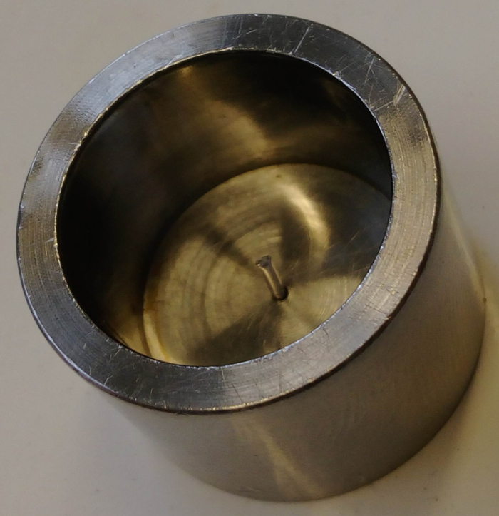
Figure 1: Custom-made Mold for Embedding Ventricles in Agarose. Please click here to view a larger version of this figure.
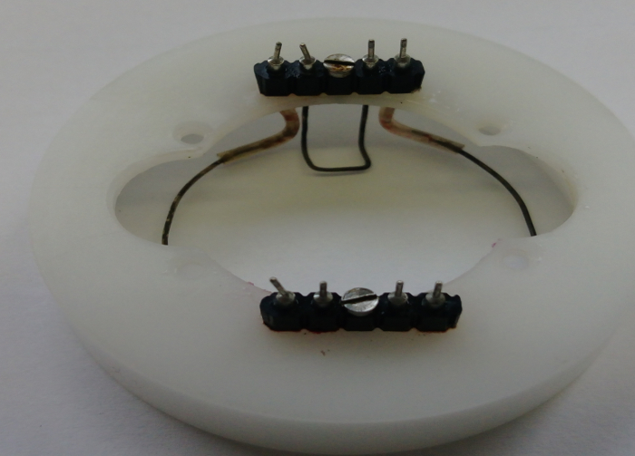
Figure 2: Custom-made Ring Electrode (ground) for Sharp Electrode Recordings. Please click here to view a larger version of this figure.
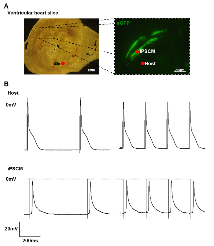
Figure 3: Electrical Integration of Transplanted iPSCM. (A) Ventricular heart slice (left) containing eGFP positive iPSCM (right). SE: Stimulation electrode. Red dots mark the location of the recordings. (B) Action potential recordings in healthy host tissue (upper traces) and transplanted iPSCM (lower traces). The slice was focally stimulated with a stimulation electrode placed in host tissue at around 2 Hz (left traces) and 5 Hz (right traces). Please click here to view a larger version of this figure.
Ventricular slices enable electrophysiological, pharmacological and mechanical studies with a preserved in vivo like tissue structure and direct access of the measurement technology to all regions of the heart. Physiological action potential properties have been demonstrated in embryonic, neonatal and adult slices 9,10,11. Vitality of the slices, except for the surface layers directly damaged by the slicing procedure, has been confirmed by vitality staining 9. Action potential recordings within the slices using sharp glass electrodes can be used to investigate integration and maturation of transplanted cardiomyocytes as described here, or for pharmacological studies after adding cardioactive compounds. Long-term recordings with duration of one hour or more in an individual cell are possible. This recording duration is sufficient to test the impact of different pharmacological compounds on one cell by sequential perfusion and washout.
Critical preparation steps
To optimize slice quality, ischemic tissue damage should be avoided. Therefore, perfusion of the heart in situ with Tyrode's solution, rapid heart resection and immediate preservation in ice cold oxygenated Tyrode's solution are considered crucial. Immobilization of the beating heart using 2,3-butanedione monoxime may further reduce susceptibility to ischemia and is required for a solid embedding in low-melt agarose. Embedding in low-melt agarose might hamper tissue supply by limiting diffusion, but is essential to stabilize the soft heart tissue before slicing. Small cavities within the specimen block, e.g. ventricular chambers not filled completely with agarose or remaining air bubbles, might hamper tissue stabilization. Unstabilized myocardial tissue does not offer sufficient resistance to the vibrating blade, which causes severe tissue disruption and damage. To ensure a clear cut and to minimize tissue damage, blades should be sharp (i.e. not reused more than three times), and forward movement of the blade through the specimen block should be as slow as possible, since dull blades or overspeeding may loosen the embedded ventricles or lacerate the tissue.
To ensure reliable and reproducible results, the quality of slices should fulfill specific criteria, including structural integrity of the tissue, which can be confirmed by microscopy, and response to electrical stimulation at physiological beating frequencies up to 10 Hz.
Slices can be cut at a thickness of 150-400 µm depending on further application. While thinner slices may prevent hypoxic conditions in the tissue core and allow for easier detection of transplanted cells with a fluorescence microscope as well as more precise positioning of recording electrodes 15, thicker slices may provide a better integrity of the tissue structure and higher stability of the tissue, which will be advantageous for force measurements 10 and further histological processing after recordings.
To prevent potential influences of high BDM concentrations on ion channels and Ca2+ handling proteins 16,17, BDM washout by incubation of slices in BDM free DMEM for at least 30 min before further use is recommended. Surprisingly, contraction and subsequent movement of the tissue after BDM washout do not hamper sharp electrode recordings.
Limitations
In contrast to common cell culture models, slices provide an intact tissue structure including preserved electrical and mechanical cell to cell connections, extracellular matrix and distribution of cardiomyocytes and non-myocytes within the native tissue 6. However, cardiac tissue slices do not exactly match the in vivo situation, since they are just 150 – 400 µm thick, supplied by artificial medium and might be damaged during the preparation process. Trypan blue staining and immunohistological evaluation of active caspase-3 and PARP p85 fragment revealed that cells within embryonic slices, except for those in the surface layers, were viable after slicing 9.
The conduction system is not preserved in ventricular slices, and excitation spread is two-dimensional rather than three-dimensional in the flat slices. Slices from embryonic hearts show spontaneous beating for up to two weeks in culture 9. Spontaneous contractions of adult slices may also occur, and it is unclear whether they are induced by cells of the conduction system or may indicate cell damage and Ca2+ overload 18.
Future applications
Besides electrophysiological studies with sharp electrodes and micro electrode arrays, which may be used for pharmacological experiments to analyze the impact of drugs on action potential properties and excitation spread 11, developmental studies 19 or characterization of transplanted cardiomyocytes 15 as shown above, ventricular slices of neonatal hearts have been used for force contraction measurements 10, which could be applied to adult slices, too. Further potential future applications include (I) long-term microscopy studies in cultivated slices, e.g. to assess structural integration of injected stem cell-derived cardiomyocytes, (II) injection of gap junction permeable dyes to investigated intercellular coupling or (III) studies of endothelial and vascular function of coronary vessels within the slices.
In conclusion, ventricular slices are a valuable multicellular tissue model with preserved in vivo like structure, which may help to overcome limitations of common cell culture models and are applicable to a wide range of pharmacological, physiological, developmental and cell therapy studies.
The authors have nothing to disclose.
We acknowledge the support provided by the workshops and the animal facility of the Institute of Neurophysiology. This work was supported by Walter und Marga Boll- Stiftung, Köln Fortune and Deutsche Stiftung für Herzforschung.
Leica VT 1000s | Leica Microsystems, Wetzlar, Germany | Microtome with vibrating blade. | |
Stainless Steel Blades | Campden Instruments, Loughborough, England | 7550-1-SS | |
Pasteur pipettes | Sigma-Aldrich, St. Louise, USA | Z627992 | |
Fine brush, e.g. size 6 (4/32") | VWR, International, Radnor, USA | 149-2125 | |
Preparation table | self made | ||
Molt for embedding ventricles in agarose | self made | ||
1 ml Syringe | Becton, Dickinson; Franklin Lakes, USA | 300013 | |
27Gx3/4“ Needles | Braun, Melsungen, Germany | 4657705 | |
20G 11/2“ Needles | 4657519 | ||
Small scissor | WPI, Sarasota, USA | 501263 | |
Tweezers #5, 0.1 x 0.06 mm tip | WPI, Sarasota, USA | 500342 | |
Oxygen gas (medical grade O2) | Linde, Munich, Germany | ||
Carbogen gas (95 % O2, 5 % CO2) | Linde, Munich, Germany | ||
NaCL | Sigma-Aldrich, St. Louise, USA | 7647-14-5 | |
KCL | Sigma-Aldrich, St. Louise, USA | 746436 | |
CaCl2 | Sigma-Aldrich, St. Louise, USA | 746495 | |
KH2PO4 | Sigma-Aldrich, St. Louise, USA | NIST200B | |
HEPES | Sigma-Aldrich, St. Louise, USA | 51558 | |
NaHCO3 | Sigma-Aldrich, St. Louise, USA | S5761 | |
D(+)-Glucose | Sigma-Aldrich, St. Louise, USA | G8270 | |
MgSO4 | Sigma-Aldrich, St. Louise, USA | M7506 | |
NaOH | Sigma-Aldrich, St. Louise, USA | S8045 | |
Cyanoacrylate glue | Henkel, Düsseldorf, Germany | ||
Low-melt Agarose | Roth, Karlsruhe, Germany | 6351.2 | |
Heparin-sodium-25000 I.E./5mL | Ratiopharm, Ulm, Germany | ||
Dulbecco's Modified Eagle Medium (DMEM), high glucose, GlutaMAX | ThermoScientific, Waltham, USA | 10566016 | |
SEC-10LX Amplifier | npi electronic GmbH, Tamm, Germany | SEC-10LX | |
EPC 9 | HEKA Elektronik GmbH, Lambrecht, Germany | ||
Zeiss Axiovert 200 | Zeiss, Oberkochen, Germany | ||
Low magnification Micromanipulator | Narashige, Tokyo, Japan | Nm-3 | |
High magnification, three-axis micromanipulator | Narashige, Tokyo, Japan | MHW-3 | |
Peristaltic perfusion pump | Multi Channel Systems, Reutlingen, Germany | PPS2 | |
2-channel temperature controller | Multi Channel Systems, Reutlingen, Germany | TCO02 | |
Square pulse stimulator | Natus Europe GmbH, Planegg, Germany | Grass SD9 | |
Glass capillaries | WPI, Sarasota, USA | 1B150F-1 |