Sialic acid is a typical monosaccharide-unit found in glycoconjugates. It is involved in a plethora of molecular and cellular interactions. Here we present a method to modify cell surface sialic acid expression using metabolic glycoengineering with N-acetylmannosamine derivatives.
Sialic acid (Sia) is a highly important constituent of glycoconjugates, such as N– and O-glycans or glycolipids. Due to its position at the non-reducing termini of oligo- and polysaccharides, as well as its unique chemical characteristics, sialic acid is involved in a multitude of different receptor-ligand interactions. By modifying the expression of sialic acid on the cell surface, sialic acid-dependent interactions will consequently be influenced. This can be helpful to investigate sialic acid-dependent interactions and has the potential to influence certain diseases in a beneficial way. Via metabolic glycoengineering (MGE), the expression of sialic acid on the cell surface can be modulated. Herein, cells, tissues, or even entire animals are treated with C2-modified derivatives of N-acetylmannosamine (ManNAc). These amino sugars act as sialic acid precursor molecules and therefore are metabolized to the corresponding sialic acid species and expressed on glycoconjugates. Applying this method produces intriguing effects on various biological processes. For example, it can drastically reduce the expression of polysialic acid (polySia) in treated neuronal cells and thus affects neuronal growth and differentiation. Here, we show the chemical synthesis of two of the most common C2-modified N-acylmannosamine derivatives, N-propionylmannosamine (ManNProp) as well as N-butanoylmannosamine (ManNBut), and further show how these non-natural amino sugars can be applied in cell culture experiments. The expression of modified sialic acid species is quantified by high performance liquid chromatography (HPLC) and further analyzed via mass spectrometry. The effects on polysialic acid expression are elucidated via Western blot using a commercially available polysialic acid antibody.
Sialic acid is a monosaccharide that can typically be found at the non-reducing termini of glycoconjugates, such as N– and O-glycans or glycolipids. Among all monosaccharides, sialic acid has some unique chemical characteristics. It has a 9 C-atom backbone, a carboxylic group in the C-1 position, that is deprotonated and thereby negatively charged under physiological conditions, and an amino function in the C-5 position. Although over 50 naturally occurring variants of sialic acid have been characterized to date1, the predominant form of sialic acid found in humans is N-acetylneuraminic acid (Neu5Ac). Other mammals also express higher amounts of N-glycolylneuraminic acid (Neu5Gc)2,3.
Due to its exposed position in glycoconjugates, sialic acid is involved in a plethora of receptor-ligand interactions, e.g., the hemagglutinin dependent binding of the influenza virus to host cells4. A sialic acid epitope with important biological functions, especially during embryogenesis and in the nervous system, is polysialic acid. Polysialic acid is a polymer of up to 200 alpha 2,8-linked sialic acids. The major protein carrier of polysialic acid is the neural cell adhesion molecule (NCAM). Polysialic acid expression modulates the adhesive property of NCAM in that polysialic acid expression decreases the adhesion and increases plasticity with the nervous system5.
Changes in the expression of (poly)sialic acid will ultimately affect a multitude of different biological interactions. This can be used to study known sialic acid dependent processes on a molecular level, to uncover novel glycoconjugate interactions, or explore possible therapeutic approaches. There are different methods available by which the expression of sialic acid on the cell surface can be modulated, for example treatment with sialic acid specific glycosidases (sialidases), inhibition of enzymes involved in the sialic acid biosynthesis6,7,8, or knocking down or changing the expression of the key enzyme of sialic acid biosynthesis9.
Another versatile method to modulate sialic acid expression is MGE (also known as metabolic oligosaccharide engineering, MOE). Herein, cells, tissues, or even animals are treated with non-natural derivatives of ManNAc that bear C2-amino modifications. Being precursor molecules for sialic acid, after cellular uptake, these ManNAc analogs are unidirectional metabolized to non-natural sialic acids and can be expressed on sialylated glycoconjugates. Cells treated with ManNAc derivatives carrying aliphatic C2-modifications, such as ManNProp or ManNBut, do incorporate N-propionylneuraminic acid (Neu5Prop) or N-butanoylneuraminic acid (Neu5But) in their glycoconjugates10,11. By using functional groups introduced to the C2-position of ManNAc, the occurring non-natural sialic acids can be coupled, e.g., via the Staudinger ligation or the azide alkine cycloaddition, with fluorescent dyes and therefore visualized on the cell surface12.
The expression of these non-natural sialic acids has intriguing effects on many biological processes, including pathogen infections, the adhesion and migration of tumor cells, general cell adhesion, as well as vascularization and differentiation (for review see: Wratil et al.13). Interestingly, MGE with N-acyl modified mannosamines can also be used to interfere with the expression of polysialic acid. Polysialic acid is generated by two different polysialyltransferases (ST8SiaII and ST8SiaIV). It has been demonstrated, that polysialyltransferase ST8SiaII is inhibited by unnatural sialic acid precursors, such as ManNProp or ManNBut14,15. In addition, it has been demonstrated in human neuroblastoma cells that ManNProp or ManNBut application also reduces sialylation in total15.
MGE with N-acyl modified mannosamines is an easy to apply method that has been successfully used, not only in mammalian and bacteria cell culture but also in entire animals of different species, such as Caenorhabditis elegans16, zebrafish17, or mice18,19,20,21. Especially ManNAc derivatives bearing aliphatic modifications, including ManNProp and ManNBut, are negligibly cytotoxic, even at millimolar concentrations in cell culture medium or blood plasma. Furthermore, they are relatively easy to synthesize.
Here, we provide details on how to use MGE with N-acetyl modified mannosamines. First, the chemical synthesis of two of the most widely used ManNAc derivatives in this field, ManNProp and ManNBut, is explained. Next, we show how MGE can be applied in an in vivo experiment. As an example, the neuroblastoma cell line Kelly was chosen to demonstrate decreased expression of the polysialic epitope by Western blot after treatment with the ManNAc derivatives. The non-natural sialic acids on the cell surface were quantified by HPLC and further analyzed via mass spectrometry.
1. Preparation of Buffers and Reagents
2. Synthesis of ManNProp and Related N-acyl-modified Mannosamines
3. Cell Culture
4. Cell Lysis for HPLC Analysis
5. Separation of the Membrane Fraction
6. Acidic Hydrolysis
NOTE: Oligo- and polysaccharides in the cell membranes are hydrolyzed to monosaccharides. Further, possible O-modifications in the monosaccharides are hydrolyzed, as well. This is necessary for quantitative HPLC analysis, because the overwhelming majority of sialic acids in the membrane fractions are naturally incorporated in glycoconjugates and might possibly bear O-acetyl or O-lactolyl modifications.
7. Fluorescent Labeling
8. HPLC Analysis
NOTE: Labeled samples are analyzed on a HPLC system equipped with a C18 RP column (110 Å, 3 µm particle size, 4.6 x 150 mm), a fluorescence detector, and a fraction collector. The solvents used are methanol, acetonitrile, and water. Make sure to degas all solvents prior to the measurements, if the HPLC system lacks an internal degasser.
9. Mass Spectrometry Analysis of Sialic Acid Monosaccharides
NOTE: HPLC retention peaks of interest can be further analyzed by liquid chromatography (LC) electrospray-ionization mass spectrometry (ESI-MS), in order to verify the unnatural sialic acid species.
10. Western Blot Analysis of Polysialic Acid in Kelly Neuroblastoma Cells
HPLC chromatograms of the fluorescent labeled Neu5Ac and the Neu5Gc standards are depicted in Figure 2. Using the herein described method, DMB-labeled Neu5Gc typically elutes between 7 – 9 min elution time, and DMB-Neu5Ac between 10 – 12 min. Several smaller peaks in the chromatogram usually appear between 2 – 6 min. These peaks represent unreacted DMB and reaction intermediates25.
Figure 3 shows representative chromatograms of cell lysates. Compared to the chromatograms of the DMB-labeled standards (Figure 2), in these chromatograms several undefined peaks are visible. This is most likely due to the internal impurity of cell lysates and to the fact that DMB not only reacts with sialic acid species, but also with other α-keto acids present in cell lysates, including pyruvate, succinate, and α-ketoglutarate26. The presence of small amounts of sialic acids bearing O-acetyl modifications that have not been cleaved by acetic hydrolysis might also be discussed as a reason for these undefined peaks23. Chromatograms from lysed untreated cells are compared with chromatograms from cells that have previously been treated with ManNAc or its analogs. As shown here, treatment with ManNAc often leads to an almost entire depletion of Neu5Gc on the cell surface. In chromatograms of lysed cells treated with Neu5Prop or Neu5But, peaks are visible that do not appear in chromatograms of untreated cells. These peaks (for ManNProp treated cells at approximately 19 min elution time and for ManNBut treated cells at 42 min) indicate the appearance of the corresponding non-natural sialic acids, Neu5Prop and Neu5Gc. As described in section 8.3 of the protocol, the HPLC chromatograms can be analyzed in order to quantify the amounts of the different sialic acid species in the cell lysates.
The fact that distinct HPLC retention peaks correspond to certain sialic acid species was verified via mass spectrometry. Representative ESI-MS data from collected HPLC peaks of interest are depicted in Figure 4: the spectrum of the collected DMB-Neu5Ac retention peak in the upper left panel, DMB-Neu5Gc in the upper right panel, and the two non-natural sialic acid species, DMB-Neu5Prop and DMB-Neu5But in the lower left and right panels. The reaction with DMB leads to an increase in the molecular mass of 116.2 Da, compared to the sialic acid species that are not labeled with this dye. In the mass spectra, when displaying the positive mass/charge ratio of the injected samples between 300 and 700, several peaks are visible. This is due to adduct formation of the respected DMB-labeled probe. Besides the protonated ion [M+H]+, the sodium adducts [M+Na]+ and [M+2Na-H]+ appear. As acetonitrile (ACN) is present during the LC analysis, it may be found as an adduct in the mass spectra (shown for DMB-Neu5Gc, Figure 4: upper right panel). Due to fragmentation caused by collisional decompensation activations generated in the electronspray transport region between the capillary and the first skimmer, dehydrated sialic acid species [M+H]+-H2O can also be observed (shown for DMB-Neu5Ac, Figure 4: upper left panel)23,27. In the LC setup, DMB-Neu5Gc elutes shortly after the unreacted or partly reacted DMB-species. Therefore, the collected DMB-Neu5Gc probe may contain impurities caused by these reaction intermediates. This serves as an explanation for the appearance of undefined peaks in the mass spectrum of DMB-Neu5Gc (Figure 4: upper right panel).
Treatment of Kelly neuroblastoma cells with ManNProp or ManNBut leads to reduced expression of polysialic acid on NCAM, as shown by Western blot analysis of the cell membrane from lysed cells (Figure 5). Polysialic acid normally appears as a smear do to its heterogeneity in size and charge of (≈ 200 kDa). Treatment with ManNAc analogs leads to the reduction of polysialic acid on the cells surface: the longer the aliphatic N-acyl side chain, the stronger the reduction28.
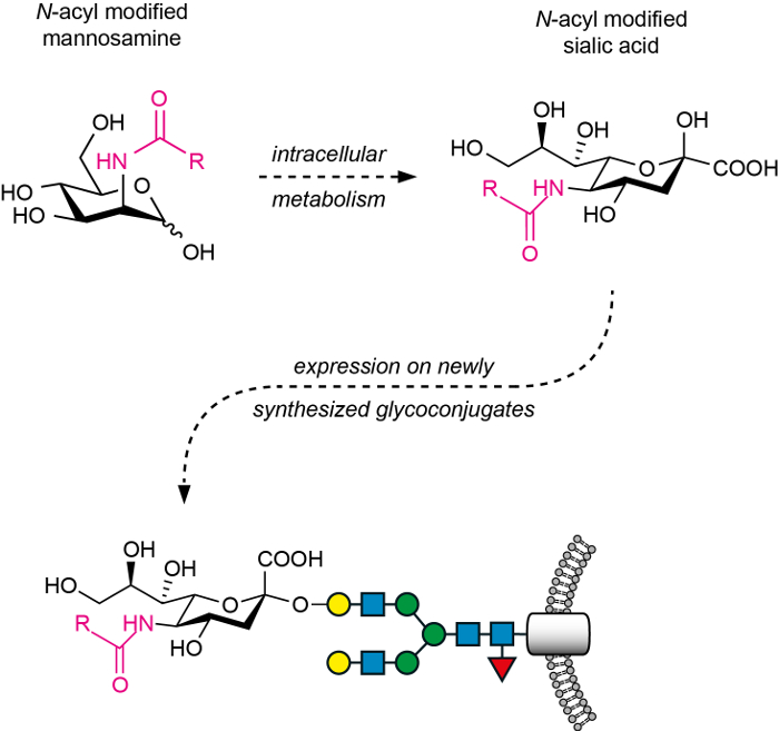
Figure 1: The general principle of MGE with N-acyl modified mannosamines. After treatment with the N-acyl modified mannosamines, these ManNAc analogs are unidirectionally metabolized (intracellular metabolism) to the corresponding non-natural sialic acids. These are transported to the Golgi, transferred to the respective oligosaccharide acceptor by sialyltransferases, and expressed on the cell surface as sialosides (here, a glycoprotein). Please click here to view a larger version of this figure.
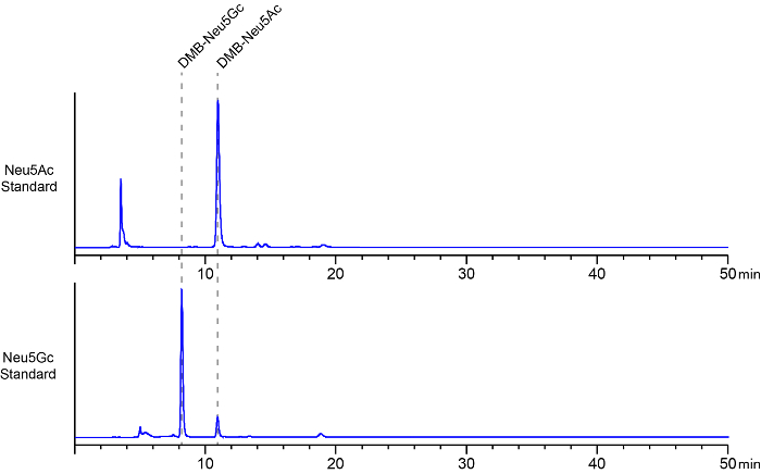
Figure 2: Representative chromatograms of DMB-labeled sialic acid standards. DMB-labeled Neu5Ac (upper panel) and Neu5Gc (lower panel) were analyzed by HPLC. Retention times (dashed lines) and peak areas of both sialic acids were determined after injecting 10 ng of the respected DMB-labeled species into the HPLC system. In the herein shown representative chromatograms, DMB-Neu5Gc eluted after approximately an 8 min retention time and DMB-Neu5Ac at 11 min, respectively. Several smaller peaks in the chromatogram usually appear between 2 – 6 min, representing unreacted DMB and reaction intermediates. A small fraction of DMB-labeled Neu5Ac appears as a minor impurity in the DMB-Neu5Gc standard (lower panel). This figure is modified from reference22. Please click here to view a larger version of this figure.
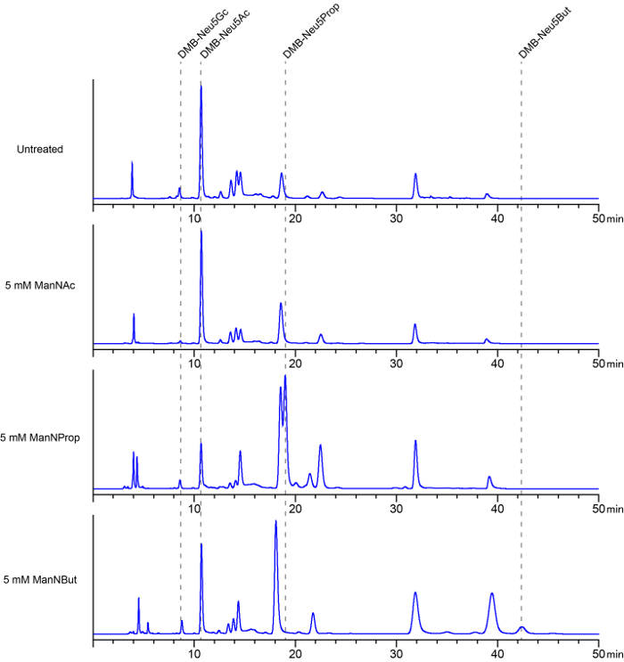
Figure 3: Characterization of membrane-bound sialic acids by DMB-HPLC. Cells were cultured in the absence (untreated, upper panel) or presence with 5 mM ManNAc (second panel from top), ManNProp (third panel from top), or ManNBut (lower panel), respectively for 7 days. The medium was changed every 24 h. Cells were harvested and membrane fractions were prepared, labeled with DMB, and analyzed by HPLC. By comparison with the chromatograms of DMB-labeled sialic acid standards (Figure 2), peaks at the retention times between 8-9 min were identified as DMB-Neu5Gc, and peaks between 10 – 11 min as DMB-Neu5Ac. Compared to the peaks obtained from injecting DMB-labeled sialic acid standards, chromatograms from cell lysates show additional, undefined peaks. This is due to the internal impurity of the probes, as well as to the fact that DMB can react not only with sialic acid present in the cell lysates, but also with other α-keto acids present in cell lysates, including pyruvate, succinate, and α-ketoglutarate. Peaks that only appear in the chromatograms of samples from cells cultured in the presence of N-acyl modified mannosamine analogs indicate the corresponding DMB-labeled non-natural sialic acids. This figure is modified from reference22. Please click here to view a larger version of this figure.
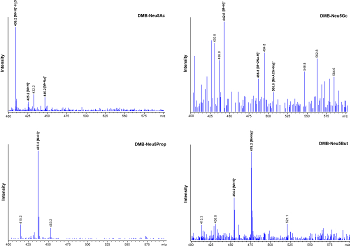
Figure 4: Mass spectra of DMB-labeled sialic acid species found in cell lysates. HPLC retention peaks of interest (see Figure 3) were collected and subsequently analyzed by ESI-MS. 20 µL of the collected retentions peaks were injected into the LC-MSD system. Reaction with DMB leads to an increase in the molecular mass of 116.2 Da compared to the corresponding unreacted sialic acid species. The positive mass/charge ratio spectrograms obtained from different sialic acid species are depicted (DMB-Neu5Ac: upper left panel, DMB-Neu5Gc: upper right panel, DMB-Neu5Prop: lower left panel, DMB-Neu5But: lower right panel). Due to adduct formation several peaks are visible. Besides the protonated ion [M+H]+, sodium adducts [M+Na]+ and [M+2Na-H]+ appear. Acetonitrile, which is present during the LC analysis, can also be found as an adduct (upper right panel). Dehydrated DMB-Neu5Ac, which is generated by fragmentation in the instrument [M+H]+-H2O can be observed (upper left panel). The collected DMB-Neu5Gc probe may contain impurities caused by unreacted DMB, as well as reaction intermediates seen as additional undefined peaks (upper right panel). ACN, acetonitrile; H, hydrogen; Na, sodium. This figure is modified from reference22. Please click here to view a larger version of this figure.
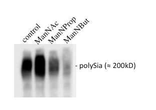
Figure 5: Analysis of the polysialylation of NCAM. Kelly neuroblastoma cells were cultured in the absence or presence of 5 mM ManNAc, ManNProp, or ManNBut for 7 days. Cells were harvested and proteins were separated on 8% SDS-acrylamide gels and analyzed by Western blot using monoclonal anti-polySia antibody 735. Note that polySia appears as a smear due to its heterogeneity in number of Sia, resulting in different sizes and charges of polySia.
If the chemically synthesized ManNAc derivatives, ManNProp and ManNBut are analyzed via mass spectrometry, only the correct mass peak for both specimens should be identified. Therefore, the products can be assumed to have a purity of over 99%. Small amounts of Neu5Gc, which is normally not found in human cells29, are detected in the membrane fractions of the lysed cells. This most likely occurs through a salvage pathway that recruits Neu5Gc from fetal bovine serum sialoglycoconjugates in the media30. Treatment with the natural precursor of sialic acid biosynthesis, ManNAc, drastically lowers the expression of the Neu5Gc epitope on the cell surface.
Application of modified mannosamines is negligibly toxic to all cells investigated so far. Cells accept increased millimolar sugar concentrations within the medium, which is necessary, since there are no specific transporters for N-acylmannosamines. It has been demonstrated that signal transduction pathways are activated upon application of N-acylmannosamines, which could affect cell growth or differentiation31,32. Whether this is due to a changed sialylation pattern or reflects direct effects on the modified sialylation is not known so far. Growth of cells in the presence of the C2-modified ManNAc analogs, ManNProp or ManNBut results in the expression of non-natural sialic acid species carrying the corresponding N-acyl substitution. The metabolic efficiency of the two tested N-acyl modified mannosamines varies. Treatment with ManNProp usually leads to expression of the corresponding Neu5Prop on the cell surface and to a decreased expression of Neu5Ac as well as Neu5Gc. Other N-acyl modified mannosamine analogs, like N-cyclopropylcarbamyl mannosamine, tend to have an even higher metabolic efficiency22.
Previous studies revealed that sialic precursors interfere with polysialylation. Polysialic acids are nearly exclusively expressed on the NCAM. Interestingly, NCAM can be polysialylated by two distinct polysialyltransferases expressed in the Golgi (ST8SiaII and ST8SiaIV). It turned out that ManNProp, ManNBut, and other N-acyl-modified mannosamines interfere with polysialylation and that this is predominantly due to interaction with ST8SiaII14. This sialyltransferase is expressed during development and is important for the development of the brain. Interestingly also, many tumors from neuronal origin re-express ST8SiaII increasing their malignancy and further making them undetectable from the immune system33. In contrast, the application of ManNAc leads to an increase of polySia, since it leads to an increase of CMP-Neu5Ac, which is the natural substrate of both polysialyltransferases.
MGE is a unique method to introduce novel sialic acids on proteins of interest (e.g., erythropoietin) and to modulate thereby its function. Furthermore, it can be used to modulate cell surface glycosylation, which might be of interest in many diseases, such as cancer, since many cancer cells try to escape the immune system by expressing high levels of sialic acid. The method presented here allows: first, to perform glycoengineering using cell cultures, and second, to analyze engineered glycans to prove successful glycoengineering. The method is very robust and up to now, no pitfalls are reported; in contrast, the method has been expanded to introduce chemical active groups to perform click-chemistry on cells13.
Here we present two ManNAc analogs with aliphatic N-acyl side chain modifications that are comparably simple to synthesize, even by laboratories with less equipment and less experience in chemical synthesis. Groups that are more familiar to organic chemistry could synthesize other ManNAc analogs with more complex structures and use these for MGE, including the so-called 'bioorthogonal' analogs, which can be utilized, for example, to visualize sialylated glycoconjugates. There are review articles giving an overview of the N-acetyl-mannosamine derivatives that have been synthesized to date13,34. N-azidoacetyl mannosamine (ManNAz), a substance that is frequently used, to visualize sialylated glycans, is commercially available. Because of the low membrane permeability of ManNAc analogs, it can be advantageous to use derivatives of these sugars with protected hydroxyl-groups, e.g., peracetylated ManNAc analogs. After entering the cytoplasm, the protecting groups are cleaved by cytoplasmic esterases, thereby releasing the active monosaccharides35,36,37. However, peracetylated ManNAc analogs expose higher cytotoxicity compared to the corresponding unprotected derivatives.
In this manuscript, we chose immortalized neuroblastoma cells for our experiments with MGE. Generally, MGE (especially with ManNProp and ManNBut) can be applied in a wide variety of cell lines, including immortalized cell lines (see Wratil et al.13 for examples) and primary cells38,39. Furthermore, MGE was successfully utilized to alter sialylation in vivo de C. elegans40, zebrafish17,41, mouse19,20,42, and rat43,44. The treatment time and the concentration of the ManNAc analogs in MGE experiments can be varied to affect the metabolic efficiency of this method. From our experience, longer treatment and higher concentrations correspond with better replacement of the naturally occurring sialic acid species in glycoconjugates by modified Neu5R. If very high concentrations of ManNAc derivatives are to be used, the cytotoxicity of the treatment should be evaluated, e.g., by the MTT assay or the resazurin reduction assay.
In this manuscript, we suggest using ultra-sonication for cell homogenization. Other cell lysis methods were tested in our laboratory, including douncing on ice (approximately 250 strokes) and french press disruption (10,000 psi, 4 passages), with comparable outcomes regarding the amount of sialic acid detected in the samples. The advantage of ultra-sound homogenization is that it needs little time and does not require the addition of DNAase to the samples prior to lysis.
We focused our analysis on the membrane fractions of cell lysates, because we wanted to show alterations in the sialylation of membrane bound glycoconjugates. Sialic acid derivatives and their concentrations can also be measured in the cytosol of cells, after the membrane fraction has been separated by high-speed centrifugation, using the same method as described above. However, the concentrations of sialic acid in the cytosol are up to 100x lower compared to sialic acid expression on the cell membrane7. Thus, larger amounts of cells are needed to generate reliable results. The pool of cytosolic, activated CMP-sialic acid can be measured indirectly by pre-treating the cytosolic fractions of cell lysates with sodium borohydride prior to hydrolysis7,45. In order to hydrolyze sialic acid in glycoconjugates and sialic acid with hydroxyl modifications, the cells were incubated with 1 M TFA. Other acidic solutions can be used for hydrolysis, as well, e.g., 2 M propionic acid, 2 M acetic acid, or 1 M HCl.
A possible encountered issue during HPLC analysis is that peaks of interest are overlapping with undefined peaks. This is especially problematic when the amount of sialic acid in a sample is to be calculated by the area under the curve of a certain peak. To separate a peak of interest from an undefined peak, the acetonitrile concentration in the HPLC solvent might be adjusted (± 3%). We suggest using steady solvent concentrations for HPLC analysis, giving the advantage of preparing the solvent mixture before chromatography. Therefore, our method requires only one HPLC pump and is feasible for a wide range of different HPLC systems. If more than one pump is available in the HPLC system, an isocratic gradient with increasing concentrations of acetonitrile can be applied, e.g., 4-9% in 35 min45. By using this technique, the retention time of certain HPLC peaks will be altered considerably. This strategy can also help to achieve better separation of overlapping peaks. Matrix-assisted laser desorption ionization mass spectrometry (MALDI-MS) was successfully established to verify the expression of modified sialic acid species on the cell surface45, and thus serves as an equivalent to the ESI-MS method presented here.
Due to the hydrophilicity of ManNAc analogs and the lack of specific transporters for the uptake of these derivates46, high concentrations are needed in MGE experiments with these compounds. Therefore, in up-scaled experiments and, especially in in vivo trials, large amounts of the respective ManNAc analogs are necessary showing a possible limitation of this technique. On the other hand, depending on the cell lines used, a certain minimal number of treated cells is necessary to obtain reliable results in the HPLC analysis. From our experience, a minimum of about 100,000 adherent cells or 150,000 suspension cells (1-2 wells of a 96-well plate with confluent cells) is sufficient. This can become a bottleneck, if the analysis is to be downsized, for example in screening approaches.
With the methods presented in this manuscript, sialic acid and its derivatives can be detected in cell lysates. However, the structure and composition of glycoconjugates in cells treated with ManNAc derivatives cannot be assessed with the herein described technique. Resolving the structure of glycoconjugates, e.g.,N– and O-glycans, generally requires greater experience and a glycomics facility47. To our knowledge, so far, no data are available on the distinct structure of glycoconjugates from cells that have been treated with ManNAc analogs.
Western blot analysis of polysialic acid is a very rapid and easy method. However, data obtained by this method are not quantitative, since the detection uses antibodies and chemiluminescence. Therefore, the use of HPLC methods is an advantage over Western blotting. Nevertheless, we suggest using the immunoblotting method, since it is easy to apply and known to work very reliably.
The expression of sialic acid on the cell surface can be altered also by methods other than MGE. As described above, cells can be treated with sialidase, an enzyme that cleaves sialic acid residues from glycoconjugates in a relatively unspecific manner. Sialidase treatment consequently induces hyposialylation in treated cells. Unfortunately, sialidase treatment is a rather limited technique, as it is cytotoxic, not feasible for long time treatment, and not applicable in in vivo experiments. Another technique to induce hyposialylation in cells is by using inhibitors of sialic acid biosynthesis. A variety of inhibitors is available that either target the key enzyme of sialic acid biosynthesis, the UDP-N-acetylglucosamine-2-epimerase/N-acetylmannosamine-kinase (GNE/MNK), or the group of sialyltransferases6,7,8,48. One of these compounds, a C3-fluorinated sialic acid analog, was shown to lower the expression of sialic acid in living mice49. Animals treated with this substance, however, showed liver impairment, irreversible kidney dysfunction, and failure to thrive49. These results confirm the crucial role of Neu5Ac in liver and kidney function and further indicate the limits of sialylation inhibitors for in vivo experiments. A third method to generate cells with altered cell surface sialylation is by interfering with the expression of enzymes that are crucial for sialic acid biosynthesis. A knock-down of the GNE/MNK in immortalized cells, for example, was shown to render these cells hyposialylated 9. The possibility to relatively easily knock-in and -out genes in cellular and in vivo using the novel and well recognized CRISPR-Cas9 technique might lead to novel discoveries regarding sialic acid biosynthesis and cell surface sialylation.
The advantage of MGE compared to the techniques described here, is that it is easy to apply and adaptable to a myriad of different experiments, from in vitro trials to cell based assays and in vivo experiments. An alternative method to measuring the expression and concentrations of different sialic acid species in cell samples is by high-performance anion-exchange chromatography (HPAEC) with pulsed electrochemical detection50. By using this method, the expression of sialic acid is measured directly and not after labeling with a fluorescent dye. The advantage of this method is that it can be utilized also to analyze monosaccharides other than sialic acid and its derivatives in cell- and protein-samples. Unfortunately, the sensitivity of HPAEC is lower compared to fluorometric detection of DMB-sialic acid. Thus, HPAEC is limited to experiments with the availability of large sample amounts51.
The herein described methods can be used to reveal unknown characteristics of glycoconjugates. A multitude of examples for the use of MGE to investigate sialic acid dependent interactions already exists13, showing the feasibility of this technique in context of a wide range of experimental setups.
Nowadays, MGE with N-acetylmannosamines is mainly used by researchers in the field of glycobiology. We hope that this technique will be recognized by a broader audience as a versatile tool to modify glycoconjugates. Other fields, including immunology, virology, neurology, or oncology will benefit by adapting this technique for their own purposes. Such cross-over research might enable the discovery of yet uncharted areas, and therefore give a better understanding of health and disease. In early studies, for example, this technique was used to produce glycoproteins with modified glycosylation, which later serve for in vivo vaccination. In certain cases, treatment with such glycoengineered vaccines lead to production of antibodies that exposed advanced avidity and toxicity to the respective targets52,53. Others successfully used MGE to develop a model for targeted tumor therapy in mice54. Another study showed that systemic in vivo injection of ManNProp promoted nerve regeneration55. In the case of polySia, it was shown that diminishing the expression of this epitope in neuroblastoma cells by the methods presented in this manuscript, increases the sensitivity of these tumor cells to radiation or treatment with anticancer drugs56.
The amounts of sialic acid measured within samples might differ depending on the method used to detach the treated cells. In this manuscript, incubation with trypsin was used to detach cells. If other techniques are to be used, such as scraping the cells or long-time treatment with PBS + EDTA, the amounts of sialic acid measured in the samples might differ. Even adapting the incubation time with trypsin can have an impact on the sialic acid concentrations measured later on. From our experience, the impact of the cell detachment method on the amounts of sialic acid measured is below 15%, but if results are to be normalized and compared, this might become an important bottleneck. Additionally, the method for lysis of the cells can affect the outcome of the HPLC analysis. Thus, we recommend the reader to standardize cell detachment and lysis in their experiments.
To achieve separation of the membrane fraction in cell lysates, we used centrifugation for 2 h at 20,000 x g and 4 °C (section 5.1). Other centrifugation protocols might affect the separation and therefore the amounts of sialic acid detected in the samples. A critical compound within this protocol is DMB, because it is very sensitive to light and degrades over time. Here we recommended to aliquot small portions of the DMB-solution and store it as stocks at – 20 °C, protected from light (section 1.6.1). This way the DMB-solution can be stored for several weeks. If the results from DMB-HPLC analysis show decreasing signals for sialic acid species or increasing signals within the first 6 min of the HPLC measurement, representing DMB and its reaction products, new DMB-solution should be prepared. Do not re-freeze and re-use DMB-solution after thawing it. After DMB labeling, all samples should be analyzed immediately. If larger numbers of samples (> 10) are to be analyzed, these samples should be divided into smaller badges and gradually labeled with DMB. Both the labeling reaction as well as the DMB-labeled probes should be protected from light at all times. After elution, the DMB-labeled sialic acid species should be analyzed immediately by ESI-MS. Direct coupling of the HPLC with the ESI-MS system (in a LC-ESI-MS setup) is not necessary, but can be advantageous to achieve higher quality of the mass spectrometry analysis.
The authors have nothing to disclose.
We thank L. D. Nguyen for proofreading the manuscript and for fruitful discussions. Furthermore, we thank J. Dernedde and H. G. Nguyen for helping us preparing the video shoot. Most scenes of the video were shot in the laboratories of R. Tauber. We also thank the Max Planck Institute for the colloids and interfaces, and for giving us free access to their mass spectrometry facility. RH was supported by the DFG (ProMoAge).
Cells | Sigma-Aldrich | 92110411 | |
RPMI medium | Sigma-Aldrich | R8758 | |
75 ml tissue culture flasks | Greiner | 690175 | |
48-well plates | Corning | 3548 | |
FCS | PAA | A15-102 | |
Pen/Strep | Gibco | 15140-122 | |
Trypsine | Gibco | 15400-054 | |
EDTA | Roth | X986.1 | |
Tris | Serva | 37190.03 | |
SDS | Roth | 2326.2 | |
SDS-PAGE equipment (tanks, glassware etc., machine | VWR | SDS Gel/Blot | |
Acrylamide | Roth | 3019.1 | |
Protein ladder | Fisher Scientific | 267620 | |
Nitrocellulose | GE Healthcare | 10600002 | |
Ponceau red | Roth | 5938.2 | |
Milk powder | Roth | T145.3 | |
ECL | Millipore | WBLUF 0500 | |
0.5 ml Centrifugal Filter Unit with 3 kDa membrane | Merck-Millipore | UFC500324 | |
15 mL centrifuge tubes | Sigma-Aldrich (Corning) | CLS430791-500EA | |
2-Mercaptoethanol | Sigma-Aldrich | M6250-10ML | |
2-Propanol | Sigma-Aldrich | 34965-1L | HPLC gradient grade |
4,5-Methylenedioxy-1,2-phenylenediamine dihydrochloride | Sigma-Aldrich | D4784-50MG | |
48 well, flat bottom tissue culture plate | Sigma-Aldrich (Corning) | CLS3548-100EA | |
50 mL centrifuge tubes | Sigma-Aldrich (Corning) | CLS430829-500EA | |
Acetonitrile | Sigma-Aldrich | 34967-1L | HPLC gradient grade |
Aprotinin from bovine lung | Sigma-Aldrich | A1153-10MG | lyophilized powder, 3-8 TIU/mg solid |
Butyryl chloride | Sigma-Aldrich | 109614-250G | |
C18 RP column | Phenomenex | 00F-4435-E0 | 110 Å, 3 µm particle size, 4.6 x 150 mm |
D-Mannosamine hydrochloride | Sigma-Aldrich | M4670-1G | |
Dulbecco`s Phosphate Buffered Salt Solution | PAN Biotech | P04-36500 | |
Ethylenediaminetetraacetic acid | Sigma-Aldrich | E9884-100G | |
Formic acid | Sigma-Aldrich | 56302-50ML-GL | |
Hydrochloric acid solution | Sigma-Aldrich | H1758-100ML | 36.5-38.0%, in water |
Leupeptin | Sigma-Aldrich | L2884-10MG | |
Methanol | Carl-Roth | T169.2 | HPLC gradient grade |
N-Acetyl-D-mannosamine | Sigma-Aldrich | A8176-250MG | |
N-Acetylneuraminic acid | Sigma-Aldrich | A0812-25MG | |
N-Glycolylneuraminic acid | Sigma-Aldrich | G9793-10MG | |
Phenylmethanesulfonyl fluoride | Sigma-Aldrich | P7626-250MG | |
Propionyl chloride | Sigma-Aldrich | P51559-500G | |
Safe-Lock Tubes, 1.5 mL, amber (light protection) | Eppendorf | 30120191 | |
Safe-Lock Tubes, 1.5 mL, colorless | Eppendorf | 30120086 | |
Sodium bisulfite solution | Sigma-Aldrich | 13438-1L-R-D | 40%, in water |
Sodium chloride | Sigma-Aldrich | 746398-500G-D | |
Sodium hydroxide | Sigma-Aldrich | 795429-500G-D | |
Sodium hydroxide solution | Sigma-Aldrich | 319511-500ML | 1.0 M, in water |
Sodium methoxide | Sigma-Aldrich | 164992-5G | |
Trifluoroacetic acid | Sigma-Aldrich | T6508-100ML-D | |
Tris hydrochloride | Sigma-Aldrich | T5941-100G | |
Trypsin 0.25 %/EDTA 0.02 % in PBS | PAN Biotech | P10-019100 | |
Water | Carl-Roth | T905.1 | HPLC gradient grade |
Silica Gel 60 | Carl-Roth | 9779.1 | |
HPLC | Shimadzu |