Here, we present a protocol to detect discrete metal oxygen clusters, polyoxometalates (POMs), at the single molecule limit using a biological nanopore-based electronic platform. The method provides a complementary approach to traditional analytical chemistry tools used in the study of these molecules.
Individual molecules can be detected and characterized by measuring the degree by which they reduce the ionic current flowing through a single nanometer-scale pore. The signal is characteristic of the molecule's physicochemical properties and its interactions with the pore. We demonstrate that the nanopore formed by the bacterial protein exotoxin Staphylococcus aureus alpha hemolysin (αHL) can detect polyoxometalates (POMs, anionic metal oxygen clusters), at the single molecule limit. Moreover, multiple degradation products of 12-phosphotungstic acid POM (PTA, H3PW12O40) in solution are simultaneously measured. The single molecule sensitivity of the nanopore method allows for POMs to be characterized at significantly lower concentrations than required for nuclear magnetic resonance (NMR) spectroscopy. This technique could serve as a new tool for chemists to study the molecular properties of polyoxometalates or other metallic clusters, to better understand POM synthetic processes, and possibly improve their yield. Hypothetically, the location of a given atom, or the rotation of a fragment in the molecule, and the metal oxidation state could be investigated with this method. In addition, this new technique has the advantage of allowing the real-time monitoring of molecules in solution.
Detecting biomolecular analytes at the single molecule level can be performed by using nanopores and measuring ionic current modulations. Typically, nanopores are divided into two categories based on their fabrication: biological (self-assembled from protein or DNA origami)1,2,3, or solid-state (e.g., manufactured with semiconductor processing tools)4,5. While solid-state nanopores were suggested as potentially more physically robust and can be used over a wide range of solution conditions, protein nanopores thus far offer greater sensitivity, more resistance to fouling, greater bandwidth, better chemical selectivity, and a greater signal to noise ratio.
A variety of protein ion channels, such as the one formed by Staphylococcus aureus α-hemolysin (αHL), can be used to detect single molecules, including ions (e.g., H+ and D+)2,3, polynucleotides (DNA and RNA)6,7,8, damaged DNA9, polypeptides10, proteins (folded and unfolded)11, polymers (polyethylene glycol and others)12,13,14, gold nanoparticles15,16,17,18,19, and other synthetic molecules20.
We recently demonstrated that the αHL nanopore can also easily detect and characterize metallic clusters, polyoxometalates (POMs), at the single molecule level. POMs are discrete nanoscale anionic metal oxygen clusters that were discovered in 182621, and since then, many more types have been synthesized. The different sizes, structures, and elemental compositions of polyoxometalates that are now available led to a wide range of properties and applications including chemistry22,23, catalysis24, material science25,26, and biomedical research27,28,29.
POM synthesis is a self-assembly process typically carried out in water by mixing the stoichiometrically required amounts of monomeric metal salts. Once formed, POMs exhibit a great diversity of sizes and shapes. For example, the Keggin polyanion structure, XM12O40q- is composed of one heteroatom (X) surrounded by four oxygens to form a tetrahedron (q is the charge). The heteroatom is centrally located within a cage formed by 12 octahedral MO6 units (where M = transition metals in their high oxidation state), which are linked to one another by neighboring shared oxygen atoms. While tungsten polyoxometalates structure is stable in acidic conditions, hydroxide ions lead to the hydrolytic cleavage of metal-oxygen (M-O) bonds30. This complex process results in the loss of one or more MO6 octahedral subunits, leading to the formation of monovacant and trivacant species and eventually to the complete decomposition of the POMs. Our discussion here will be limited to the partial decomposition products of 12-phosphotungstic acid at pH 5.5 and 7.5.
The goal of this protocol is to detect discrete metal oxygen clusters at the single molecule limit using a biological nanopore-based electronic platform. This method allows the detection of metallic clusters in solution. Multiple species in solution can be discriminated with greater sensitivity than conventional analytical methods33. With it, subtle differences in POM structure can be elucidated, and at concentrations markedly lower than those required for NMR spectroscopy. Importantly, this approach even allows the discrimination of isomeric forms of Na8HPW9O341.
Note: The protocol below is specific to the Electronic BioSciences (EBS) Nanopatch DC System. However, it can be readily adapted to other electrophysiology apparatus used to measure the current through planar lipid bilayer membranes (standard lipid bilayer membrane chamber, U-tube geometry, pulled microcapillaries, etc.). The identification of commercial materials and their sources is given to describe the experimental results. In no case does this identification imply recommendation by the National Institute of Standards and Technology, nor does it imply that the materials are the best available.
1. Solution and Analyte Preparation
2. Test Cell Assembly
3. Lipid Bilayer Formation
4. αHL Pore Formation
5. Metallic Cluster Partitioning in the Nanopore
6. Ion Channel Recordings and Data Analysis
Over the past two decades, membrane-bound protein nanometer-scale pores have been demonstrated as versatile single-molecule sensors. Nanopore-based measurements are relatively straightforward to execute. Two chambers filled with electrolyte solution are separated by a nanopore embedded in an electrically insulating lipid membrane. Either a patch-clamp amplifier or an external power supply provides an electrostatic potential across the nanopore via Ag/AgCl electrodes immersed in the electrolyte reservoirs. The electric field drives individual charged particles into the pore, which produces transient reductions in the ionic current that depend on the size, shape, and charge of the particles. A computer program controls the applied voltage and monitors, in real time, the ionic current blockades caused by molecules reversibly partitioning into the pore. The current is amplified and converted to voltage with a low-noise, high impedance field-effect transistor and digitized using a data acquisition card.
Here, we provide a general procedure for detecting polyoxometalates with a biological nanopore. As seen in Figure 2, prior to the addition of POMs the unobstructed channel has a mean open channel current of ~ 100 pA at an applied potential of -120 mV. The addition of POMs produces transient blockades and decreases the ionic current by approximately 80%. As expected, because these particles are negatively charged, the blockades are not observed when the polarity of the applied potential is reversed. Note that if the POMs didn't interact with the pore wall, they would diffuse through the pore's in about 100 ns, which is far too brief to be detected with a conventional patch clamp amplifier. Thus, most of the time a given particle spends in the pore is a direct consequence of the interaction between the particle and the pore. The duration of an ionic current blockade event is defined as the residence time, tau (τ).
To illustrate the utility of this method, we discuss the use of an αHL nanopore to monitor the decomposition of 12-phosphotungstic acid (PTA, H3PW12O40) at pH 5.5 and pH 7.5. This decomposition can be observed with 31P NMR measurements, but the concentration needed is 2 mM while nanopore measurements need less than 30 μM, because of the nanopore measurement sensitivity. At pH 5.5, [PW11O39]7- is the predominant species30.
The data analysis is performed by calculating a histogram of the relative blockade depth ratio (i.e.,<i>/<io>, where <i> is the mean current with the POM in the pore and <io> is the mean open channel current). The histogram of the mean current blockade depth ratios at -120 mV and pH 5.5 exhibits a minor peak at <i>/<io> ≈ 0.06 and major peak at <i>/<io> ≈ 0.16 (Figure 3, green). We assume these peaks correspond to [P2W5O23]6- and [PW11O39]7-, respectively, based on 31P NMR. 31P NMR studies suggest that increasing the pH changes the relative concentration of these two species, and this is borne out by the change in the area of the two peaks shown in Figure 3.
When the POM solution is titrated to pH 7.5 ex situ, the total POM concentration decreases due to the partial degradation of the two-principal species to inorganic salts (i.e., free phosphate, HxPO4−3+x and tungstate, WO42− ions). The histogram of the relative blockade depth ratio also shows two principal peaks (Figure 3, orange), but with 20-fold fewer events (which suggests the total POM concentration at pH 7.5 is approximately 20-fold less than that at pH 5.5, if the nanopore's capture efficiency for POMs is the same at the two pH values). It is interesting to note that at pH 7.5 and greater, the POM species observed here were not detected in the 31P NMR spectrum due to their low concentration caused by their dissociation into phosphate and tungstate ions.
Each event's residence time in the pore is defined by the duration of the individual ionic current blockades. The distribution of residence times provides insight into the different species that are present. It was shown earlier that for blockades caused by a differently-sized polymers of poly(ethylene glycol), the residence time distribution for each size of that polymer is well described by a single exponential. That result suggests the interaction of that polymer is a simple reversible chemical reaction12,13,20.
Figure 4 illustrates that the residence time distributions for the two peaks were well differentiated at pH 5.5 and 7.5. Two features are clear. First, under all conditions, multiple exponentials are required to fit each of the distributions, which suggests there are variations of the POMs within each species. Second, the residence times of the POMs in the pore are much shorter at pH 7.5 compared to those at pH 5.5, which suggests a weakening of the interaction between the pore and POMs. It has been shown previously that a change in pH alters the relative number of fixed charges in or near the αHL channel lumen. These changes will directly alter the interactions with partitioning POMs inside the pore and therefore modify their residence times34,35.
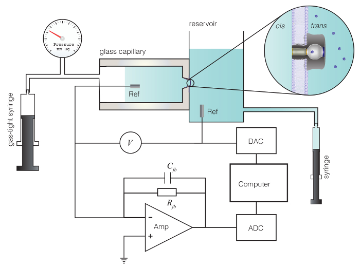
Figure 1: Schematic diagram of the experimental setup. Method for nanopore-based characterization of individual polyoxometalate molecules. A protein nanopore that self-assembles in a 4 nm thick lipid bilayer membrane is bathed by aqueous electrolyte solutions in a glass capillary and larger reservoir. A pressure is applied to the glass capillary with a gas tight syringe to aid nanopore incorporation. A potential V is applied across the membrane with a matched pair of Ag/AgCl electrodes and drives an ionic current (e.g., Na+ and Cl–) through the pore. The current is converted to voltage with a high impedance amplifier, digitized with an analog to digital converter (ADC) and stored on a computer. Computer software controls the applied potential through a digital to analog converter (DAC) and monitors, in real time, the transient current blockades caused by single molecules that partition into the pore. Please click here to view a larger version of this figure.
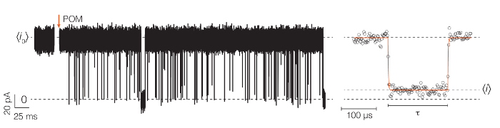
Figure 2: Nanopore-based detection of individual metallo-nanoparticles. An illustration of ionic current time series traces that occur before and after the addition of a POM solution to the nanopore apparatus. The partitioning of individual anionic POMs into the pore causes transient current reductions in the mean open pore current, <io>. (Right) A typical event, illustrating the mean current of the blockade (<i>) and the residence time (τ) of the particle in the pore. The applied potential was -120 mV, and the solutions contained 1 M NaCl, 10 mM NaH2PO4 at pH 5.5. The cis compartment also contained 30 μM of 12-phosphotungstic acid. The current blockade depth ratio (<i>/<io>) and the residence times (τ) provide information about which POM species are present in solution. Under the conditions we used here, the αHL channel does not gate (spontaneously close) when POMs are not present. Please click here to view a larger version of this figure.
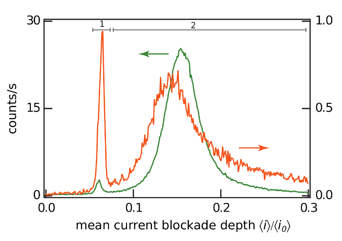
Figure 3: Histograms of the current blockade depth ratio at pH 5.5 and 7.5. Histograms of the POM-induced ionic current blockade depth ratio at pH 5.5 (green) and 7.5 (orange) with an applied potential V = -120 mV. The two peaks present at each pH value correspond to the known predominate POM species in solution under those conditions. The current blockade depth ratios of 0 and 1 correspond to a fully blocked and open pore, respectively. The histograms were created with a bin width of 0.001 and normalized to counts/s by dividing by the data acquisition time. Please click here to view a larger version of this figure.
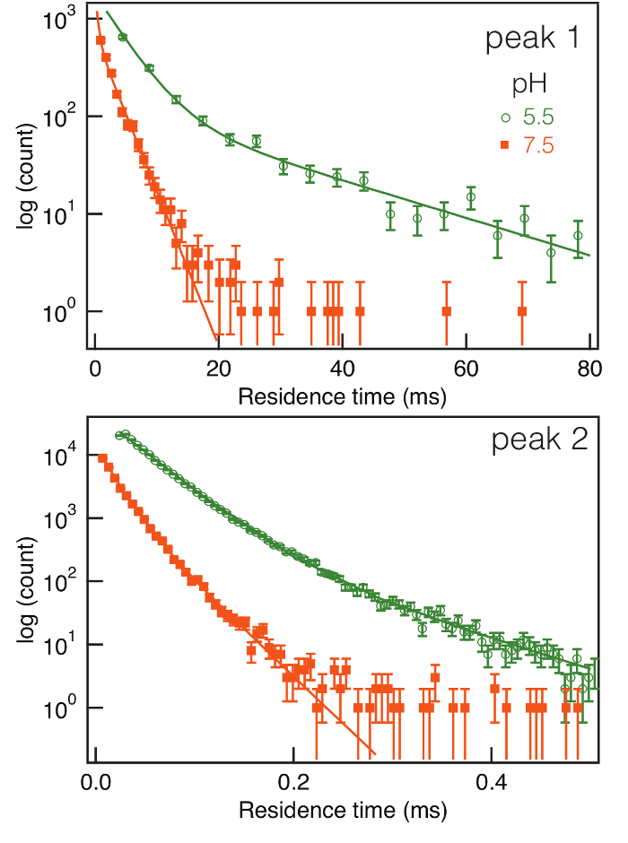
Figure 4: Residence time distribution and fitting with several exponentials. The distribution of residence times for POM-induced current blockades caused by the two-principal species (peaks 1 and 2 in Figure 4) observed at pH 5.5 and 7.5 in a semi-log plot. For both species, the residence times are markedly shorter at the higher pH value, which suggests the interaction between the pore and POMs changed. The solid lines are fits of an exponential mixture model to the data. Please click here to view a larger version of this figure.
Due to their anionic charge, POMs likely associate with organic counter cations through electrostatic interactions. Therefore, it is important to identify the proper solution conditions and the right electrolyte environments (especially cations in solution) to avoid complex formation with POMs. Particular care is required in the buffer choice. For example, the capture rate of POMs with tris(hydroxymethyl)aminomethane and citric acid-buffered solutions is significantly lower than that in phosphate buffered solution, likely because either of the first two buffers form a complex with the POM that doesn't strongly interact with the nanopore. Moreover, the NaCl electrolyte was purposely used instead of KCl (as well as the other alkali metals) to avoid the precipitation of [PW11O39]7- by K+.
Critical to the accurate measurement of the residence time distributions is the ability to measure the current at a sufficiently high bandwidth. For instance, with exponentially-distributed residence times there are far more blockades with short than long residence times, and an accurate estimation of the residence time distributions is better achieved by collecting a great deal of data (i.e., acquiring it at as high as a bandwidth the system's electrical capacitance allows). To achieve this condition in nanopore spectroscopy, the system capacitance (membrane and stray capacitance) should be minimized. Stray capacitance is reduced by decreasing the length of all connecting cables and using high quality electrical contacts. The membrane capacitance is minimized by decreasing the surface area of the bilayer, increasing the thickness of supporting materials (i.e., quartz, polytetrafluoroethylene, etc.), and decreasing the area of exposed supporting materials to the electrolyte. In practice, a typical instrument's stray capacitance (≈ 2 pF) will limit the noise for membranes ≈ 1 micron to 5 micron in diameter. This constitutes the method's limitation. For example, the detection of small and highly charged single molecules can be challenging due to their relatively short residence times.
The mechanism by which pressure enables control of channel insertion is not completely understood. The quartz microcapillaries have a very small diameter on which the membrane is formed. Applying pressure will cause the membrane to bulge (thereby increasing the membrane surface area) and possibly thin the membrane. Both effects would increase the rate at which channels will form in the membrane. When a single channel spontaneously forms, reduce the pressure to prevent the insertion of additional channels. The removal of non-inserted αHL from the bulk aqueous phase is not required if the αHL concentration is sufficiently low.
The structures and charges of polyoxometalates are currently studied using traditional analytical chemistry techniques, including NMR, Ultraviolet-visible, Infrared and Raman spectroscopy, mass spectrometry, and X-ray diffraction. We expect that nanopore measurements will complement the characterization of these and other physical properties of POMs, as well as the study of their speciation at low concentration, which will help better understand the synthetic pathway of polyoxometalates formation. It was also shown previously that the αHL pore can even distinguish between 2 isomers of the trivacant Keggin form Na8HPW9O3430.
In summary, we have shown that a membrane-bound protein nanopore can be used to detect and characterize tungsten oxide metallic clusters (heteropolytungstates) in solution using a simple high-resolution electrical measurement. The sensitivity afforded by this novel approach permits the tracking of subtle differences in POM structure that arise at different pH values at concentrations that are substantially lower (> 70-fold) than required for traditional methods such as NMR spectroscopy. Due to the single molecule detection capability of nanopores, the actual limit of detection in the method can be made much lower by measuring the current for longer times (the capture rate scales in proportion to the POM concentration).
The authors have nothing to disclose.
We are grateful for financial support from the European Molecular Biology Organization for a postdoctoral fellowship (to J.E.) and a grant from the NIH NHGRI (to J.J.K.). We appreciate the help of Professors Jingyue Ju and Sergey Kalachikov (Columbia University) for providing heptameric αHL, and for inspiring discussions with Professor Joseph Reiner (Virginia Commonwealth University).
Nanopatch DC System | Electronic Biosciences, Inc., EBS | ||
Millipore LC-PAK | Millipore vacuum filter | ||
1,2-Diphytanoyl-sn- Glycero-3-Phosphocholine (DPhPC) | Avanti Polar Lipids, Alabaster, AL | 850356P | |
Decane, ReagentPlus, ≥99%, | Sigma-Aldrich | D901 | |
αHL | List Biological Laboratories, Campbell, CA | ||
Ag wire | Alfa Aesar | ||
2 mm Ag/AgCl disk electrode | In Vivo Metric | E202 | |
High-impedance amplifier system | Electronic Biosciences, San Diego, CA | ||
quartz capillaries | |||
custom polycarbonate test cell | |||
Data Processing and Analysis MOSAIC | https://pages.nist.gov/mosaic/ | ||
Phosphotungstic acid hydrate | Sigma-Aldrich | 455970 | |
Sodium Chloride | Sigma-Aldrich | S3014 | |
sodium phosphate monobasic monohydrate | Sigma-Aldrich | 71507 |