Here, we present a protocol to produce an adipose tissue-derived stromal vascular fraction and its application to improve knee functions by regenerating cartilage-like tissue in human patients with osteoarthritis.
Osteoarthritis (OA) is one of the most common debilitating disorders. Recently, numerous attempts have been made to improve the functions of the knees by using different forms of mesenchymal stem cells (MSCs). In Korea, bone marrow concentrates and cord blood-derived stem cells have been approved by the Korean Food and Drug Administration (KFDA) for cartilage regeneration. In addition, an adipose tissue-derived stromal vascular fraction (SVF) has been allowed by the KFDA for joint injections in human patients. Autologous adipose tissue-derived SVF contains extracellular matrix (ECM) in addition to mesenchymal stem cells. ECM excretes various cytokines that, along with hyaluronic acid (HA) and platelet-rich plasma (PRP) activated by calcium chloride, may help MSCs to regenerate cartilage and improve knee functions. In this article, we presented a protocol to improve knee functions by regenerating cartilage-like tissue in human patients with OA. The result of the protocol was first reported in 2011 followed by a few additional publications. The protocol involves liposuction to obtain autologous lipoaspirates that are mixed with collagenase. This lipoaspirates-collagenase mixture is then cut and homogenized to remove large fibrous tissue that may clog up the needle during the injection. Afterwards, the mixture is incubated to obtain adipose tissue-derived SVF. The resulting adipose tissue-derived SVF, containing both adipose tissue-derived MSCs and remnants of ECM, is injected into knees of patients, combined with HA and calcium chloride activated PRP. Included are three cases of patients who were treated with our protocol resulting in improvement of knee pain, swelling, and range of motion along with MRI evidence of hyaline cartilage-like tissue.
Mesenchymal stem cells (MSCs) are known to have the capability to regenerate cartilage1,2,3,4,5,6. They can be easily obtained from various sources: bone marrow, cord blood, and adipose tissue among many others. Among these sources, adipose tissue is the only source where a sufficient number of MSCs can be obtained without any culture expansion to regenerate cartilage in clinical settings7,8. Autologous bone marrow stromal vascular fraction (SVF) can be easily obtained as well. However, the number of stem cells contained in the non-culture expanded marrow is very low7,8. Cord-blood may contain a sufficient number of MSCs. However, cord blood is not a readily available source of autologous SVF.
Numerous methods of processing adipose tissue to obtain SVF are available for clinical applications. Among these, the method of obtaining MSCs from adipose tissue using collagenase, developed and confirmed by Zuk et al.5,6, is very well accepted. This method of using collagenase has been modified for clinical applications in orthopedics. In order to be applied to clinical settings, the system must be a closed system to maintain the sterility while keeping the convenience. One particular modification presented in this article involves homogenization of the lipoaspirates. Small sized lipoaspirates are digested relatively faster than the larger ones that are resulting in the uneven breakdown of adipose tissue. Furthermore, these larger sized lipoaspirates may produce fibrous tissues that can clog up the syringes and needles while performing joint injections9,10. In order to prevent these issues, the lipoaspirates may be homogenized by cutting and mincing the lipoaspirates before the incubation with collagenase. The resulting adipose tissue-derived SVF may contain more uniform extracellular matrix (ECM) compared to lipoaspirates that are not homogenized11. The broken-down ECM contained in the SVF may work as a scaffold12.
In 2009, autologous adipose tissue-derived SVF has been allowed by the Korean Food and Drug Administration (KFDA) when processed within a medical facility with minimal processing by a physician13. Afterwards, autologous adipose tissue-derived SVF has been utilized as a potential agent to improve knee functions in osteoarthritis (OA) patients by potentially regenerating cartilage-like tissue10,14,15,16,17,18.
In 2011, Pak showed for the first time that adipose tissue-derived stem cells (ASCs) contained in the adipose tissue-derived SVF can improve knee functions potentially regenerating cartilage-like tissue in human OA patients when injected with platelet-rich plasma (PRP) 14. In addition, Pak et al. have reported safety data in 2013 involving 91 patients. The mean efficacy rate reported in this safety data was 67%15. Subsequently, additional studies by Pak et al. showed improved knee functions potentially due to cartilage-like tissue regeneration in patients with a meniscus tear and chondromalacia patellae10,16,17,18. Based on articles reported, it is known that the number of stem cells contained in 100 g of adipose tissue processed by the protocol presented in this article may range from 1,000,000 – 40,000,000 depending on patients' characteristics8,19,20,21,22,23.
Here, we present a clinical protocol of human knee OA by using autologous adipose tissue-derived SVF with HA and PRP activated with calcium chloride. The first version of this clinical protocol, involving a closed, manual system to maintain the sterility, was reported in 201114. The identical protocol was optimized, maintaining sterility, and was reported in 2013 and 201610,15. Here, the optimized protocol is presented. The schematic overview of the protocol is presented in Figure 1.
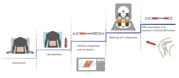
Figure 1: The schematic overview of the protocol. Please click here to view a larger version of this figure.
The approval and consent to report following case reports were waived by Myongji University Institutional Review Board committee (MJUIRB). Further, this clinical protocol was in compliance with the Declaration of Helsinki and regulation guidelines of the KFDA. For the procedures, informed consents were obtained from the patients.
1. Liposuction
NOTE: Perform with sterile technique.
2. Preparation of ASC/ECM Mixture with Sterile Closed System
3. PRP Preparation with Sterile Technique
4. ASC/ECM Mixture-Based Treatment
5. Post-Treatment Follow Up
Three patients (one 87-year-old female with stage 3 OA, one 68-year-old male with stage 3 OA, and one 60-year-old female with stage 3 OA) without any significant past medical history presented to the clinic with persistent knee pain and desired for potential autologous adipose tissue-derived SVF treatment. All three patients had their knee examined by an orthopedic surgeon and were offered to have total knee replacement (TKR) and were reluctant to have the surgery. Prior to the procedure, all three patients had received multiple injections of steroids and HA without any prolonged improvement.
The 87-year-old Korean female patient, at the time of examination, complained of severe pain (VAS score of 8; Figure 2A) while on rest. She described the pain to be increasing ([FRI: 37; Figure 2A) when walking up and down stairs. On physical exam, mild joint swelling with decreased ROM and tenderness with flexion (Figure 2B) were noted. However, no ligament laxity was appreciated. McMurray's and Apley's tests were negative. A before-treatment MRI demonstrated a decreased size medial meniscus along with deformation and maceration. (Figure 2C, 2E, 2G, and 2I).
The second patient, a 68-year-old Korean male, complained of severe left knee pain (VAS score: 7; Figure 3A) while on rest. The pain (FRI: 33; Figure 3A) was described as increasing when walking up and down stairs. On physical exam, there was mild deformity with mild joint swelling. ROM (Figure 3B) was decreased. Further, no ligament laxity was appreciated. McMurray's and Apley's tests were negative. A before-treatment MRI showed cartilage thinning along with a decreased size and deformed medial meniscus secondary to the previous meniscectomy (Figure 3C, 3E, 3G, and 3I). The patient was diagnosed to have stage 3 OA.
The third patient, a 60-year-old Korean female, also reported severe pain (VAS score: 8; Figure 4A) on rest. The pain (FRI: 36; Figure 4A) was described as increasing when walking. The patient also had mild knee swelling and decreased ROM (Figure 4B). No ligament laxity was appreciated. McMurray's and Apley's tests were negative. A before-treatment MRI showed decreased size medial meniscus with deformation and maceration, and there was cartilage thinning (Figure 4C, 4E, 4G, and 4I).
Treatment Plan. All three representative patients were restricted from taking steroids, aspirin, non-steroidal anti-inflammatory drugs (NSAIDs), and Asian herbal medications at least for 1 week prior to the procedure. After taking MRI imaging studies, lipoaspirates were obtained and processed as referenced above. Afterwards, the autologous adipose tissue-derived SVF containing ASCs, ECM, HA, and calcium-chloride-activated autologous PRP were injected to the knees at day 0. There was no complication due to liposuctions and joint injections. Subsequently, the patients returned to the clinic in one week, then 2 weeks, and then 3 weeks for additional injections of HA and autologous PRP activated with calcium chloride.
Outcome. After the second week of the ASC/ECM mixture injection, the 87-year-old female patient's pain and ROM improved (Figure 2A and 2B). By the 16th week, the patient's pain and ROM significantly improved by more than 70% (Figure 2A and 2B). Post-treatment MRI taken after the 16th week showed the increased thickness of hyaline cartilage-like tissue on the medial side of the knee (Figure 2D, 2F, 2H, and 2J). As a comparison, the mean efficacy rate was 67% in data using this clinical protocol, involving 91 patients, and reported in 201315.
Figure 2: Outcome of pain measurements (A) and range of motion (B); and MRI sagittal (C-F) and coronal (G-J) sequential T2 views of the knee from the patient case 1.* indicates a statistically significant finding (p < 0.05). Pre-treatment MRI scans (C: sequential image, 5/20; E: 6/20; G: 10/20; and I: 11/20) show cartilage lesions (arrows). Post-treatment MRI scans at 16 weeks (D: 6/20; F: 7/20; H: 10/20; and J: 11/20) indicate cartilage-like tissue regeneration (arrowhead) that has been repaired by ASC/ECM mixture-based treatment. This figure has been modified from the previous report of Pak et al.10. Please click here to view a larger version of this figure.
The 68-year-old male patient's pain and ROM improved after the second week of the ASC/ECM mixture injection (Figure 3A and 3B). By the 18th week, his pain and ROM significantly further improved to 80% (Figure 3A and 3B). Repeated MRI taken after 18th week showed an increase in the height of hyaline cartilage-like tissue on the anterior medial side of the knee (Figure 3D, 3F, 3H, and 3J).
Figure 3: Outcome of pain measurements (A) and range of motion (B); and MRI sagittal (C-F) and coronal (G-J) sequential T2 views of the knee from the patient case 2.* indicates a statistically significant finding (p < 0.05). Pre-treatment MRI scans (C: sequential image, 6/20; E: 7/20; G: 13/20; and I: 14/20) show cartilage lesions (arrows). Post-treatment MRI scans at 16 weeks (D: 6/20; F: 7/20; H: 13/20; and J: 14/20) indicate cartilage-like tissue regeneration (arrowhead) that has been repaired by ASC/ECM mixture-based treatment. This figure has been modified from the previous report of Pak et al.10. Please click here to view a larger version of this figure.
The 60-year-old female patient's pain and ROM improved approximately 50% after the second week of the ASC/ECM mixture injection, (Figure 4A and 4B). By the 22nd week, the pain and ROM significantly improved over 80% (Figure 4A and 4B). Repeated MRI taken after the 22nd week showed an increase in the height of the hyaline cartilage-like tissue on the medial side of the knee (Figure 4D, 4F, 4H, and 4J).
Figure 4: Outcome of pain measurements (A) and range of motion (B); and MRI sagittal (C-F) and coronal (G-J) sequential T2 views of the knee from the patient case 3.* indicates a statistically significant finding (p < 0.05). Pre-treatment MRI scans (C: sequential image, 4/20; E: 5/20; G: 10/20; and I: 11/20) show cartilage lesions (arrows). Post-treatment MRI scans at 16 weeks (D: 4/20; F: 5/20; H: 10/20; and J: 11/20) indicate cartilage-like tissue regeneration (arrowhead) that has been repaired by ASC/ECM mixture-based treatment. This figure has been modified from the previous report of Pak et al.10. Please click here to view a larger version of this figure.
In 2001, Zuk et al. isolated stem cells from adipose tissue by breaking down the collagen matrix with collagenase6. Afterwards, the group showed that these stem cells isolated from the adipose tissue could transform into cartilage and other tissues of mesoderm in origin, proving that these stem cells were mesenchymal in origin.
Likewise, the procedure presented in this article is a modified protocol to apply the similar method to human patients. The main modification of the protocol is the incorporation of closed system syringes in obtaining and processing the lipoaspirates in order to minimize the air contact while maintaining the ease of performing the procedure. Using such closed system syringes, adipose tissue was obtained via liposuction. Then, the lipoaspirates were mixed with collagenase within the closed system syringes. Afterwards, the collagenase was removed from the mixture by diluting the collagenase concentration within the closed system syringes. The end result is a sterile adipose tissue-derived SVF obtained with ease of the process.
By using the similar method, Pak showed in 2011, for the first time, that autologous adipose tissue-derived SVF can be used to treat OA patients by potentially regenerating cartilage-like tissue. Later, Pak et al. also demonstrated that autologous adipose tissue-derived SVF can be used to treat meniscus tear and chondromalacia of patellae by regenerating fibrous cartilage-like tissue and hyaline cartilage-like tissue, respectively. In 2013, Pak et al. reported a safety study involving 91 patients treated with autologous adipose tissue-derived SVF. The mean efficacy of the study reported was 67%, paving the road to the possibility of applying autologous adipose tissue-derived SVF in general patient population with cartilage damage.
One of the most common side effects reported by Pak et al. in 2013 safety study was post-treatment knee joint swelling, which can be explained by 1) stem cell death, 2) inflammation by RBCs contained in the PRP, and/or 3) residual collagenase that has remained in the final volume of adipose tissue-derived SVF. Since MSCs exist within ECM of adipose tissue11,12, digesting the lipoaspirates with collagenase to break down the matrix to lease the stem cells is a mandatory process12. However, after breaking down the matrix to release the stem cells, the remaining residual collagenase should be fully removed from the final volume in order to prevent any potential inflammation that can be produced by the residual collagenase28. Although collagenase can cause inflammation, an insignificant amount of collagenase in the final volume of adipose tissue-derived SVF may not trigger a joint swelling. Such final volume of autologous adipose tissue-derived SVF with an insignificant amount of collagenase has been used safely for the last few years to treat OA of knees by regenerating cartilage-like tissue.
Recently two non-enzymatic methods of obtaining adipose tissue-derived SVF have been introduced29,30. These methods utilize either mechanical force or ultrasound (vibrational) force, instead of collagenase. In 2018, D'Ambrosi et al. reported a result of treating four osteochondral lesions of the talus of the ankle with micro-fractured, purified autologous adipose tissue that was obtained by using mechanical force to breakdown the matrix29. No collagenase was used to break down the matrix. Four patients were treated with the micro-fractured adipose tissue (i.e., mechanically broken-down adipose tissue), for their talus29. All these four patients showed clinical improvement six months after the procedure. None had any complications. However, on this study29, there was no report of potential regeneration of cartilage. Another study published this year involving vibrational energy showed that vibrational energy in certain frequency was not sufficient to isolate stem cells30.
As shown above, different groups have different protocols for using an autologous adipose tissue to treat various degenerative joint disorders. The protocol can vary from the size of the cannula used for liposuction, the amount of adipose tissue used, type and amount of collagenase (if any), method of injections, and so on. Since MSCs exist within ECM of adipose tissue, the effect of collagenase, or purification method using mechanical forces, may play an important role. Especially, the efficacy of collagenase may depend on factors such as the size of the adipose tissue, amount of the adipose tissue, concentration of collagenase, time and temperature of incubation with collagenase, and the process of removing the leftover collagenase, which can cause cell damage28.
It should be noted that collagenase, as it is an important component of this procedure to produce stem cell containing adipose tissue-derived SVF, may have a negative effect on the final volume of adipose tissue-derived SVF. In this procedure, collagenase is mixed with condensed lipoaspirates, homogenized, incubated, and then washed off. Throughout this process, stem cells are exposed to collagenase, which can have a detrimental effect on the survival of the stem cells28. Too much concentration or prolonged exposure of collagenase may lower the stem cell viability in the final volume28. Further, too much of collagenase left in the final volume may cause inflammation of the joint since collagenase may break down the cartilage matrix of the joint injected28.
In addition to the potential negative effects of collagenase, liposuction can carry other potential complications. In order to obtain adipose tissue-derived SVF, liposuction must be performed first. Performing liposuctions, as other medical procedures, carries few potential complications such as irregularities in the skin contour, seromas, infection, perforation of the abdominal wall, necrotizing fasciitis, fat embolism, and pulmonary embolism. Among these complications, skin contour irregularities are a very common side effect that can be easily prevented by using small cannulas and avoiding superficial liposuctions31,32. Seromas, which is a collection of serous fluid in the suctioned area, can also be a result of aggressive liposuction33. Other potential complications such as infection, necrotizing fasciitis, perforation of abdominal walls, fat embolism, or pulmonary embolism are all possible. However, these complications can also be prevented by using strict sterile techniques and by improving patient compliance34.
Further, all three patients included in the study initially presented with a mild joint effusion, a clear sign of synovitis. After the treatment with adipose tissue-derived SVF, all these patients joint swelling improved. For this reason, it is also reasonable to assume that the improvement of clinical symptoms is not due to the possible cartilage regeneration, but instead, due to the modulatory and anti-inflammatory effect of stem cells and/or PRP on synovial membrane35,36. Alternatively, the symptom improvement can be contributed to both modulatory and anti-inflammatory effect of stem cells and/or PRP on synovial membrane along with possible regeneration of cartilage-like tissue. Although more research is necessary to delineate the true mechanism of symptom improvement, this percutaneous intra-articular injection of cell therapy in the form of autologous, homogenized adipose tissue-derived SVF with autologous PRP activated with calcium chloride and /or HA may offer an alternative treatment to the current strategy of treating knee OA.
As for the addition of autologous PRP is concerned, it is widely accepted that PRP contains various growth factors and differentiating factors for injected MSCs to grow and differentiate37,38,39. PRP also has shown to have a collagenase neutralizing effect40. Regarding the addition of hyaluronic acid (HA), HA has been shown to have a potential role as a scaffolding material, due to its high affinity to cartilage tissue, and a potential role to assist stem cells in penetrating the cartilage matrix41.
As more new processes of obtaining adipose tissue-derived SVF are available, an optimization of a method of processing lipoaspirates is necessary to standardize the process. However, this may be a difficult task due to the number of potential variations in the process of obtaining and processing autologous adipose tissue-derived SVF. For example, differences in the texture of subcutaneous adipose tissue in each individual patient, due to aging or the degree of obesity, may have different responses to collagenase activities, resulting in a different number of ASCs in the autologous adipose tissue-derived SVF42. In addition, the number of stem cells in each gram of adipose tissue may differ from one patient to another as shown by the great variation of reported stem cell numbers in numerous publications7,19,20,21,22,23.
This protocol presented in this article is a novel, innovative procedure attempting to improve the current strategy of treating human knee OA by potentially regenerating cartilage-like tissue with percutaneous injections of autologous adipose tissue-derived SVF. The correct use of collagenase is an important and critical step in this protocol. Using closed system syringes to keep the sterility while maintaining the convenience of performing the procedure is the major modification of the protocol developed by Zuk et al.5 In addition, for human application, the residual amount of collagenase contained in the final adipose tissue-derived SVF should be insignificant to prevent any joint inflammation. There are limitations of this protocol: the collagenase residues in the final volume (although this may be negligible), no control groups that singly injected PRP and HA, no biopsies, no evidence excluding the improvement of clinical symptoms due to PRP, the small number of patients (pilot study), no defying components of the adipose tissue-derived SVF, and no specification of the number of stem cells in each of the 3 patients. Applying adipose tissue-derived SVF for disorders such as a meniscus tear, chondromalacia patellae, spinal disc disorders, a delayed healing (or non-healing) bone fracture, a non-healing skin ulcer, breast reconstruction, and other medical disorders may improve the clinical outcome. Although these are potential clinical applications of adipose tissue-derived SVF, more thorough and vigorous clinical researches are necessary for incorporating adipose tissue-derived SVF in actual clinical settings.
The authors have nothing to disclose.
The author acknowledges the support from the staff of Mipro Medical Clinic and the figure design by Jaepil/David Lee. This work was supported by research grants from the Bio & Medical Technology Development Program of the NRF funded by the MSIT (number NRF-2017M3A9E4078014); and the National Research Foundation of Korea (NRF) funded by the Ministry of Science and ICT (numbers NRF-2017R1A2B4002315 and NRF-2016R1C1B2010308).
Material | |||
5% Betadine (povidone-iodine) | Firson Co., Ltd. | 657400260 | |
2% Lidocaine | Daehan Pharmaceutical Co. | 670603480 | |
Tumescent solution | Myungmoon Pharm. Co. Ltd. | N01BB01 | The solution was composed of 500 mL normal saline, 40 mL 2% lidocaine, 20 mL 0.5% marcaine, and 0.5 mL epinephrine 1:1000. |
Liberase TL and TM research grade | Roche Applied Science | 5401020001 | |
D5LR | Dahan Pharm. Co., Ltd. | 645101072 | Dextrose 5% in lactated Ringer's solution |
Anticoagulant citrate dextrose solution | Fenwal, Inc. | NDC:0942-0641 | The solution was composed of 0.8% citric acid, 0.22% sodium citrate, and 0.223% dextrose. |
3% (w/v) Calcium chloride | Choongwae Pharmaceutical Co. | 644902101 | |
0.5% (w/v) HA (Hyaluronic acid ) | Dongkwang pharm. Co., Ltd. | 645902030 | |
0.25% Ropivacaine | Huons Co., Ltd. | 670600150 | |
Equipment | |||
3.0 mm Cannula | WOOJU Medical Instruments Co. | ML30200 | |
60-mL Luer-Lock syringe | BD (Becton Dickinson) | 309653 | |
Centrifuge Barrel Kit | CPL Co., Ltd. | 30-0827044 | |
Tissue homogenizer that contains blades | CPL Co., Ltd. | 30-0827045 | |
Rotating incubator mixer | Medikan Co., Ltd | MS02060092 | |
Centrifuge | Hanil Scientific Inc. | CE1133 | |
Magnetic Resonance Imaging | Philips Medical Systems Inc. | 18068 | |
Ultrasound Imaging System | Samsung Medison co., Ltd | CT-LK-V10-ICM-09.05.2007 |