This article provides an efficient and feasible method for constructing multilayered stem cell sheets with favorable stem cell property.
Stem cell therapy shows a promising future in regenerating injured organ and tissues, and the cell sheet technique has been developed to improve the low cell retention and poor survival within the target zone. However, during the in vitro construction process, a solution for maintaining stem cell bioactivity and increasing the cell amount within the cell sheet is urgently needed. Here, this protocol presents a method for constructing a multilayered cell sheet with favorable stem cell bioactivity and optimal operability. Decellularized porcine pericardium (DPP) is prepared by phospholipase A2 (PLA2) decellularization method as the cell sheet scaffold, and rat bone marrow mesenchymal stem cells (BMSCs) are isolated and expanded as the seeded cells. The temporary multilayered cell sheet structure is constructed by using RAD16-I peptide hydrogel. Finally, the cell sheet is cultured with a dynamic perfusion system to stabilize the three-dimensional (3D) structure, and the cell sheet could be obtained following a 48-hour culture in vitro. This protocol provides an efficient and feasible method for constructing a multilayered stem cell sheet, and the cell sheet could be developed as a favorable stem cell therapy product in the future.
Stem cell therapy has been reported as an effective treatment for many diseases; however, low cell retention and poor survival within the target zone remain critical issues following traditional stem cell injection. To solve this problem, tissue engineering scientists developed the cell sheet technique. A monolayered cell sheet with intact extracellular matrix was firstly prepared by using the temperature-response culture dish1, and its follow-up studies reported the significant improvements of stem cell retention and survival within the infarcted area2,3. Among the methods, constructing the multilayered cell sheet has been reported as an effective strategy for improving the cell survival and the cell sheet therapeutic effect3,4. Since then, scientists have worked on developing different cell sheet construction methods in order to increase the cell amount, stem cell property, and mechanical property of the cell sheets. So far, certain types of cell sheet have been constructed and studied in the treatment of myocardial infarction5, cartilage injury6, and skin wound7.
The bioactivity of stem cells before transplantation showed an emerging influence on injured tissue regeneration, and different cell sheet construction strategies have different effects on the stem cells. On one hand, confluent cell sheets only consisted of high-density stem cells, and natural extracellular matrices could be acquired by stacking monolayered cell sheets8 or by using magnetic tissue engineering techniques9. On the other hand, researchers developed different scaffolds to provide adequate mechanical strength and support cell growth10,11,12, which allowed a low stem cell seeding density to ensure the nutrition supply. However, despite these approaches, the low efficient nutrition supply within the multilayered cell sheet structure remains a major concern during the in vitro construction. Therefore, an efficient and feasible cell sheet construction system is urgently required.
This protocol describes the steps to prepare a multilayeredmesenchymal stem cell (MSC) cell sheet. In this construction system, the cell sheet mechanical strength is provided by a DPP. Based on this scaffold, the 3D cell structure can be quickly constructed with RAD16-I peptide hydrogel, and a dynamic perfusion system is used to culture the multilayered cell sheet, in order to stabilize the 3D cell sheet structure and provide sufficient nutrition supply for the cells. Using this system, a multilayered BMSC sheet was successfully prepared and exhibited an optimal therapeutic effect on the rat myocardial infarction model13.
All stem cell and animal experiment procedures were conducted according to the ethical guidelines of the National Guide for the Care and Use of Laboratory Animals and approved by the Jinan University Animal Care and Use Committee (Guangzhou, China).
1. Preparation of the DPP Scaffold with the PLA2 Decellularization Method14
Note: See Figure 1A for a schematic of the PLA2 decellularization method.
2. Preparations for the Cell Sheet Construction
3. Preparation of the Cells for Cell Sheet Construction
Note: This protocol is for cell culture using a 100 mm dish. See Figure 1B for a schematic of the construction of the multilayered cell structure.
4. Preparation of the BMSCs and the RAD16-I Peptide Hydrogel Mixture
Note: See Figure 1B for a schematic of the construction of the multilayered cell structure.
5. In Vitro Culture of a 3D Multilayered Cell Sheet Using a Dynamic Culture System
NOTE: See Figure 1C for a schematic of the 3D dynamic system.
6. Obtaining the Multilayered MSC Cell Sheet
The schematic of the multilayered stem cell sheet construction is shown in Figure 1. Preparing the cell sheet scaffold by the PLA2 decellularization method is the first step. Based on the scaffold, a temporary 3D cell structure is constructed by mixing the stem cells with the RAD16-1 peptide hydrogel. In order to obtain a multilayered cell sheet with favorable stem cell bioactivity and optimal mechanical strength, the cell sheet is cultured in a dynamic perfusion system. Under the dynamic nutrition supply, the stem cells are allowed to proliferate and establish cell contacts within the multilayered cell sheet, and the final stable multilayered cell sheet product can be obtained after a ~24- to 72-hour cultivation.
In this case, the cell sheet scaffold DPP is prepared by the PLA2 decellularization method. The appearance of dried DPP is flat, smooth, and semitransparent (Figure 3A). Owing to the specific lyse effect of PLA2, the heterogeneous cells can be completely removed while the ultrastructure of the natural collagen within the DPP scaffold is well-preserved (Figure 3B), and this is important for maintaining the mechanical strength and biocompatibility of the scaffold. Additionally, the scaffolds can be modified as a growth factor control release system to support stem cell growth and improve the in vivo regeneration13.
When the stem cells reach ~80% – 90% confluence, the cells are isolated from the culture dish and washed with a 10% sucrose solution. After centrifugation, the cells are mixed with the RAD16-I peptide hydrogel and added to the rehydrated DPP scaffold. A temporary multilayered structure is formed following a two-hour static culture. Finally, the multilayered BMSC sheet product (Figure 4) is acquired following a 48-hour culture in the dynamic perfusion system. With the support of the DPP scaffold, the cell sheet can be easily manipulated with forceps, and it can be temporarily preserved in culture medium in the 1.5 mL tube at 4 °C for 4 hours before examination or transplantation (Figure 4). As the immunofluorescence staining result shows, the BMSCs are highly positive for the stem cell markers CD90 and CD29. After the cell sheet construction, the BMSCs within the multilayered cell sheet show high levels of CD29 and CD90 (Figure 5).
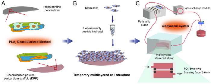
Figure 1: The flowchart of constructing the multilayered stem cell sheet. (A) By using the PLA2 decellularized method, the heterogeneous cells within the FPP are destroyed while the natural extracellular matrices are well-preserved in the DPP scaffold. (B) Based on the DPP scaffold, the temporary multilayered cell structure is constructed by mixing the stem cells and self-assembling peptide hydrogel. (C) To follow, the cell sheet is cultured in a 3D dynamic system, and the stem cells are expected to proliferate and establish cell contacts under the dynamic nutrition supply. Please click here to view a larger version of this figure.
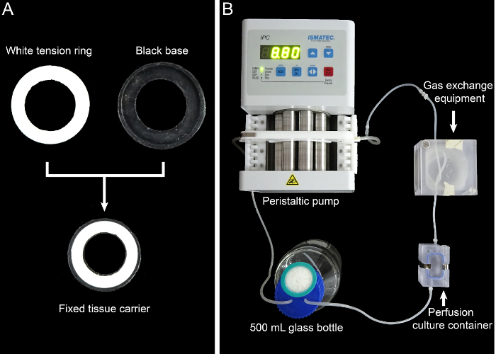
Figure 2: The tissue carrier and the dynamic perfusion system. (A) This panel shows the 13 mm-diameter tissue carrier. (B) This panel shows the assembly of the dynamic perfusion system. Please click here to view a larger version of this figure.
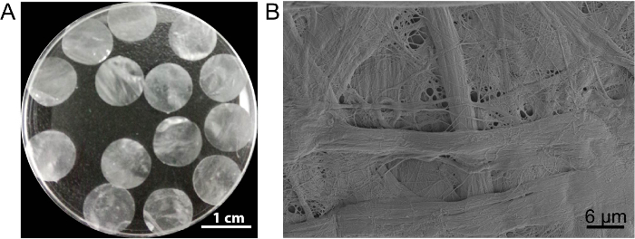
Figure 3: The appearance and ultrastructure of DPP. (A) This panel shows the appearance of the 10.5 mm-diameter DPP scaffolds. (B) This panel shows a representative image of the scanning electron microscope (SEM) result of the DPP scaffold. Please click here to view a larger version of this figure.
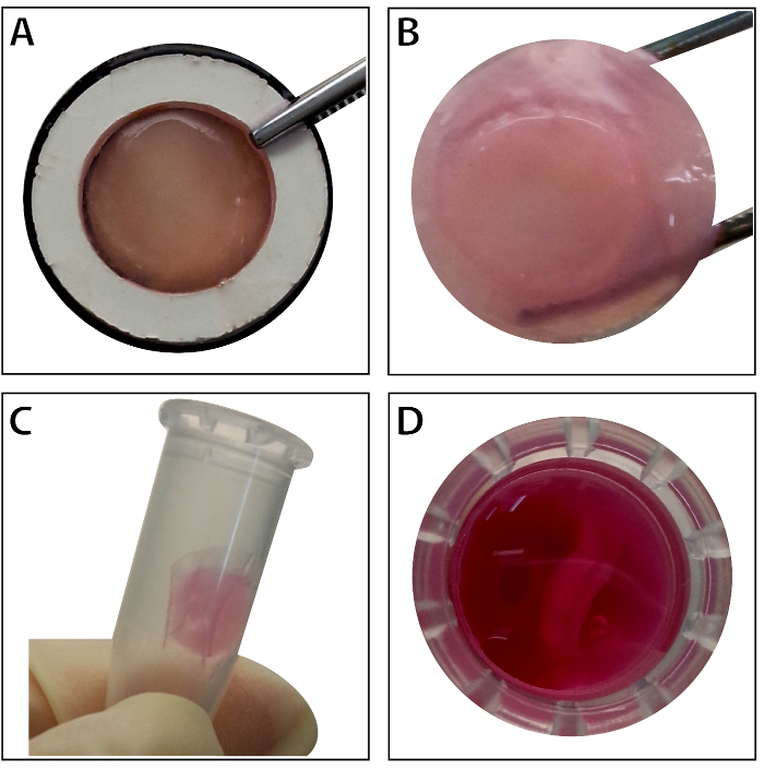
Figure 4: The appearance of the multilayered BMSC sheet. (A) This panel shows the appearance of the multilayered BMSC sheet within the tissue carrier. (B) The intact multilayered BMSC sheet is held by forceps. (C – D) The multilayered cell sheet can be preserved temporarily in the 1.5 mL tube before use. Please click here to view a larger version of this figure.
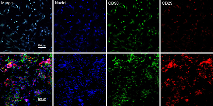
Figure 5: Immunofluorescence staining results of BMSC markers expression. (A) This panel shows immunofluorescence staining results of BMSCs before cell sheet construction. (B) This panel shows immunofluorescence staining results of the multilayered BMSC sheet section. CD90 (green) and CD29 (red) were positively expressed in the BMSCs and the cell sheet. Please click here to view a larger version of this figure.
The present protocol reports an efficient method for constructing a multilayered MSC sheet. This cell sheet exhibits optimal mechanical strength, high cell seeding density, and favorable stem cell bioactivity. Using BMSCs as an example, the 3D cell structure is quickly constructed with RAD16-I peptide hydrogel. After being cultured in the dynamic perfusion system, the multilayered BMSC sheet is successfully obtained and the BMSCs maintain a high expression of stem cell markers.
Constructing the temporary multilayered cell structure is the critical step of the protocol. The RAD16-I is a commercial hydrogel peptide, and it consists of 1% amino acid and 99% water. Several studies reported that this peptide hydrogel can mimic the natural ECM environment and is beneficial for stem cell proliferation and survival15,16,17. In the present protocol, a three million MSC suspension (in 20 µL of 10% sucrose solution) was mixed with 20 µL of RAD16-I peptide hydrogel. The volume ratio of the cell suspension and the peptide hydrogel was 1:1. This peptide hydrogel is sensitive to the environmental pH value, and the peptide molecules would automatically form the 3D network when the pH value changes from acid to neutral. Because the cell surface contains charged particles, the cell mixture changed from liquid to hydrogel in a short time, which has influences the even mixing of the cells. A favorable cell-hydrogel should be an even mixture of the cell suspension and the peptide hydrogel and enables the cell mixture to be evenly added onto the scaffold. The researchers can optimize the mixture condition by altering the seeded cell number, sucrose solution volume, and the peptide hydrogel volume according to their actual need. It is worthwhile to notice that washing the cells with 10% sucrose solution and evenly mixing the cell-hydrogel mixture are the critical steps of the protocol, and an uneven mixture could cause great cell loss and an unstable temporary multilayered structure.
After adding the cell-hydrogel mixture onto the DPP scaffold, the mechanical strength of the multilayered cell sheet structure is weak because the peptide hydrogel network is not strong enough to maintain the long-term multilayered cell structure, and cell connections and ECM secretions are needed to enhance the stability of the cell sheet. Moreover, the dynamic infiltration of the culture medium can facilitate the stem cells to proliferate and establish cell contacts within the multilayered cell structure, while an insufficient nutrition supply will cause cell apoptosis and reduce the cell density of the cell sheet13. Therefore, the dynamic perfusion system is important for stabilizing the multilayered cell sheet structure. In addition, the appropriate flow rate of the culture medium should be adjusted according to the specific stem cell type and cell seeding density. Also, the weak mechanical connection between the DPP scaffold and the multilayered cell structure remains the limitation of the present construction method, which may cause the division of the multilayered cell layers and the scaffold. Therefore, further studies are required to enhance the mechanical biocompatibility of the 3D hydrogel scaffold and the DPP scaffold.
So far, tissue engineering scientists have been focusing on establishing efficient nutrition supply systems in vitro, such as coculturing endothelial cells18 and using a porous scaffold19. However, the nutrition permeability within the 3D structure is low in the traditional static 3D culture system, and the stem cell viability will be greatly affected. In this case, using the dynamic perfusion system can provide enough nutrition supply to maintain stem cell viability. Using this protocol, a multilayered BMSC sheet improved the cardiac function and angiogenesis in a rat myocardial infarction model13. Constructing a stem cell sheet product with a high cell load and favorable stem cell property is significant to the tissue regeneration. Using this efficient constructed method, different kinds of multilayered stem cell sheets could be constructed by altering the seeded stem cell types, such as epithelial stem cell sheet, neural stem cell sheet, or cardiac stem cell sheet. Further explorations of and alternatives to the multilayered stem cell sheet are expected to expand the applications for more tissue regeneration.
The authors have nothing to disclose.
This work was supported by the National Natural Science Foundation of China (grant number 31771064); the Science and Technology Planning Project of Guangdong Province (grant numbers 2013B010404030, 2014A010105029, and 2016A020214012); the Science and Technology Planning Project of Guangzhou (grant number 201607010063); and the Undergraduate Innovation and Entrepreneurship Training Program (grant number 201610559028); the National Science Foundation for Young Scientists of China (grant number 31800819).
Phospholipase A2 | Sigma-Aldrich | P6534 | |
Sodium deoxycholate | Sigma-Aldrich | D6750-100G | |
Phosphate buffer | Gibco BRL | 89033 | |
Penicillin streptomycin / amphotericin | Gibco BRL | 15640055 | |
Buffer bicarbonate | Sigma-Aldrich | C3041 | |
Table concentrator | Changzhou Aohua Instrument Co. | KT20183 | |
Dulbecco's Modified Eagle Medium(DMEM) | Corning Cellgro | 10-014-CVR | |
South American fetal bovine serum | Gibco BRL | 10270-106/P30-3302 | |
L-Glutamine | Corning Cellgro | 25-005-CI | |
0.25% Trypsin/2.21 mM EDTA | Corning Cellgro | 25-053-CI | |
Biosafety cabinet | Esco,Singapore | AC2-2S1 | |
Constant temperature incubator | Esco,Singapore | CLS-170B-8 | |
Centrifuge tube | Corning | 430790 | |
EP tube | Axygen | 31617934 | |
Centrifugal machine | TOMOS | 1-16R | |
Sucrose | Sigma-Aldrich | S9378-500G | |
Pura Matrix | BD | 354250 | |
Dynamic perfusion culture system | Minucells and Minutissue | D-93077 | |
Peristaltic pump | Ismatec | IPC N8 | |
Pump tubing | Ismatec | Nr.1306 | |
MINUSHEET 1300 | Regensburg | tissue carrier components | |
MINUSHEET | Regensburg | dynamic perfusion system | |
MINUSHEET 0006 | Regensburg | gas exchange equipment | |
MINUSHEET 0002 | Regensburg | 500 mL glass bottle | |
MINUSHEET 1301 | perfusion culture container |