Vascularized composite allograft offers life-altering benefits to transplant recipients, but the biological causes of graft rejection and vasculopathy remain poorly understood. The rodent surgical model presented here offers a reproducible, clinically relevant model of transplantation, allowing researchers to evaluate rejection events and potential therapeutic strategies to prevent their occurrence.
Vascularized composite allotransplantation (VCA) is a relatively new field in the reconstructive surgery. Clinical achievements in human VCA include hand and face transplants and, more recently, abdominal wall, uterus, and urogenital transplants. Functional outcomes have exceeded initial expectations, and most recipients enjoy an improved quality of life. However, as clinical experience accumulates, chronic rejection and complications from the immunosuppression must be addressed. In many cases where grafts have failed, the causative pathology has been ischemic vasculopathy. The biological mechanisms of the acute and chronic rejection associated with VCA, especially ischemic vasculopathy, are important areas of research. However, due to the very small number of VCA patients, the evaluation of proposed mechanisms is better addressed in an experimental model. Multiple groups have used animal models to address some of the relevant unsolved questions in VCA rejection and vasculopathy. Several model designs involving a variety of species are described in the literature. Here we present a reproducible model of VCA heterotopic hindlimb osteomyocutaneous flap in the rat that can be utilized for translational VCA research. This model allows for the serial evaluation of the graft, including biopsies and different imaging modalities, while maintaining a low level of morbidity.
Reconstructive surgery for the catastrophic tissue loss from amputation, blast injuries, malignancies, and congenital defects are limited by the availability of tissue from the patient and the additional morbidity caused at the donor site. In some cases, such as burn victims or quadrilateral amputees, viable tissue for reconstruction is not available from the patient. In 1964, the first modern hand transplant was performed in Ecuador. While this was a technical success, immunosuppression available at the time was insufficient to prevent rejection, and the graft was lost in less than 3 weeks1. In 1998 and 1999, the first hand transplants in the modern era of immunosuppression were performed in Lyon, France2 and Louisville, Kentucky, USA3. For the first time, reconstructive surgeons could replace like with like. Face transplantation was first performed in 20054, and a number of other VCA grafts are now routinely being performed, such as abdominal wall5, uterine, and urogenital transplants6.
Unlike solid organ transplantation, most VCA techniques involve the presence of the highly antigenic donor skin. Clinical experience has determined that the acute rejection of the skin is relatively easy to control but may contribute to the chronic rejection of the underlying tissues and vessels, which do not respond well to treatment7. The vascular dysfunction associated with an alloimmune response is a more ominous obstacle for the field of VCA7. Macrovasculopathies lead to perfusion deficits, delayed healing, and proinflammatory conditions. Both confluent aggressive large-vessel vasculopathy and focal intimal hyperplasia occur in hand transplant recipients7. Additionally, microvasculopathies likely contribute to VCA complications as well and may even lead to rejection events. While both immune and nonimmune factors likely play a role in the vasculopathy of hand transplant recipients, the specific mechanisms promoting distal vessel dysfunction in VCA are not known, particularly in the context of low-grade, chronic rejection. These unanswered questions necessitate the development of an animal VCA model that will allow for the serial assessment of the graft during the clinical course of VCA rejection/maintenance and vasculopathy. Such a model will offer insights into the rejection and vasculopathy in the face of immunosuppression, infectious challenge, and/or other postoperative traumatic injury8,9.
Presented here is an allogeneic rat VCA heterotopic hindlimb osteomyocutaneous flap model. Based on previously published VCA models, this procedure is technically easy to perform, reproducible in a large number, and exhibits minimal morbidity and discomfort to the recipient animal. This model was designed to allow clinical and histopathological assessments of VCA acceptance vs. rejection, and provides an opportunity to evaluate underlying immune and nonimmune mechanisms involved in rejection.
All animal surgeries were performed in accordance with protocols approved by the University of Louisville's Institutional Animal Care and Use Committee (IACUC-approved protocol 18198) and the National Institutes of Health (NIH) Guide for the Care and Use of Laboratory Animals10. Four-month-old male Brown-Norway (RT1.An) and 4-month-old male Lewis (RT1.Al) rats were used as VCA donor and recipients, respectively.
1. Donor Allograft Harvest
2. Recipient Transplantation Surgery
3. VCA recipient Monitoring
4. Histology
The rat VCA heterotopic hindlimb osteomyocutaneous flap model allows for long-term allograft survival under immunosuppression. The model is reliable, reproducible, and simple to perform. The flap is well hidden in the groin area and constitutes minimal morbidity and discomfort to the animal. The skin presentation is a clinical manifestation of the allograft’s survival and rejection (Figure 1). The flap design allows for gross clinical monitoring and creates an opportunity for various imaging techniques, such as laser Doppler (Figure 2). Serial biopsies of the skin, muscle, and arteries make it possible to achieve histopathological follow-up and analysis at different rejection stages (Figure 3).
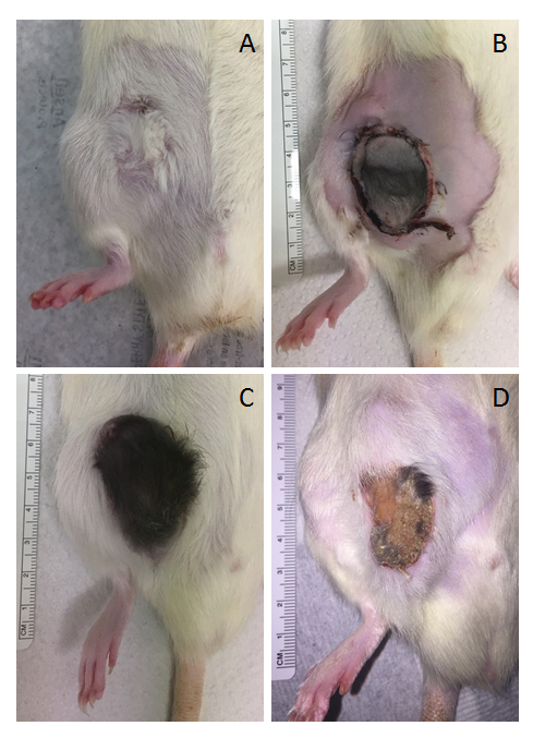
Figure 1: Representative images from transplanted animals. (A) Syngeneic VCA long-term survival, without immunosuppression treatment, on postoperative day 45 (POD 45); note the difference in direction of fur growth due to the graft’s inverted orientation. (B) Allogeneic VCA, treated daily with an immunosuppressant drug, on POD 5. (C) Allogeneic VCA long-term survival, treated daily with an immunosuppressant drug, on POD 40; note normal fur growth indicating proper perfusion of the graft, without signs of rejection. (D) Allogeneic VCA in rejection on POD 33. Immunosuppression treatment was stopped completely on POD 14; note the clinical signs of rejection (skin atrophy, desquamation, loss of fur). Please click here to view a larger version of this figure.
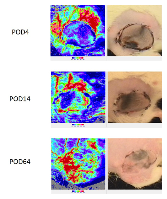
Figure 2: Laser Doppler imaging system to monitor superficial skin revascularization of the allograft. The allograft presented was monitored on postoperative days 4, 14, and 64. The panels on the left show blood perfusion as measured by Doppler imaging, while the panels on the right show the area being imaged by the Doppler. Note the shift from minimal blood perfusion immediately post-VCA to full revascularization of the flap on day 64. This allograft was kept under proper immunosuppression without signs of rejection. Please click here to view a larger version of this figure.
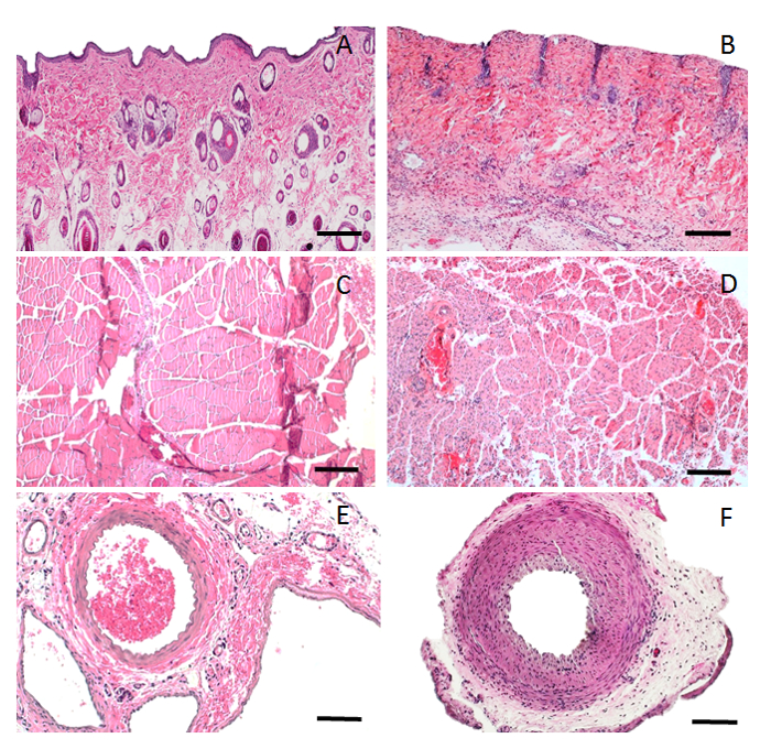
Figure 3: H&E histopathology of allograft in syngeneic vs. allogeneic transplants. (A) Skin biopsy of a syngeneic allograft on POD 45 (10x magnification); note the normal morphology of the skin components (epidermis, adnexa, and no sign of mononuclear cell infiltration). (B) Skin biopsy of an allogeneic allograft in rejection on POD 75, treated daily with a lower dose of an immunosuppressant (10x magnification); note epidermal atrophy, adnexa atrophy, mononuclear cell infiltration, perivascular infiltration, and capillary thrombosis. (C) Muscle biopsy of a syngeneic allograft on POD 45 (10x magnification); note the normal morphology of the striated muscle. (D) Muscle biopsy of an allogeneic allograft in rejection on POD 98, treated daily with a lower dose of an immunosuppressant (10x magnification); note the muscle atrophy and mononuclear cell infiltration. (E) Femoral artery biopsy of a syngeneic allograft on POD 45 (20x magnification); note the normal morphology of the artery. (F) Femoral artery biopsy of an allogeneic allograft in rejection on POD 98, treated daily with a lower dose of an immunosuppressant (20x magnification); note the intimal hyperplasia, narrow lumen, and perivascular infiltration. Scale bar = 200 µm (A–D); 100 µm (E and F). Please click here to view a larger version of this figure.
In developing this model of VCA, several key issues were considered. First, it was important to include intact bone (tibia and fibula), bone marrow, and skin in the graft. While clinical hand transplants from adult donors do not transfer significant amounts of actively hematopoietic marrow, studies of the role of the bone marrow niche are better mirrored using an intact, vascularized bone rather than a cut long bone, which results in fibrosis of the exposed marrow. Moreover, the closed bone osteomyocutaneous flap design reduces the risk of infection and bleeding. Both bone marrow and skin are highly immunogenic tissues, which may be used to trigger an immune response if desired. Second, it was not necessary for the graft to be functional, eliminating the need for an orthotopic model that requires complex osteosynthesis and re-enervation of the graft. This also prevents some of the well-known troublesome consequences of orthotopic models, such as a prolonged surgical procedure and animal discomfort11,12. However, it is important to note that the heterotopic design does not allow functional outcome measures of bone and cartilage, as well as muscle function, all of which are of significant interest in VCA research. Third, the graft needed to be accessible to imaging systems, clinical follow-up, and serial biopsies. Finally, for throughput purposes, the grafting surgeries needed to be readily performed without complications. With these considerations in mind, a modified rat osteomyocutaneous model of VCA was developed in which the distal hindlimb, between the knee and ankle of the donor (Brown-Norway), including the overlying skin and associated vasculature, was transplanted into the inguinal region on the recipient (Lewis). In this case, the vascular supply to the graft occurred via the femoral artery and vein anastomoses.
Because the skin is an important key factor to monitor VCA rejection, specific care was taken in preparing the graft in order to preserve the small artery perforators supporting skin perfusion. When establishing this model, we performed preliminary experiments using indocyanine green (ICG) angiography (results not shown) to confirm the model’s skin perfusion design.
Since the graft is oriented upside down, such that the distal part of the graft is superior and the proximal part of the graft is inferior, a long pedicle is required in order to avoid kinking. Therefore, it should be emphasized that the donor femoral vessels should be divided as proximally as possible and that the recipient femoral artery should be divided as distal as possible.
The simultaneous participation of two surgeons is recommended during the final preparation/isolation of the donor graft; one surgeon should finish the graft isolation, while the other surgeon anesthetizes and intubates the recipient animal and begins preparing the vessels for anastomoses. If the space and equipment are available, a third surgeon could prepare a second recipient animal and both donor legs may be used for VCA grafts. Surgeons must coordinate with one another to ensure minimal graft ischemic time prior to anastomoses. In our experience, most of the postoperative mortalities are attributed to anesthesia technique. If possible, we recommend that a different member of the team should be in charge of anesthesia monitoring during the surgery. It goes without saying that, in order to perform this model successfully, a trained surgeon with basic microvascular techniques is required. Depending on the surgeon’s experience, the model can be achieved successfully following two to six surgery attempts.
Syngeneic rats may be used as a control group to account for healing dynamics unrelated to rejection. The contralateral leg of the recipient rat may also be used as a control, especially when performing imaging and biopsies.
Fur regrowth over the transplanted skin is one of the best indications of successful allograft perfusion. On the other hand, fur loss, skin erythema, and de-epithelization may indicate a rejection event and decreased blood supply to some parts of the flap. In a very advanced rejection stage, the skin may show necrosis and exfoliation. A decrease in the allograft muscle mass is shown in an advanced stage because of denervation atrophy. The animals usually lose body weight (up to 10%) in the first 7–10 days, but then recover and thrive. We recommend adding nutritionally fortified water gel (e.g., DietGel Recovery) in the first few postoperative days to support the recipient rat nutrition. In a very small number of animals (two out of over 50 experimental animals), we witnessed skin infection and autophagy.
In conclusion, the modified model of heterotopic, allogeneic hindlimb osteomyocutaneous VCA graft presented here offers a reproducible, versatile transplantation paradigm. Serial biopsies and imaging offer information on the time course of rejection events. The variety of clinical symptoms that may be studied with this method make it a highly adaptable translational model with the potential for numerous insightful discoveries in the years to come.
The authors have nothing to disclose.
This work was supported by the Office of the Assistant Secretary of Defense for Health Affairs through the Congressionally Directed Medical Research Program under Award No. W81XWH-13-2-0057. Opinions, interpretations, conclusions, and recommendations are those of the authors and are not necessarily endorsed by the Department of Defense.
Acepromazine | Henry Schein | 5700850 | |
Adventitia Scissors | ASSI | SAS15R8 | |
Approximator Clamp (Double) | ASSI | ABB2V, ABB22V | |
Approximator Clamp (single) | FST | 00398-02 | |
Clamp Applying Forceps | ASSI | CAF4 | |
Dissecting Scissors | ASSI | SDS18R8 | |
Flushing blunt needle 27G | SAI | ||
Heparin Sodium | Sagent | 25021-400-30 | |
Isoflurane | Patterson Veterinary | 14043-704-06 | |
Jewelers Bipolar | ASSI | 103000BPS03 | |
Jewelers forceps #3 | FST | 11231-30 | |
Ketamine HCl 100 mg/ml | Zoetis | 043-304 | DEA License required |
Lactated Ringer Solution | Hospira | 0409-7953-03 | |
Lactated Ringer Solution + 5% Dextrose | Hospira | 0409-7953-09 | |
Meloxicam | Henry Schein | 11695-6925-2 | |
Micro forceps | ASSI | JFAL3 | |
Micro needle holder | ASSI | B138 | |
Prograf (Tacrolimus) 5 mg/ml | Astellas | 0469-3016-01 | |
Suture, 10-0 Prolene | Ethicon | W2790 | or 10-0 Ethilon (2830) |
Suture, 4-0 Coated Vicryl | Ethicon | J714D | |
Vessel Dilator Forceps | ASSI | D5AZ | |
Xylazine | VetOne | 13985-612-50 |