All animal experiments were performed according to the guidelines and regulations approved by the Institutional Animal Care and Use Committee of Nanyang Technological University, Singapore (Animal Protocol Number ARF-SBS/NIE-A0331).
1. System description
- Mount the PLD laser into the circular scanner and mount the optical diffuser (OD) in front of the PLD exit window to make the output beam homogeneous, as shown in Figure 1A. Connect the PLD to the laser driver unit (LDU).
NOTE: The PLD generates ~816 nm wavelength pulses, pulses of ~107 ns in duration, and up to a 2 KHz repetition rate with a maximum pulse energy of ~3.4 mJ. The LDU consists of chiller, 12 V power supply, variable high voltage power supply to control the laser power, and function generator to change the pulse repetition rate. - Mount all eight SUTRs on each SUTR holder one-by-one such that the surface of each acoustic reflector faces towards the center of the scanning area, as shown in Figure 1B. Connect each SUTR cable to the low-noise signal amplifier with the help of connecting cables.
NOTE: The central frequency of the ultrasound transducer is 5 MHz and has a 13 mm diameter active area. Two amplifiers each of 24 dB gain are connected in series for each channel. - Switch on the power supply of the chiller, then turn on the switch of the chiller to set the temperature between 20 °C and 25 °C.
- Switch on the supply of the low voltage power supply and slowly turn the current control to set the current limit at 0.3 A. Set the voltage to 12 V. Verify that the current does not exceed 0.1 A.
- Switch on the supply of the high voltage power supply. Press the “Preset” button and set the current to 1 A and voltage to 0 V. Enable the “Output” button: 0 V/0 A.
- Switch on the power supply of the function generator. Press the “Recall” button and choose a 2 KHz configuration to generate the laser pulses at this repetition rate.
- Place acrylic tank inside the scanner as shown in Figure 1A and fill the tank with water such that the detecting surface of the SUTRs are immersed completely inside water.
- Make sure all the SUTRs detecting surfaces are inside the water medium. Switch on the power supply of the low-noise-signal amplifier.
2. Animal preparation for rat brain imaging
NOTE: Healthy female rats (see Table of Materials) were used to demonstrate the above described desktop PLD-PAT system for imaging small animal cortical vasculature.
- Hold the animal on its back by arresting the head and body motion. Anesthetize the animal by intraperitoneal injection of a mixture of 2 mL of ketamine (100 mg/mL), 2 mL of xylazine (20 mg/mL), and 1 mL of saline (dosage of 0.2 mL/100 g).
NOTE: After the injection, the animal’s toe is pinched to test for any positive reflexes such as leg or body movements, vocalization, or marked increases in respirations. An absence of such reflex actions confirms successful anesthetization of the animal. - To prevent dryness due to anesthesia and laser illumination, very carefully apply artificial tear ointment to the rat eyes. Place the animal in prone position on the working bench and remove the fur on the scalp of the animal using a hair trimmer and gently apply hair removal cream to the shaved area and remove the fur completely.
- After 4–5 min, remove the applied cream using a cotton swab.
- Mount the custom-made animal holder (see Table of Materials) equipped with a breathing mask (see Table of Materials) on a lab-jack.
- Place the animal in prone position on the holder so that the head rests on the horizontal platform of the holder. Use surgical tape to secure the animal to the holder.
- Ensure that the breathing mask covers the nose and mouth of the rat to deliver anesthesia mixture. The breathing mask is customized to suit the imaging window. 10% of the commercially available nose cone is cut and then connected to a piece of glove.
- Connect the breathing mask to the anesthesia machine before switching it on.
- Switch on the anesthesia machine and set it to deliver anesthetic mixture containing 1.0 L/min of oxygen with 0.75% isoflurane to the animal breathing mask.
- Clamp the pulse oximeter to one of the animal’s hind legs to monitor its physiological condition.
- Apply a layer of colorless ultrasound gel to the scalp of the rat using a cotton tipped applicator. Adjust the lab-jack position to the center of the scanner and adjust the height of the lab-jack manually so that the imaging plane is at the center of the acoustic reflector.
3. Dynamic in vivo imaging of uptake and clearance process of ICG in rat brain
- Set the parameters in the data acquisition software (see Table of Materials) for a 360° acquisition scan.
- Turn on the PLD laser emission by enabling the output of the function generator (laser emission will start). Then, slowly increase the voltage of the variable high voltage power supply to 120 V for maximum per pulse energy.
- Run the data acquisition software (see the Table of Materials) program to rotate all eight SUTRs in 360° over a 4 s scan time.
NOTE: For example, if the SUTRs are rotated for 4s, the PLD delivers 8,000 (= 4 x 2,000) pulses and each SUTR collects 8000 A-lines. These 8,000 A-lines are reduced to 400 by averaging over 20 signals (after averaging A-lines = 8,000/20 = 400). A reconstruction program based on delay-and-sum back projection algorithm is used to find out the scanning radius of each SUTR. - Disable the output of the function generator to turn off the laser emission.
- Using the reconstruction algorithm in data processing software (see Table of Materials) find out the scanning radius of all eight SUTRs by trial-and-error, using the back-projection algorithm.
- Set the parameters in the data acquisition software (see Table of Materials) for 45° acquisition over a 0.5 s scan time.
NOTE: For example, if the SUTRs are rotated for 0.5s, the PLD delivers 1,000 (= 0.5 x 2,000) pulses and each SUTR collects 1000 A-lines. These 1,000 A-lines are reduced to 400 by averaging over 20 signals (after averaging A-lines = 1,000/20 = 50). - Enable the output of the function generator to turn on the laser emission.
- Run the data acquisition software (see Table of Materials) program to rotate all eight SUTRs in 45° to obtain initial control data before administering ICG.
- Disable the output of the function generator to turn off the laser emission.
- Identify the tail vein of the animal and inject 0.3 mL of ICG (see Table of Materials) (323 μM) into the tail vein of the rat.
4.
NOTE: 1.25 mg of ICG powder was weighed using a micro-weighing machine and mixed with 5 mL of distilled water to obtain a concentration of 323 μM for the ICG solution.
- Enable the output of the function generator to turn on the laser emission.
- Run the data acquisition software (see Table of Materials) program to acquire A-lines over a 0.5 s scan time in 45° rotation.
5.
NOTE: A-lines acquired during a 0.5 s scan time are used to generate one cross-sectional image. There is time gap of ~0.4–0.6 s between each scan.
- After the data acquisition is over, using the back-projection algorithm in data processing software (see Table of Materials), reconstruct the cross-sectional brain image from the saved A-lines.
- Turn off the laser and then turn off anesthesia machine, lower the lab-jack and remove the animal from the stage. Return the animal to the cage and monitor until it regains consciousness.
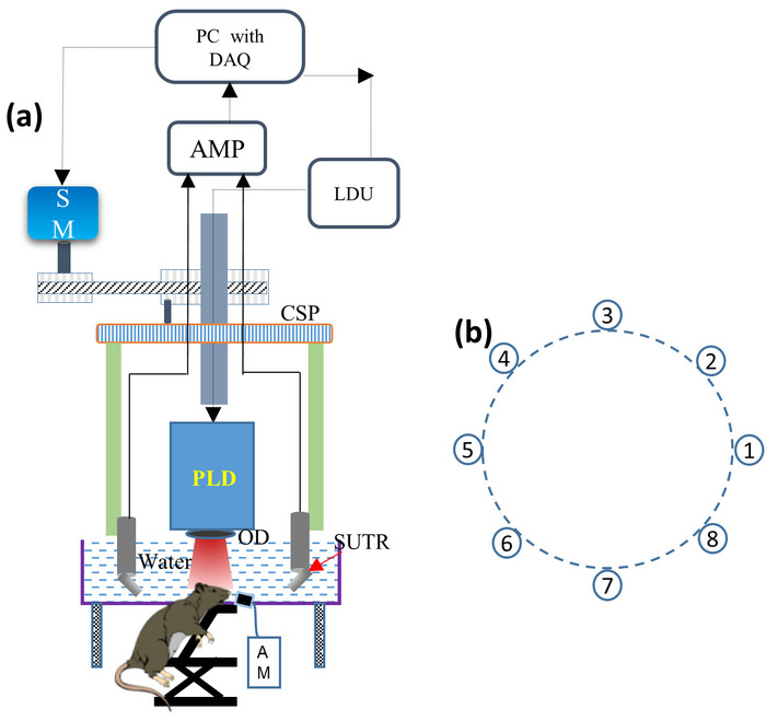
Figure 1: Schematic of the desktop PLD-PAT system. (A) Schematic of the desktop PLD-PAT set up. PLD: pulsed laser diode, OD: optical diffuser, SUTR: acoustic reflector based single-element ultrasound transducer, AM: anesthesia machine, CSP: circular scanning plate, SM: stepper motor, LDU: laser driving unit, AMP: amplifier, DAQ: data acquisition card. (B) Circular arrangement of eight SUTRs around the scanning center. Please click here to view a larger version of this figure.
The potentiality of the described desktop PLD-PAT system for dynamic in vivo brain imaging has been showcased in this protocol with corresponding results. High-speed imaging capability of the desktop PLD-PAT system was demonstrated by performing in vivo brain imaging of healthy female rats. PA signals were collected using eight SUTRs rotating in 360° and 45° around the rat brain at scan speeds of 4 s and 0.5 s, respectively. Figure 2A,B show brain images of a female rat (98 g) at scan speeds of 4 s and 0.5 s, respectively. Sagittal sinus (SS) and transverse sinus (TS) are clearly visible in both the images. Figure 2C,D show photographs of the rat brain before and after removing the scalp over the brain area, respectively. PAT imaging was done non-invasively with intact skin and skull.
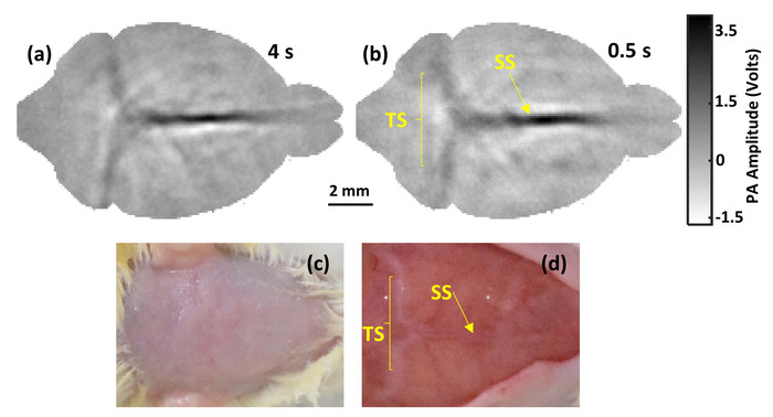
Figure 2: Non-invasive in vivo desktop PLD-PAT images. In vivo images of cortical vasculature at scan times of (A) 4 s and (B) 0.5 s. SS: sagittal sinus, TS: transverse sinus. (C) and (D) are photographs of the rat brain before and after removing the scalp, respectively. Please click here to view a larger version of this figure.
Before injecting ICG into the tail vein of the same rat, control data was acquired. After injecting ICG, PA data was acquired continuously for first 5 min with a 0.5 scan time. Then, PA data was acquired at ~2-3 min intervals with 0.5 s scan times each for the next 15-20 min. Figure 3 shows the plot representing the increases in average PA signal in the sagittal sinus (SS) due to increases in optical absorption by ICG at 816 nm wavelengths, and subsequently, decreases over time.
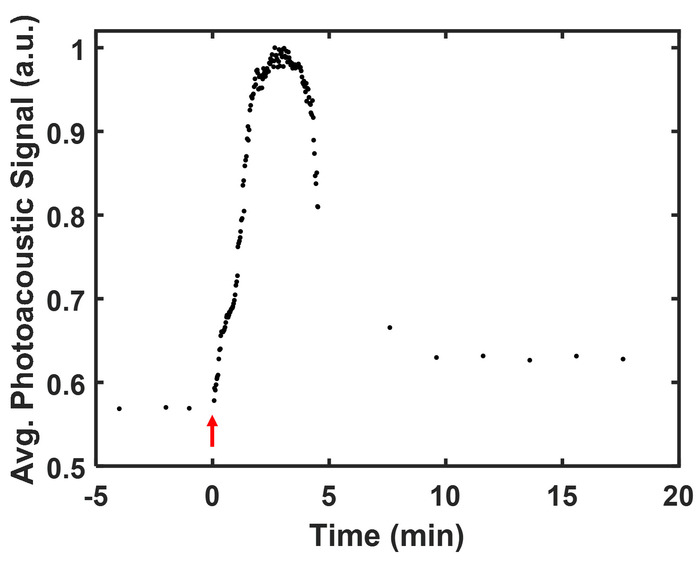
Figure 3: Pharmacokinetics of ICG. Pharmacokinetics of ICG showing the uptake and clearance process. The red arrow mark shows the time of injection of ICG into the tail vein. Please click here to view a larger version of this figure.
