The earliest models of medicine used in societies were based on natural products (NPs). Since then, humans have been searching for new chemicals in nature that can be transformed into drugs1. This search caused a continuous improvement of technologies and methods for ethnobotanical screening1,2,3. NPs offer a rich source of structurally diverse substances, with a wide range of biological activities useful for developing alternative or adjuvant therapies. However, the inherent complex plant matrix makes the isolation and identification of the active compounds a challenging and time-consuming task4.
NPs-based drugs or formulations can be used to prevent and/or treat several conditions affecting oral, including dental caries4. Dental caries, one of the most prevalent chronic diseases globally, derives from the interaction of sugar-rich diet and microbial biofilms (dental plaque) formed on the tooth surface that leads to demineralization caused by organic acids derived from microbial metabolism, and if not treated, leads to teeth loss5,6. Although other microorganisms may be associated7, Streptococcus mutans is a critical cariogenic bacterium because it is acidogenic, aciduric, and an extracellular matrix builder. This species encodes multiple exoenzymes (e.g., glycosyltransferases or Gtfs) that use sucrose as a substrate8 to build the extracellular matrix rich in exopolysaccharides, which are a virulence determinant9. Also, the fungus Candida albicans can drive up the production of that extracellular matrix7. Albeit fluoride, administered in various modalities, remains the basis for preventing dental caries10, new approaches are needed as adjuvants to increase its effectiveness. In addition, the available anti-plaque modalities are based on the use of broad-spectrum microbicidal agents (e.g., chlorhexidine)11. As an alternative, NPs are potential therapies for controlling biofilms and preventing dental caries12,13.
The further advance in the discovery of new bioactive compounds from plants includes necessary steps or approaches such as: (i) the use of reliable and reproducible protocols for sampling, considering that plants often show intraspecific variability; (ii) the preparation of comprehensive extracts and their respective fractions in small scale; (iii) the characterization and/or dereplication of their chemical profiles thought the acquisition of multidimensional data such as GC-MS, LC-DAD-MS, or NMR, for example; (iv) the use of viable and high-yield models to assess bioactivity; (v) the selection of potential new hits based on multivariate data analysis or other statistical tools; (vi) to perform the isolation and purification of the targeted compounds or promising candidates; and (vii) the validation of the corresponded biological activities using the isolated compounds2,14.
Dereplication is the process of rapidly identifying known compounds in crude extract and allows differentiating novel compounds from those that have already been studied. Besides, this process prevents isolation when bioactivity has already been described for certain compounds, and it is particularly helpful to detect “frequent hitters”. It has been used in different untargeted workflows ranging from major compound identification or the acceleration of activity-guided fractionation up to the chemical profiling of collections of extracts. It can be fully integrated with metabolomic studies for the untargeted chemical profiling of CE or the targeted identification of metabolites. All of this ultimately leads to prioritizing extracts before the isolation procedures1,15,16,17.
Therefore, in the present manuscript, we describe a systematic approach to identify antimicrobial and antibiofilm molecules from plant extracts and fractions. It includes four multidisciplinary steps: (1) collection of plant material; (2) preparation of crude extracts (CE) and fractions (CEF), followed by their chemical profile analysis; (3) bioassays; and (4) biological and chemical data analyses (Figure 1). Thus, we present the protocol developed to analyze of the antimicrobial and antibiofilm activities of Casearia sylvestris extracts and fractions against Streptococcus mutans and Candida albicans13, as well as the procedures for the phytochemical characterization and data analysis. For simplicity, the focus here is to demonstrate the approach for screening natural compounds using the bacterium.
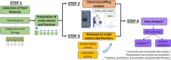
Figure 1: Flow-chart of the Systematic Approach to identify active molecules from plants extracts and fractions. Please click here to view a larger version of this figure.
We provide an example of using a systematic approach to screen the biological activity of plant extracts and fractions to identify potentially active molecules for possible new anti-caries therapies: antimicrobial and antibiofilm activities of Casearia sylvestris extracts from distinct Brazilian biomes against Streptococcus mutans and Candida albicans13.
Background
Complex interactions between specific oral microorganisms-host factors-diet rich in sucrose and starch can modulate the formation of pathogenic biofilms and initiate a cariogenic process30,31. S. mutans orchestrates the pathogenicity of biofilms associated with the development of dental caries because it produces Gtfs responsible for exopolysaccharides synthesis, besides its acidogenicity and aciduricity31. In addition, Gtfs enables the adhesion of Candida albicans (and other microorganisms), increasing the virulence of the biofilm32,33. We conducted a screening of the antimicrobial and antibiofilm activities of C. sylvestris leaf extracts and fractions from different Brazilian biomes, belonging to the lingua and sylvestris varieties against S. mutans and C. albicans13. C. sylvestris (“guaçatonga”) is part of popular and traditional use in Brazil, and other countries of South America and Asia34,35. This plant is cited in the “National List of Medicinal Plants of Interest to SUS” (RENISUS), which contains 71 species that could treat the diseases with a high incidence in Brazil36. The chemical profile of leaf extracts of var. sylvestris presents a rich phytochemical composition, with abundant diterpenes35, while phenolic compounds (mainly flavonoids) predominate in var. lingua18.
We use the approach described in the protocol to identify which extracts and fractions of C. sylvetris are most active for the microorganisms evaluated, and, based on the results using simplistic models, we select which treatments will be tested in vitro complex models (hydroxyapatite discs, microcosms)37,38. Here, we present the results of the screening of the twelve CE against S. mutans. The focus is to demonstrate the usefulness of this approach for screening natural compounds instead of interpreting and discussing the data.
We collected the leaves of individuals from the two varieties of C. sylvestris from twelve different populations in Brazil, comprising different formations of Brazilian biomes (please, see details in Ribeiro et al. 201913). The collection was carried out between June and September 2012 and 2013 (SisGen; Register #A00892A). We recommend collecting representative samples, including individuals of different chemotypes and from different biomes, to address the chemical variability of secondary metabolites. If available, at least 3 to 5 individuals should be collected. Previous information concerning plant infraspecific chemical variability should also be considered as described by Ribeiro et al. 2019 and Bueno et al. 201513,18. The chemical composition of the CE was examined by the chromatographic cited in Step 2 and we provide the chemical profile in Figure 7. The analysis of the chemical profile is essential to integrate the interpretation of data obtained in biological screening.
The CEs were fractionated in Hex, AcOEt, and MeOH fractions. The fractionation of CE allows the simplification of the mixture to increase the concentration of the potentially active compounds and decrease the possibilities of synergisms and antagonisms between compounds. Additionally, in more simple mixtures (fractions), it is easier to obtain spectral data of the compounds than in CE and to perform dereplication analysis2. Usually, fractionation can be done by liquid-liquid extraction or solid-phase extraction cartridges (SPE) containing preferentially reversed-phase adsorbent like C18 (40 µm, 100 Å). Other adsorbents or mixtures of adsorbents can be chosen, depending on the study purposes or chemical nature of the desired compounds. If the chosen technique is the SPE, the cartridges must be previously activated with pure organic solvent (e.g., EtOH) and conditioned with the initial eluent. Standardized protocols are available. Thus, the reader can consult and adapt them according to the intended study and plant material of interest.
The twelve CE were solubilized with 84.15% EtOH and 15% DMSO to achieve 6 mg/mL (stock solution). Before the screening tests, we tested the diluent concentration (vehicle) that does not interfere with microbial growth. This step is important because it prevents the antimicrobial and antibiofilm actions of the solvents from affecting the results when testing treatments. The tests can be performed on a 96-well plate by treating the culture of the microorganism of interest with different concentrations of solvents (associated and/or isolated). Thus, we began our screening with CE at 0.5 mg/mL and vehicle at a concentration of 7% EtOH and 1.25% DMSO.
For screening antimicrobial and antibiofilm activity, 96-well plates were treated as described above. For this purpose, the volume of 16.67 µL of stock solution CE (6 mg/mL) was added to test each CE at the concentration of 0.5 mg/mL. The biofilms formed were processed as described in Step 3. The extracts effective against S. mutans (IC50 or 3 logs) were used to evaluate the "adhesion strength" of this bacterium to the salivary pellicle and initial glucan matrix, as described in Step 3.
The raw data obtained from biological assays were organized in Excel (as described in Step 4) and analyzed with appropriate statistical treatment13. The cutoff point to identify the extracts with the best activity was the IC50 inhibition (3 logs). From this parameter, four extracts showed a favorable response (Figure 8). The chromatographic data of these four extracts show the simultaneous presence of clerodane-type diterpenes and glycosylated flavonoids. In addition, they include the same biome (Atlantic Forest) and variety (sylvestris). To help interpret the biological data, we compared the chromatographic profile of the four extracts with the best activity with the other screened extracts. Compared to the others, the extracts with the best activity have a higher amount of clerodane-type diterpenes and, simultaneously, glycosylated flavonoids. This observation indicates that it is likely that the effectiveness of these extracts is due to a synergistic interaction between the two secondary metabolites, thus increasing their biological activity. That is, the combined effect of clerodane-type diterpenes and glycosylated flavonoids is greater than the sum of their separate effects13.
To confirm the data obtained in the screening, we evaluated the detachment of S. mutans after adhesion to the salivary pellicle and glucans treated with selected CE. The assays use biofilm models of in vitro single-species to evaluate better the biological activity of the selected crude extracts and identify possible action targets. The first analysis verifies whether the treatments used are capable of inhibiting the adherence of S. mutans to the salivary pellicle, but mainly, whether the cells of the microorganism that have adhered to the treated pellicle can be removed from the surface more easily by the mechanical stimulus, thus interrupting the first stage of biofilm formation. The addition of CE (with better activity) during the synthesis of glucans did not modify the salivary pellicle because no CE significantly affected the removal of cells adhered to the salivary pellicle (Figure 9A).
The adhesion of the initial glucan matrix (gsHA) investigates whether the treatments can inhibit the adhesion of S. mutans to the initial glucan matrix. Still, this methodology verifies if the microorganism cells that have adhered to the treated glucans can be removed by the mechanical stimulus more easily of the surface, thus interrupting the stage biofilm formation. Three CE affected the quality of glucans formed by GtfB and therefore weakened the adhesion of S. mutans to the initial glucan matrix (most S. mutans cells were removed after adhesion for glucans; Figure 9B). We believe that this behavior is related to the synergism between the secondary metabolites13.
The Systematic Approach has helped us identify and select active crude extracts to halt the formation of cariogenic biofilms. Once selected and based on the chromatographic profile, we have the basis for elucidating the molecular mechanisms of action in complex models.
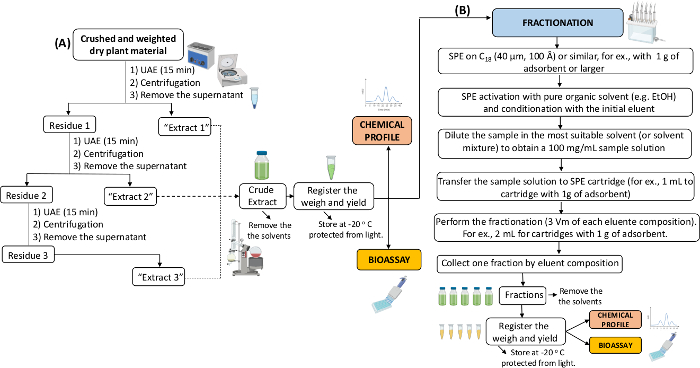
Figure 2: Flow-chart of the plant material extraction and fractionation. The illustration shows the experimental design to prepare the crude extracts (A) and fractionation of crude extracts (B). UAE: Ultrasound Assisted Extraction; SPE: Solid Phase Extraction. Please click here to view a larger version of this figure.
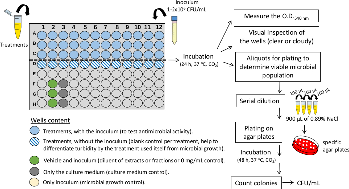
Figure 3: Experimental design for assessing antimicrobial activity in 96-well plates. The illustration depicts treatments (crude extracts or fractions) and controls. For the screening of multiple treatments, use a single concentration (mg/mL) in each well. CFU/mL: colony forming units per milliliter. O.D.: optical density. Please click here to view a larger version of this figure.
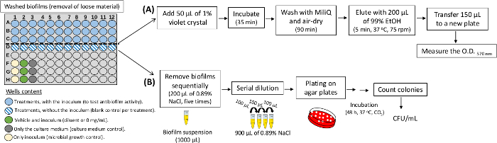
Figure 4: Experimental design for antibiofilm assay in 96-well plates. The illustration shows treatments (crude extracts and fractions) and controls. For the screening of various treatments, use a single concentration (mg/mL) in each well. In A, the steps to quantify biomass of treated biofilms are illustrated. In B, is shown the steps to determine the population (CFU/mL) of the treated biofilms. Please click here to view a larger version of this figure.
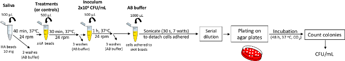
Figure 5: Experimental design to evaluate the adhesion to the salivary pellicle, followed by the detachment of adhered cells. The illustration shows the steps to be performed. Treatments: selected based on biological screening. Please click here to view a larger version of this figure.
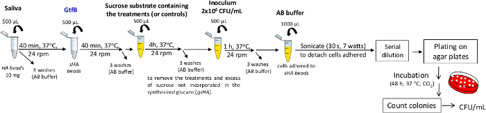
Figure 6: Experimental design to evaluate the adhesion to the initial glucan matrix (gsHA) followed by the detachment of adhered cells. The illustration shows the steps to be performed. Treatments: selected based on biological screening. Sucrose substrate: 100 mmol of sucrose. Please click here to view a larger version of this figure.
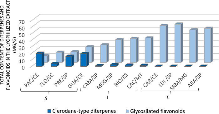
Figure 7: Quantity of clerodane-type diterpenes and glycosylated flavonoids in C. sylvestris extracts from Brazilian biomes. The letters S, I, and L indicate the varieties sylvestris, intermediate, and lingua, respectively. Personal communication by Dr. Paula Carolina Pires Bueno. This figure has been modified from Ribeiro et al.13. Please click here to view a larger version of this figure.
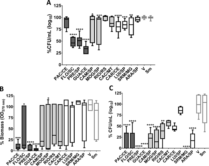
Figure 8: Antimicrobial and antibiofilm activity of C. sylvestris crude extracts from Brazilian biomes against S. mutans. A. % CFU (log10) of treated planktonic cells; B. % biomass of treated biofilms. C. % CFU (log10) of treated biofilm. The data described are median (traces) and interquartile (boxes). The error bars represent the maximum and minimum values. The asterisks denote a statistically significant difference of a specific extract versus vehicle control (V), where: ****p ≤ 0.0001; ***p ≤ 0.001; ** p ≤ 0.01; and *p ≤ 0.05 (Kruskal-Wallis test, followed by Dunn’s multiple comparisons test). Each species growth control (without treatment) is represented as Sm for S. mutans. The colors of the bars in each graph represent the variety to which the extracts belong, being, in dark gray color: var. sylvestris; light gray: var. intermediate and white: var. lingua. This figure has been modified from Ribeiro et al.13. Please click here to view a larger version of this figure.
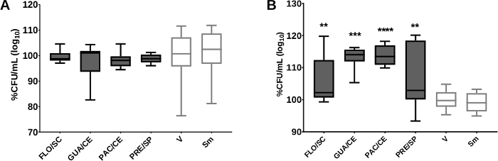
Figure 9: S. mutans detachment after adhesion to the treated salivary pellicle and initial matrix of glucans. Post-release data of S. mutans to the treated salivary pellicle and glucans are shown in (A) and (B), respectively. There was no difference between the control vehicle (V), and the extracts tested for both analyses. The percentage of CFU/mL was obtained considering the vehicle control (V) as 100%. The data described are median (traces) and interquartile (boxes). The error bars represent the maximum and minimum values. The asterisks denote a statistically significant difference of a specific extract versus vehicle control (V), where ****p = 0.0001 and **p < 0.0031 (Kruskal-Wallis test, followed by the multiple comparison test of Dunn). The growth control is represented by Sm for S. mutans. The colors of the bars of the graph represent the variety to which the extracts belong, being the color dark gray to var. sylvestris. This figure has been modified from Ribeiro et al.13. Please click here to view a larger version of this figure.
