Here, we present a new protocol to study and map the targeted deposition of drug carriers to endothelial cells in fabricated, real-sized, three-dimensional human artery models under physiological flow. The presented method may serve as a new platform for targeting drug carriers within the vascular system.
The use of three-dimensional (3D) models of human arteries, which are designed with the correct dimensions and anatomy, enables the proper modeling of various important processes in the cardiovascular system. Recently, although several biological studies have been performed using such 3D models of human arteries, they have not been applied to study vascular targeting. This paper presents a new method to fabricate real-sized, reconstructed human arterial models using a 3D printing technique, line them with human endothelial cells (ECs), and study particle targeting under physiological flow. These models have the advantage of replicating the physiological size and conditions of blood vessels in the human body using low-cost components. This technique may serve as a new platform for studying and understanding drug targeting in the cardiovascular system and may improve the design of new injectable nanomedicines. Moreover, the presented approach may provide significant tools for the study of targeted delivery of different agents for cardiovascular diseases under patient-specific flow and physiological conditions.
Several approaches have recently been applied utilizing 3D models of human arteries1,2,3,4,5. These models replicate the physiological anatomy and environment of different arteries in the human body in vitro. However, they have been mainly used in cell biology studies. Current studies on vascular targeting of particles to the endothelium include in silico computational simulations6,7,8, in vitro microfluidic models9,10,11, and in vivo animal models12. Despite the insights they have provided, these experimental models have failed to accurately simulate the targeting process that occurs in human arteries, wherein blood flow and hemodynamics constitute dominant factors. For example, the study of particle targeting to atherosclerotic regions in the carotid artery bifurcation, which are known for their complex recirculation flow pattern and wall shear stress gradient, may impact the journey taken by the particles before they reach the endothelium13,14,15,16. Therefore, these studies must be performed under conditions that replicate the physiological environment, i.e., size, dimension, anatomy, and flow profile.
Recently, this research group fabricated 3D-reconstructed human arterial models to study the deposition and targeting of particles to the vasculature17. The models were based on geometrical 3D replicas of human blood vessels, which were then cultured with human ECs that subsequently lined their inner walls. In addition, when subjected to a perfusion system that produces physiological flow, the models accurately replicated physiological conditions. The perfusion system was designed to perfuse fluids at a constant flow rate, using a peristaltic pump in both closed and open-circuit configurations (Figure 1). The system can be used as a closed-circuit to map particle deposition and targeting to the cells seeded inside the carotid model. In addition, it can be used as an open circuit to wash out non-adherent particles at the end of the experiments and to clean and maintain the system. This paper presents protocols for the fabrication of 3D models of the human carotid bifurcation, design of the perfusion system, and mapping of the deposition of targeted particles inside the models.
NOTE: This protocol describes the fabrication of a 3D model of the carotid artery and can be applied to generate any other artery of interest by simply modifying the geometric parameters.
1. Design and fabrication of a 3D bifurcation of the human carotid artery model
2. Cell culture and seeding in models
3. Design of the perfusion system
4. Closed-circuit configuration: perfusion experiment and imaging
5. Open-circuit configuration: the washing step
6. Data acquisition and analysis
This paper presents a new protocol to map the deposition of particles inside real-sized 3D human artery models, which may provide a new platform for drug delivery research. Using a 3D printing technique, a model of the human carotid bifurcation artery was fabricated (Figure 2). The model was made of silicone rubber and seeded with human ECs (Figure 3). Importantly, this protocol enabled the replication of physiological conditions, especially with respect to fluid dynamics. A perfusion system was designed to infuse particles to the carotid bifurcation under constant flow at the magnitude of the physiological waveform characteristic of the carotid. Figure 1 presents the perfusion system, which consists of the peristaltic pump, an oscillation damper, the cultured bifurcation model, tubing, and fluid containers.
To map the deposition and adhesion of the perfused particles, the arterial model was imaged under a stereomicroscope, both at the end of the experiment and after washing (step 5.3). The images were captured using 2x objective and tiled together to form a whole image of the model. Then, the number of adhered particles was calculated using a customized software code. To examine the formation of the recirculation pattern at the bifurcation, 10 µm fluorescent glass beads were infused into the model. Figure 4A shows the recirculation, which suggests that the conditions inside the model mimic physiological conditions.
To map the deposition of particles inside the model, 2 µm fluorescent carboxylated PS particles were infused, and their adhesion to the ECs was imaged (Figure 4B,C). These particles adhered differently at various regions along the model-more adhesion was observed out of the recirculation area, where wall shear stress is high. These results have been previously discussed to show that the adhesion of particles is a function of the model's geometry, particle surface characteristics, and shear stress17. These deposition maps are relatively simple and may be quickly obtained for screening drug carriers' affinity and targeting under physiological conditions in patient-specific models.
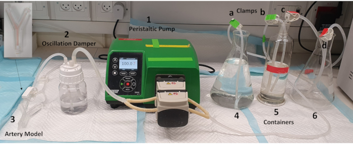
Figure 1: The perfusion system. A perfusion system was designed to perfuse fluids under constant flow. It is comprised of (1) a peristaltic pump, (2) an oscillation damper, (3) the cultured 3D arterial model, and three glass containers: two with a 1 L capacity (4 and 6) and a third that can hold 300 mL fluid (5). The system can operate in two configurations: (i) an open circuit, in which clamps a+d are open and b+c are closed, or (ii) a closed circuit, in which clamps a+d are closed and b+c are open. Please click here to view a larger version of this figure.
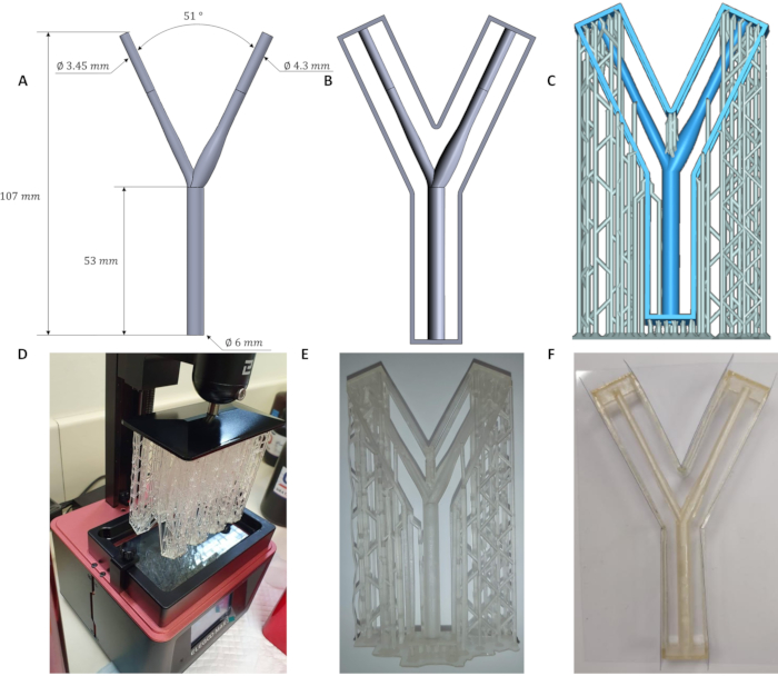
Figure 2: Fabrication process of a 3D carotid artery bifurcation model. (A–C) Human carotid bifurcation, the mold frame around the artery, and temporary printing supports were designed. (D, E) The geometries were printed using a 3D printer. (F) The temporary printing supports were cut, and the model was sanded and sprayed with lacquer. Then, transparent rectangular slides were glued to the frame from all sides. Silicone rubber was cast when the glue was dry. Abbreviation: 3D = three-dimensional. Please click here to view a larger version of this figure.
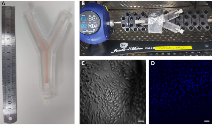
Figure 3: Seeding of ECs inside 3D models of the carotid artery. (A) Real-sized 3D model of the human carotid bifurcation made of silicone rubber. The model was cultured with human ECs and filled with cell medium. (B) The cultured model was placed on a rotator at 37 °C for 48 h. (C) Images of the cultured ECs inside the 3D model in brightfield and (D) with DAPI for nuclear staining in blue. Scale bars = 10 µm. Abbreviations: ECs = endothelial cells; DAPI = 4′,6-diamidino-2-phenylindole; 3D = three-dimensional. Please click here to view a larger version of this figure.
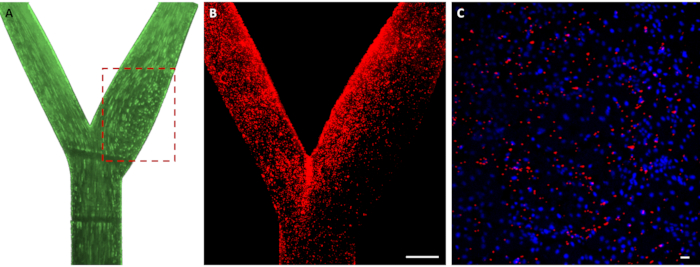
Figure 4: Perfusing and mapping the adhesion of particles. (A) Streak-line image of streamlines and recirculation (dashed rectangle) generated upon perfusion of 10 µm fluorescent glass particles at a constant flow of 400 mL/min through the model. (B) Deposition map of the 2 µm fluorescent carboxylated PS particles (in red) inside the 3D-cultured model. Scale bar = 2 mm. (C) Adhesion of the particles (in red) to the cultured ECs (in blue-DAPI) inside the model at a 10x magnification. Scale bar = 10 µm. Abbreviations: PS = polystyrene; 3D = three-dimensional; ECs = endothelial cells; DAPI = 4′,6-diamidino-2-phenylindole. Please click here to view a larger version of this figure.
Supplemental Information: Please click here to download this File.
Current approaches to study vascular targeting of particles fall short in replicating the physiological conditions present in the human body. Presented here is a protocol to fabricate 3D-reconstructed models of human arteries to study particle targeting to the ECs lining the artery under physiological flow applied using a customized perfusion system. When choosing the material for 3D printing, it is best to use a clear plastic to avoid pigment transfer to the silicone model, which should be as transparent as possible. In addition, it is important to choose a material that does not dissolve in acetone, but instead becomes soft and brittle and can subsequently easily be removed from the model.
The presented 3D models are made of silicone rubber, a transparent silicone, mixed with its curing agent. It is important to ensure that the mixture is always at room temperature or below, otherwise the crosslinking between the silicone and the curing agent will begin before the degassing and casting onto the molds. Although polydimethylsiloxane can also be used to fabricate such models (1:10 ratio with its crosslinker), silicone rubber is more durable. After immersing the model in acetone to dissolve the plastic, it is crucial to incubate it at 60 °C for at least 4 days to ensure full evaporation of any acetone residue. If any acetone remains trapped inside the model, the cells will not grow properly. Changing the medium after 24 h and fixation of the cells after 48 h are the two steps that involve manual injection of fluid using a 10 mL syringe. It is therefore important to perfuse slowly, otherwise the cells might be washed out.
The perfusion system has two inlets and two outlet tubes. Each tube has a plastic tube clamp for flow control. Most of the tubing system is comprised of 4 mm inner dimeter (ID) tubes, except for the tubes that are clamped in the pump, which are 6 mm ID tubes. The ID of the tubes clamped in the pump will determine the maximum flow rate that can be achieved in the system. This perfusion system can also generate a pulsatile waveform by superposing oscillations on the constant mean flow17 by connecting the outlet of the damper to an oscillator assembly, which superposes the oscillatory part of a desired waveform on the constant flow rate produced by the peristaltic pump. This configuration enables operation under oscillatory flow or under constant flow conditions when the oscillator is turned off.
In this paper, the perfusion system was customized based on experiments with the 3D human carotid artery bifurcation. Therefore, if other arterial models or other tubing are used, the amounts of fluids and flow rate may require adjustments. In such cases, the system and flow rate will have to be calibrated, while ensuring no cell detachment from the model walls. It is very important to gradually increase the flow rate in the peristaltic pump to guarantee that the cells are not washed away with the flow. Moreover, it is crucial to ensure that the entire system, including the tubing, the model, as well as the container are filled with fluids (e.g., in this case, it was filled with a total volume of 300 mL of fluid).In addition, before and after each experiment, the system should be washed with distilled water in an open-circuit configuration.
Blood can also be perfused into the models using the perfusion system17. In this case, extra caution must be exercised to prevent any leakage, especially if human blood is used. Moreover, the washing step is crucial as bleach must be perfused at the end of the experiment to ensure full wash out of the blood. After the bleach, water should be perfused as mentioned in the protocol. It is important to note that in this paper, carboxylated PS particles were used, which have a uniform composition and a narrow size distribution. Moreover, these particles adhere to the cells primarily through electrostatic interactions. However, other drug nanocarriers may be used, and specific targeting should be examined as well with ligand-labeled particles, e.g., to anti-intercellular adhesion molecule 1 and anti-platelet endothelial cell adhesion molecule 1, which will increase particle accumulation to ECs at the site of interest.
In addition, in this protocol, ECs were fixed prior to connecting the models to the perfusion system and injection of the particles. The adhesion of particles to fixed cells represents the first stage in the binding process and therefore, experiments with live cells need to be performed, where internalization of particles may occur during later stages of the adhesion process. This protocol can be used to fabricate 3D arterial models for the study of drug carriers under physiological conditions. The outlined approach may assist in the study of delivery of agents under patient-specific conditions.
The authors have nothing to disclose.
This work was supported by the Israel Science Foundation (ISF grant # 902/18). Maria Khoury's scholarship was supported by The Baroness Ariane de Rothschild Women Doctoral Program.
3D printer | FormLabs | PKG-F2-REFURB | |
Acetone, absolute (AR grade) | |||
Connectors | Nordson Medical | FTLL013-1 | Female Luer |
FTLL230-1 | Female Luer | ||
FTLL360-1 | Female Luer | ||
LP4-1 | Male Luer Integral Lock | ||
Damper | Thermo-Fisher Scientific | DS2127-0250 | Nalgene Polycarbonate, Validation Bottle |
Damper Cover | Thermo-Fisher Scientific | 2162-0531 | Nalgene Filling/Venting Closures |
Elastosil Elastosil RT 601 A | Wacker | 60003805 | |
Elastosil RT 601 B | Wacker | 60003817 | The crosslinker |
Endothelial Cell Media | ScienCell | 1001 | |
Fibrontectin | Sigma Aldrich | F0895-5mg | |
HUVEC | Lonza | CC-2519 | |
Isopropyl alcohol, AR grade 99.5% | Remove plastic dust from the sanded model | ||
Lacquer | Rust-Oleum | 2X-Ultra cover Gloss Clear | |
Matlab | Mathworks | https://www.mathworks.com/products/matlab.html | |
Microscope | Nikon | SMZ25 | |
Microscope Camera | Nikon | DS-Qi2 | |
Peristaltic pump | Watson Marlow | 530U IP31 | With 2 pumpheads: 313D |
Plastic tube clamp | Quickun | 1-2240-stopvalve-2pcs | |
Polystyrene Particles | Thermo-Fisher Scientific | F8827 | Diameter = 2 µm |
Printer resin | FormLabs | RS-F2-GPCL-04 | |
Rotator | ELMI Ltd. | Intelli-Mixer RM-2 | |
Solidworks | SolidWorks Corp., Dassault Systèmes | https://www.solidworks.com/ | |
Tubing | Watson Marlow | 933.0064.016 | Tubing for the pump: 6.4 mm ID |
All the other tubing: Silicon tubing: 4 mm ID |