We present a method for isolating endothelial cells and nuclei from the lumen of mouse carotid arteries exposed to stable or disturbed flow conditions to perform single-cell omics experiments.
Atherosclerosis is an inflammatory disease of the arterial regions exposed to disturbed blood flow (d-flow). D-flow regulates the expression of genes in the endothelium at the transcriptomic and epigenomic levels, resulting in proatherogenic responses. Recently, single-cell RNA sequencing (scRNAseq) and single-cell Assay for Transposase Accessible Chromatin sequencing (scATACseq) studies were performed to determine the transcriptomic and chromatin accessibility changes at a single-cell resolution using the mouse partial carotid ligation (PCL) model. As endothelial cells (ECs) represent a minor fraction of the total cell populations in the artery wall, a luminal digestion method was used to obtain EC-enriched single-cell preparations. For this study, mice were subjected to PCL surgery to induce d-flow in the left carotid artery (LCA) while using the right carotid artery (RCA) as a control. The carotid arteries were dissected out two days or two weeks post PCL surgery. The lumen of each carotid was subjected to collagenase digestion, and endothelial-enriched single cells or single nuclei were obtained. These single-cell and single-nuclei preparations were subsequently barcoded using a 10x Genomics microfluidic setup. The barcoded single-cells and single-nuclei were then utilized for RNA preparation, library generation, and sequencing on a high-throughput DNA sequencer. Post bioinformatics processing, the scRNAseq and scATACseq datasets identified various cell types from the luminal digestion, primarily consisting of ECs. Smooth muscle cells, fibroblasts, and immune cells were also present. This EC-enrichment method aided in understanding the effect of blood flow on the endothelium, which could have been difficult with the total artery digestion method. The EC-enriched single-cell preparation method can be used to perform single-cell omics studies in EC-knockouts and transgenic mice where the effect of blood flow on these genes has not been studied. Importantly, this technique can be adapted to isolate EC-enriched single cells from human artery explants to perform similar mechanistic studies.
This laboratory previously demonstrated that induction of d-flow leads to quick and rugged atherosclerosis development in hyperlipidemic mice1,2. The novel mouse model of d-flow-induced atherosclerosis was possible using partial carotid ligation (PCL) surgery 3. PCL surgery induces low and oscillatory blood flow condition or d-flow in the ligated left carotid artery (LCA). In contrast, the contralateral right carotid artery (RCA) continues to face stable laminar flow (s-flow). Previously, to understand the effect of d-flow on endothelial cells, the carotid arteries were dissected out after partial ligation surgery and flushed with a phenol and guanidine isothiocyanate-based lysing agent (luminal RNA/DNA flushing method)2,4, which provided endothelial-enriched "pooled bulk" RNAs or DNAs. These pooled bulk RNAs or DNAs were then processed for transcriptomic studies or epigenomic DNA methylome studies, respectively4,5,6. These studies helped discover multiple flow-sensitive genes and microRNAs whose roles in endothelial biology and atherosclerosis were extensively investigated4,6,7.
However, despite endothelial enrichment, these bulk RNA/DNA studies could not distinguish the specific role of each cell type in the artery wall in d-flow-induced atherosclerosis. Endothelial-enriched single-cell (sc) isolation and scRNA and scATAC sequencing studies were performed to overcome this limitation8. For this, C57Bl6 mice were subjected to the PCL surgery to induce d-flow in the LCA while using the s-flow-exposed RCA as control. Two days or two weeks after the PCL surgery, the mice were sacrificed, and the carotids were dissected and cleaned up. The lumen of both LCAs and RCAs were infused with collagenase, and the luminal collagenase digests containing ECs as a significant fraction and other arterial cells were collected. The single-cell suspension (scRNAseq) or single-nuclei suspension (scATACseq) were prepared and barcoded with unique identifiers for each cell or nucleus using a 10x Genomics setup. The RNAs were subjected to cDNA library preparation and sequenced.
The scRNAseq and scATACseq datasets were processed using the Cell Ranger Single-Cell Software and further analyzed by Seurat and Signac R packages9,10. Each cell and nucleus was assigned a cell type from these analyses and clustered into the cell type based on the marker genes and unique gene expression patterns. The results of the scRNAseq and scATACseq demonstrated that these single-cell preparations are enriched with ECs and also contain smooth muscle cells (SMCs), fibroblasts, and immune cells.
Further analysis revealed that the EC population in the luminal digestion is highly divergent and plastic (8 different EC clusters) and responsive to blood flow. Most importantly, these results demonstrated that d-flow reprograms ECs from an athero-protected anti-inflammatory phenotype to pro-atherogenic phenotypes, including pro-inflammatory, endothelial-to-mesenchymal transition, endothelial stem/progenitor cell transition, and most surprisingly, endothelial-to-immune cell-like transition. In addition, scATACseq data reveal novel flow-dependent chromatin accessibility changes and transcription factor binding sites in a genome-wide manner, which form the basis of several new hypotheses. The methodology and protocol for preparing single endothelial cells for single-cell multi-omics studies from the mouse carotid arteries are detailed below.
All animal procedures described below were approved by the Institutional Animal Care and Use Committee at Emory University. Non-hypercholesterolemic, age- and sex-matched C57BL/6 mice were used to mitigate sex-dependent variation and offset any complication of hypercholesterolemic conditions.
1. Partial carotid ligation (PCL) surgery
NOTE: Partial carotid artery ligation of LCA was carried out as previously described and demonstrated3.
2. Isolation of carotid arteries post sacrifice
3. Endothelial-cell-enriched single-cell isolation from mice carotids
NOTE: The reagents described below can be prepared in advance and stored at 4 °C until use: 1x and 0.1x single-nuclei lysis buffers with the reagents listed in Table 1; single-nuclei wash buffer with the reagents listed in Table 1; single-nuclei Nuclei Buffer recipe with the reagents listed in Table 1. The working stocks of these buffers are to be prepared following the manufacturer's protocol. Digestion buffer composition: Collagenase Type II 600 U/mL and DNase I 60 U/mL in 0.5% fetal bovine serum (FBS)-containing phosphate-buffered saline (PBS).
4. Flushing the carotid arteries
5. Single-cell and single-nucleus analyses
Partial carotid ligation surgeries were performed on 44 mice, and the onset of d-flow in the LCA was validated by performing ultrasonography one day post partial ligation surgery. Successful partial ligation surgery causes reduced blood flow velocity and reverses blood flow (disturbed flow) in the LCA3. The carotid arteries were dissected out either at two days or at two weeks post ligation. The lumen of each carotid was subjected to collagenase digestion, and endothelial-enriched single-cells or single-nuclei suspensions were prepared. Single-cell suspensions were pooled from 10 RCAs and LCAs to increase the cell yield. The cells/nuclei prepared were subsequently barcoded and sequenced. For the sc-RNAseq study, the number of single cells obtained was ~9,700. The distribution of cells from 4 samples is shown in Table 2.
Likewise, for the scATACseq study, single cells were isolated from 12 RCAs and LCAs that were prepared, subjected to transposase treatment, barcoded, and sequenced. Sequencing was performed for 18,324 single nuclei, which were pooled from 1,291 (2-day RCA [2D-R]), 5,351 (2-day LCA [2D-L]), 5,826 (2-week RCA [2W-R]), and 5,856 (2-week LCA [2W-L]) (Table 2). Representative single-cell and single-nuclei preparations as visualized by brightfield microscopy and phase-contrast microscopy are shown in Figure 2A,B. The efficiency of single-nuclei preparation from single-cell suspension is shown in Figure 2C.
The nuclei (~7,000 each) from 2D (RCAs and LCAs) and 2W (RCAs and LCAs) samples were incubated with a Transposition Mix (Tn5 transposase enzyme and buffer) see the Table of Materials) for 60 min at 37 °C following the manufacturer's protocol. Based on the manufacturer's recommendation, a mild detergent condition helped keep the nuclei intact during tagmentation. A master mix, consisting of a Barcoding Reagent, Reducing Agent B, and Barcoding Enzyme, was then loaded onto a microfluidic cell/nuclei encapsulation platform to prepare single-nuclei gel emulsions with barcoding according to the manufacturer's instructions.
Post sequencing, the Cell Ranger Single-Cell Software suite was used for demultiplexing, barcode processing alignment, and initial clustering of the raw scATACseq and scRNaseq profiles. For the scRNAseq study, the distribution of genes per cell, unique molecular identifier (UMI) per cell, mitochondrial reads per cell, and sequencing saturation information are shown in Figure 3A-C. Likewise, for the scATACseq study, quality control metrics showing the insert size distribution (nucleosome banding pattern) and normalized TSS enrichment score are shown in Figure 3D–F. Additionally, the percent fragment reads in the peaks, peak region fragments, TSS enrichment score, ratio of reads in blacklist genomic sites, and nucleosome signal ratio are shown in Figure 3G–K.
Endothelial enrichment was quantitated by comparing this method to that of enzymatic digestion of the whole carotid artery12. The endothelial cell count from the complete carotid artery digestion was 3-5% of the total cells obtained, whereas this method allowed enrichment of endothelial cells to >50% 8. Similarly, another single-cell study that used the whole mouse aorta showed that endothelial fraction was <7% of the total cell count. For an in-depth single-cell RNAseq and single-cell ATACseq analysis, readers are requested to refer to 8.
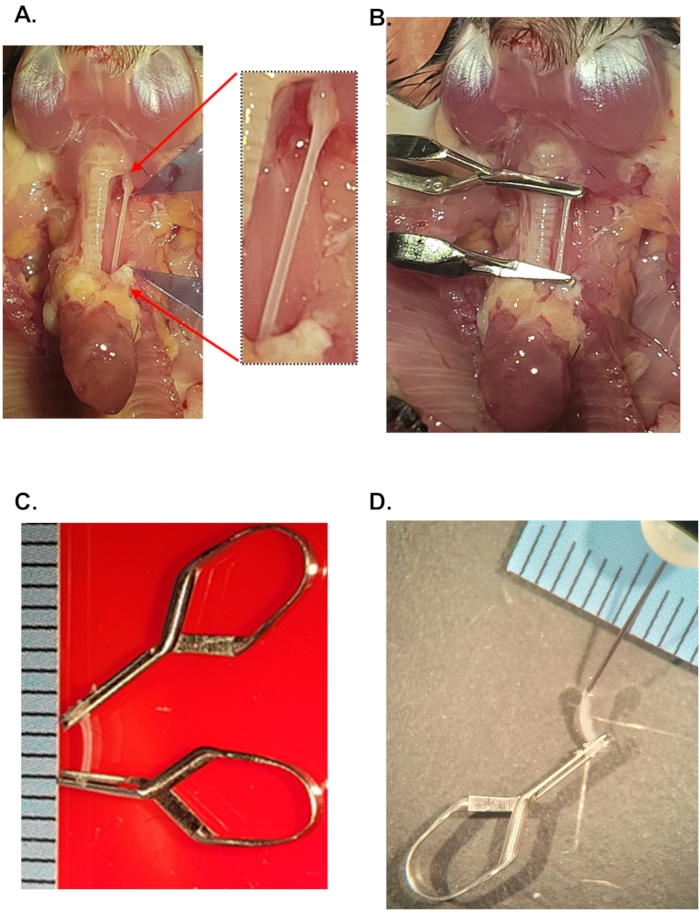
Figure 1: Isolation of carotid arteries for single-cell preparation. (A) Anatomical view of the carotid arteries in mice. Red arrows and inset show the isolated left carotid artery after clean up of periadventitial fat. For a schematic of the carotid anatomy and ligations, refer to Figure 1 in Nam et al3 (B) Image shows the location of micro-clips after filling the carotid artery with the digestion buffer. (C) Explanted carotid artery containing digestion buffer. Scale: distance between black lines = 1 mm. (D) A 29 G needle in the lumen of the mouse carotid artery. This step helps replenish the carotid artery with digestion buffer if needed. Please click here to view a larger version of this figure.
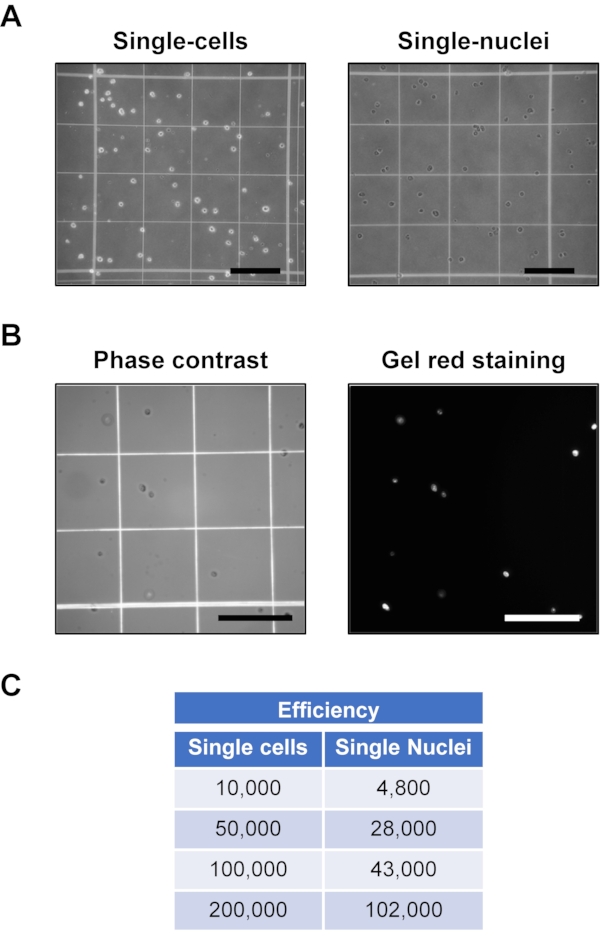
Figure 2: Single-cell and single-nuclei preparation from luminal enzymatic digestion of mouse carotid artery. (A) Representative single-cell and single-nuclei preparations. Scale bars = 0.25 mm (B) Representative phase-contrast and Gel-Red images of single-nuclei preps. Scale bars = 0.25 mm. (C) Efficiency of single-nuclei preparation in the left column shows the number of single cells at the start while the right column shows the number of single nuclei after processing with nuclei isolation buffer in different steps. Please click here to view a larger version of this figure.
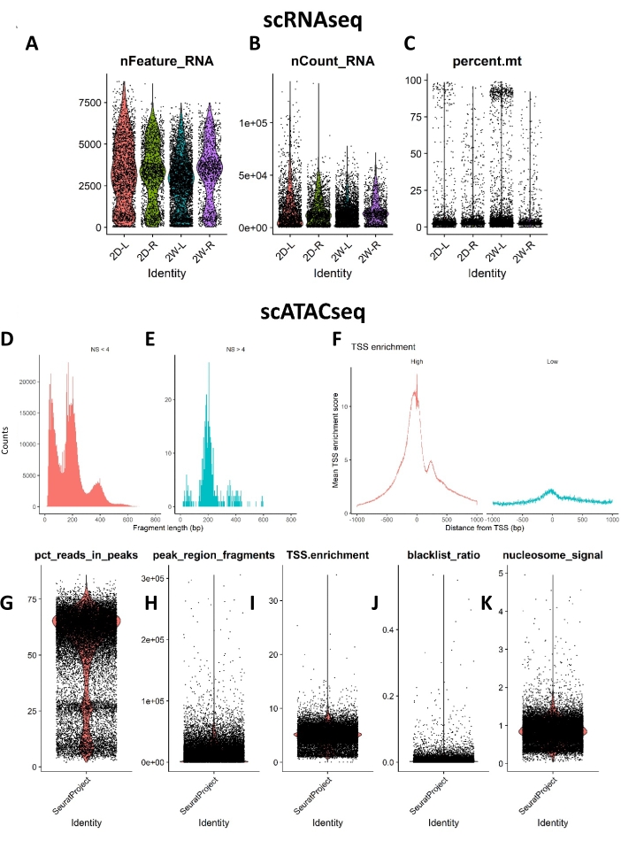
Figure 3: Standard QC metrics for the scRNAseq and scATACseq study. Violin plots show (A) the distribution of genes per cell (nFeature RNA), (B) UMI per cell (nCount_RNA), (C) mitochondrial reads per cell (percent mt) for the scRNAseq data. (D and E) show the nucleosome banding pattern for the scATACseq study. The histogram of DNA fragment sizes exhibits a strong nucleosome banding pattern corresponding to the length of DNA wrapped around a single nucleosome. (F) Normalized TSS enrichment score at each position relative to the TSS. The scatter/violin plots show (G) percent fragment reads in the peaks, a measure of sequencing depth, (H) peak region fragments showing the number of fragments overlapping peaks, (I) TSS enrichment score, a ratio between the aggregate distribution of reads centered on TSSs and that flanking the corresponding TSS, (J) blacklist ratio, a ratio of reads in blacklist genomic sites, and (K) nucleosome signal, a ratio of mononucleosomal to nucleosome-free fragments. Abbreviations: scRNAseq = single-cell RNA sequencing; scATACseq = single-cell Assay for Transposase Accessible Chromatin sequencing; QC = quality control; UMI = unique molecular identifier; TSS = transcription start site. Please click here to view a larger version of this figure.
Table 1: Composition of single-nuclei lysis and wash buffers. Please click here to download this Table.
Table 2: Single-cell and single-nuclei count from mouse carotid artery luminal digestion. The table also shows means reads/cell and number of genes/cell for scRNAseq data. For the scATACseq data, mean fragments/cell and total reads obtained per sample are shown8. Abbreviations: scRNAseq = single-cell RNA sequencing; scATACseq = single-cell Assay for Transposase Accessible Chromatin sequencing; 2D = 2-day; 2W = 2-week; R/RCA = right carotid artery; L/LCA = left carotid artery. Please click here to download this Table.
This paper provides a detailed protocol to isolate single-cell preparations from the mouse carotid arteries. The influence of d-flow on the endothelial cells can be accurately studied if the PCL surgery is performed correctly. It is crucial to correctly identify the branches of the common carotid, such as the external carotid, internal carotid, occipital artery, and superior thyroid artery. Validation of flow patterns by ultrasonography further validates the successful onset of d-flow conditions. Although PCL surgery can be performed on mice irrespective of their age, the preferred age is 10 ± 2 weeks. Older mice and mice with hypercholesterolemic backgrounds generally tend to have more periadventitial fat and stiffer skin.
It is essential to carefully dissect the carotid artery endothelial-enriched single-cell preparations free of the surrounding tissue and periadventitial fat. The lumen of the carotids must be perfused thoroughly to avoid contaminating blood cells in the single-cell preparations. Improper tissue identification and labeling can result in high standard deviation. Careful planning and time management are required for this protocol. A skilled surgeon and operator can take 15-20 min to perform one PCL surgery. Sacrificing, carotid isolation, and luminal enzymatic digestion from each mouse would take an additional 35-40 min. The hands-on processing time from endothelial cell flushing to single-cell/single-nuclei prep is an additional 2 h. Meticulous planning and teamwork are highly recommended.
As with any experimental protocol, some disadvantages and limitations should be considered. The current protocol does not incorporate steps to avoid artifactual activation of immediate early response during the luminal tissue dissociation. It has been reported in the literature that enzymatic digestion can lead to stress signaling events, such as inflammation and apoptosis. This can be addressed by incorporating appropriate control groups in the experimental design. In addition, wet laboratory methods that can minimize the artifactual activation of immediate response during enzymatic digestion, such as cold-active proteases, should be considered13,14.
This protocol can be used for any mouse strain irrespective of its genetic background. However, the endothelial-enriched preparations can best answer research questions pertaining to the changes in endothelium and are therefore well suited for EC-specific knockouts and EC-specific transgenic mice. This single-cell isolation method can be adapted to perform single-cell multi-omics studies and other flow cytometry-based assays such as Imaging Flow Cytometry. The luminal digestion approach could be used for freshly obtained mouse aortas, arteries from large animals (rabbits and pigs), as well as for freshly obtained human vascular explants. Collectively, this method would allow us to fully understand the precise role of blood flow on endothelial function and reprogramming at a single-cell resolution.
The authors have nothing to disclose.
This work was supported by funding from National Institutes of Health grants HL119798, HL095070, and HL139757 to HJ. HJ is also supported by the Wallace H. Coulter Distinguished Faculty Chair Professorship. The services provided by the Emory Integrated Genomics Core (EIGC) were subsidized by the Emory University School of Medicine and were also partly supported by the Georgia Clinical and Translational Science Alliance of the National Institutes of Health under award no. UL1TR002378. The content provided above is solely the authors' responsibility and does not reflect the official views of the National Institutes of Health.
Chemicals, Peptides, and Recombinant Proteins | |||
1x PBS (Cell Culture Grade) | Corning | 21040CMX12 | |
1.5 mL Protein LoBind Microcentrifuge Tubes | Eppendorf | 022-43-108-1 | |
15 mL Centrifuge Tube – Foam Rack, Sterile | Fablab | FL4022 | |
50 mL SuperClear Centrifuge Tubes | Labcon | 3191-335-028 | |
6-0 Silk Suture Sterile | Covidien | s-1172 c2 | |
70 µm Cell Strainer, White, Sterile, Individually Packaged | Thermo Fisher Scientic | 08-771-2 | |
Accutase solution,sterile-filtered | Sigma-Aldrich | A6964-100ML | or equivalent |
ATAC Buffer (Component I of Transposition Mix) | 10x Genomics | 2000122 | |
ATAC Enzyme (Component II of Transposition Mix) | 10x Genomics | 2000123/ 2000138 | |
Bovine Serum albumin | Sigma-Aldrich | A7906-500G | |
Buprenorphine | Med-Vet International | RXBUPRENOR5-V | |
Chromium Controller & Next GEM Accessory Kit | 10X Genomics | 1000204 | |
Chromium Next GEM Single Cell 3' Reagent Kits v3.1 | 10X Genomics | 1000121 | |
Chromium Next GEM Single Cell ATAC Reagent Kits v1.1 | 10X Genomics | 1000175 | |
Collagenase II | MP Biomedicals | 2100502.5 | |
Digitonin | Sigma-Aldrich | D141-100MG | |
Dissecting Forceps | Roboz Surgical Instruments Co | RS-5005 | |
Dnase1 | New England Biolabs Inc | M0303S | |
Centrifuge (Benchtop-Model # 5425) | Eppendorf | 22620444230VR | |
Fetal Bovine Serum – Premium Select | R&D systems | S11550 | |
Fixed Angle Rotor | Eppendorf | FA-45-24-11-Kit Rotor | |
HEPES buffered saline | Millipore Sigma | 51558 | |
Insulin syringe (3/10 mL 29 G syringe) | BD | 305932 | |
Isoflurane | Patterson vet | 789 313 89 | |
MACs Smart Strainers (30 µm) | Miltenyi Biotec | 130-098-458 | |
MACS SmartStrainers (100 µm) | Miltenyi Biotec | 130-098-463 | |
Normal Saline (0.9% sodium chloride) | Baxter International Inc | 2B1323 | |
Nuclei Buffer (20x) | 10x Genomics | PN 2000153/2000207 | |
PBS (10x), pH 7.4 | Thermo Fisher Scientic | 70011-044 | |
Small scissors | Roboz Surgical Instruments Co | RS-5675 | |
Stainless Steel Micro Clip Applying Forceps With Lock | Roboz Surgical Instruments Co. | RS-5480 | or similar |
Tissue Mend II | Webster Veteinanry | 07-856-7946 | |
Type II Collagenase | MP biomedicals | 2100502.1 | |
Deposited Data | |||
scATACseq FastQ files | NCBI | www.ncbi.nlm.nih.gov/bioproject Accession # PRJNA646233 | |
scRNAseq FastQ files | NCBI | www.ncbi.nlm.nih.gov/bioproject Accession # PRJNA646233 | |
Software and Algorithms | |||
Cell Ranger 3.1.0 | 10X Genomics | https://support.10xgenomics.com/ single-cell-gene-exp | |
Cicero | Pliner et al., 2018 | https://cole-trapnell-lab.github.io/cicero-release/ | |
Ggplot2 v3.2.1 | Hadley Wickham | https://cran.r-project.org | |
Harmony | Korsunsky et al., 2019 | https://github.com/immunogenomics/harmony | |
ImageJ | Schneider et al., 2012 | https://imagej.nih.gov | |
Monocle 2.8.0 | Qiu et al., 2017 | https://github.com/cole-trapnell-lab/ monocle-release | |
R version 3.6.2 | R Foundation | https://www.r-project.org | |
Seurat 3.1.3 | Stuart et al., 2019 | https://github.com/satijalab/seurat | |
Signac 0.2.5 | Stuart et al., 2019 | https://github.com/timoast/signac |