This article presents a guide for sampling six significant and diverse organs in adult Xenopus that can be rapidly and easily accessed: the heart ventricle, liver lobe, pancreas, fat bodies, paired kidneys, and skin.
Xenopus has been a powerful model organism for understanding vertebrate development and disease for over a hundred years. While experimental analysis and dissection techniques of the embryo have been well documented, descriptions of adult Xenopus structures and organs, together with techniques for working with adults, have not been updated to take into consideration the requirements of such modern approaches as quantitative proteomics and single-cell transcriptomics. The cell-type and gene-centric perspectives require contrasting observations in embryonic stages to those in adult tissues. The organs of the larva undergo significant changes in their overall structure, morphology, and anatomical location all along the larval to adult transition, most notably during massive metamorphosis remodeling. Establishing robust standards for organ identification and dissection is crucial to ensure datasets resulting from studies performed at different laboratories can be consistent. The present protocol identifies six of the organs in the adult Xenopus, demonstrating methods for dissection and sampling of the heart ventricle, liver, fat body, pancreas, paired kidney, and skin of the adult Xenopus. Depending on the preservation methods, the dissected organs can be used for quantitative proteomics, single cell/nuclei transcriptomics, in situ hybridization, immunohistochemistry, histology, etc. This protocol aims to standardize tissue sampling and facilitate multi-lab investigations of the adult organ systems.
Though the "digital dissection" of adult Xenopus” is available1, replicable organ and tissue sampling of adult Xenopus remains challenging without the detailed instruction available for other adult models (e.g. mice2,3,4). This article aims to provide clear guidance for accurate and replicable organ sampling of adult Xenopus similar to what is currently available for their larvae5. An emphasis is placed on ease of completion to maintain maximum freshness and make the protocol accessible to all users.
Though there is a thorough dissection guide for Rana sp.6, as well as numerous classroom dissection guides for other anurans7, no Xenopus dissection and sampling guide is currently available. For those not familiar with sampling practices or amphibian anatomy, the small differences between Xenopus and other anurans render these resources suboptimal for replicable tissue sampling.
Many valuable tissues are not included and are even discarded in the present guide; this is to ensure tissue freshness. Six samples are limited enough to ensure that these tissues can be collected in under an hour after the heart starts beating, regardless of the experience or skill level of the user. More advanced and detailed guides for collecting many other tissues are under preparation as separate companion papers.
For less experienced users, it is always recommended that this protocol be first attempted on animals being euthanized for reasons other than experimentation before sampling any animals that are challenging to replace (i.e., transgenics, animals of advanced age, etc). Ideally, all animals sampled will be healthy and, if female, will not have been ovulated in the past two weeks.
All experiments were performed in accordance with the rules and regulations of the Harvard Medical School IACUC (Institutional Animal Care and Use Committee) (IS 00001365_3). The representative results are shown for both a perfused and unperfused mature albino male Xenopus laevis.
1. Experimental preparation
NOTE: If perfusion protocol8 is being followed before sampling, skip to step 2.2.
2. Sampling
NOTE: If the animal has been perfused, skip to step 2.2.
By utilizing Figure 1 à Figure 20 and following all steps of this protocol, the heart ventricle, the left lobe of the liver, the pancreas, the left fat bodies, paired kidneys, and a flap of skin were cleanly excised within an hour of euthanasia. Within this time, the samples are rinsed and trimmed so that they will appear, as shown in Figure 21.
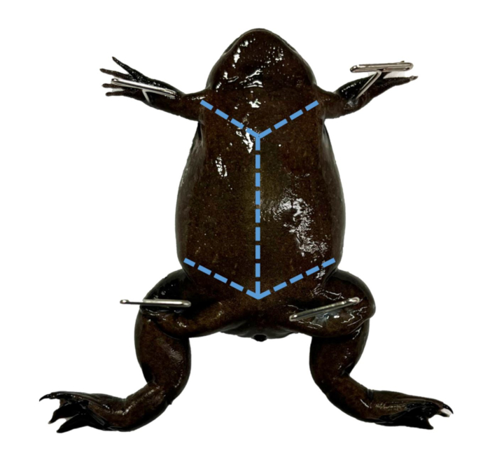
Figure 1: Pinned Xenopus. A mature female X. tropicalis pinned through each limb. Please click here to view a larger version of this figure.
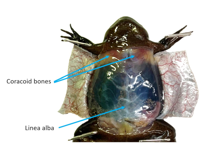
Figure 2: Abdominal wall. The ventral skin of an X. tropicalis female is cut into flaps, making the linea alba and coracoid bones visible. Please click here to view a larger version of this figure.
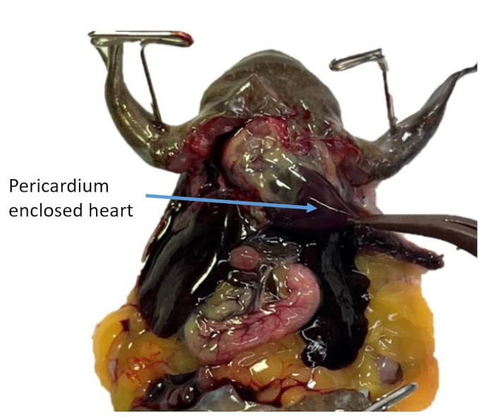
Figure 3: Pericardium enclosed heart. The apex of the heart ventricle is grasped through the pericardium. Please click here to view a larger version of this figure.
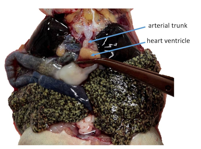
Figure 4: Heart ventricle and arterial trunk. The ventricle of a perfused X laevis, being grasped, showing its attachment to the arterial trunk. Please click here to view a larger version of this figure.
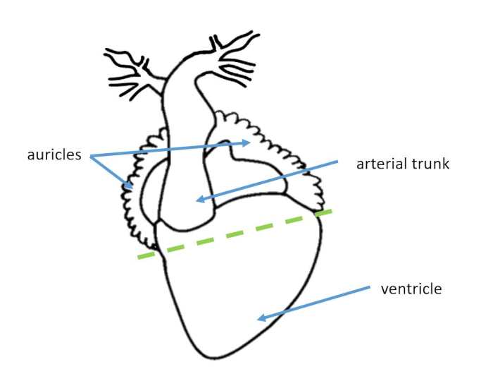
Figure 5: Heart diagram. A diagram of the relevant structures of the heart with a dashed line indicating where to sample the ventricle. Please click here to view a larger version of this figure.
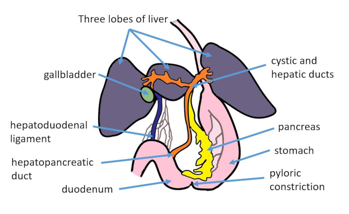
Figure 6: Hepatopancreatic diagram. A diagram of the 3 lobes of the liver, pancreas, and associated organs. Please click here to view a larger version of this figure.
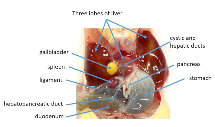
Figure 7: Hepatopancreatic organs. A perfused, albino X. laevis male with 3 lobes of liver, pancreas, and associated organs labeled. Please click here to view a larger version of this figure.
Figure 8: Cystic and hepatic ducts. The left lobe of the liver is being lifted to show the cystic and hepatic ducts in perfused X. laevis. Please click here to view a larger version of this figure.
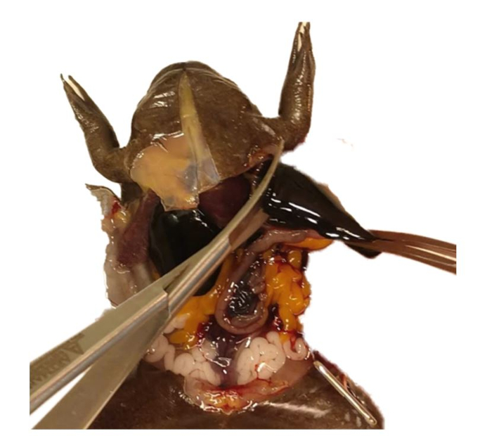
Figure 9: Liver sampling. The left liver lobe of an unperfused X. tropicalis is severed under the attachments of the hepatic ducts. Please click here to view a larger version of this figure.
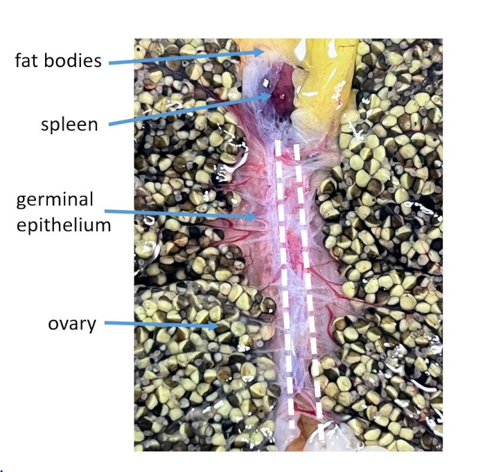
Figure 10: Ovary attachment. With the ovary lobes on their respective sides, the continuity of the germinal epithelium to the peritoneal wall (over the kidneys) is visible. Two white dashed lines indicate where to sever these attachments. Please click here to view a larger version of this figure.
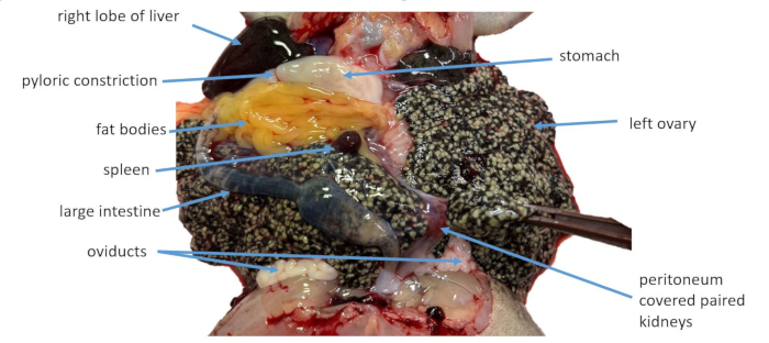
Figure 11: Ovary removal. The ovary of an unperfused X. laevis, is pulled away from the paired kidneys. Please click here to view a larger version of this figure.
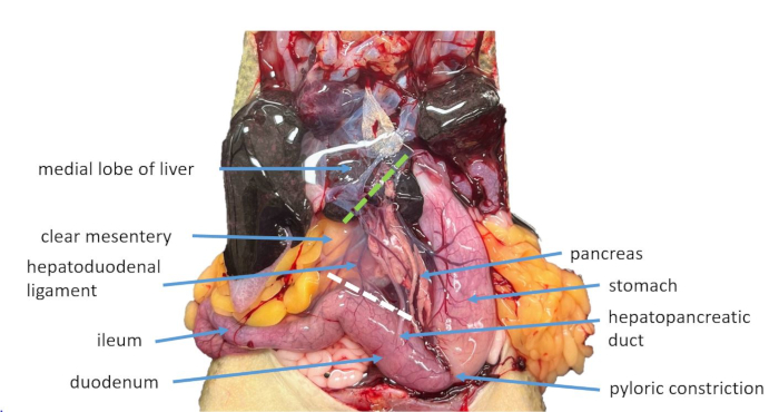
Figure 12: Mesentery incisions. The coelomic cavity of an unperfused X. laevis, following the sampling of the heart ventricle and left lobe of the liver as well as the removal of the ovary. A white dashed line indicates where to sever the hepatopancreatic ligament and duct, while a green dashed line indicates where to sever the pancreas from the medial lobe of the liver. Please click here to view a larger version of this figure.
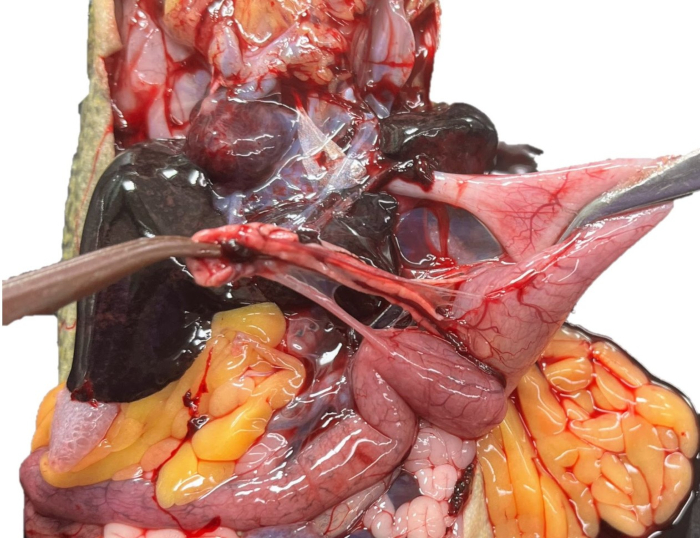
Figure 13: Pancreas sampling. The pancreas of an unperfused X. laevis is being teased off of the stomach. Please click here to view a larger version of this figure.
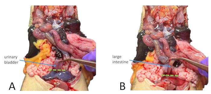
Figure 14: Organ removal. (A) The urinary bladder of an unperfused X. laevis is pulled away from the cloaca with a dashed line indicating where to cut it. (B) The large intestine of an unperfused X. laevis, is being pulled away from the cloaca with a dashed line indicating where to sever it. Please click here to view a larger version of this figure.
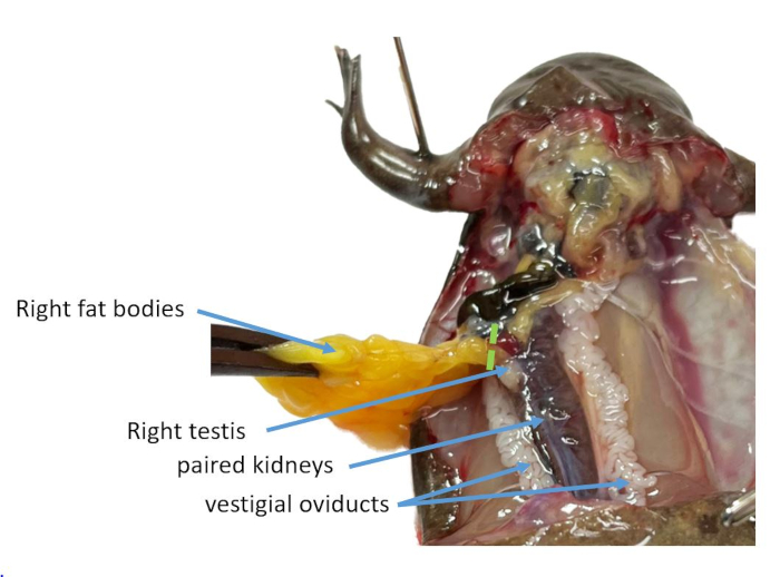
Figure 15: Fat body sampling. The fat bodies, attached to the peritoneum at the superior end of the paired kidneys, are pulled out of the coelomic cavity with a dashed line showing where to cut them. Note that adjacent to this attachment, this male X. tropicalis has 1 testis as well as a pair of distinct vestigial oviducts. Please click here to view a larger version of this figure.
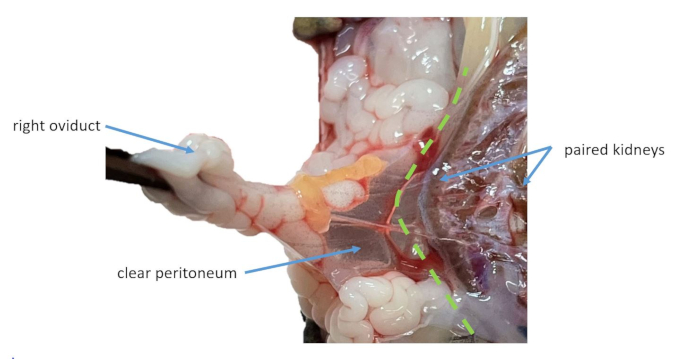
Figure 16: Oviduct removal. The oviduct of a perfused X. laevis is tugged away from the paired kidney, making the clear peritoneum visible. A dashed line indicates where to incise the peritoneum. Please click here to view a larger version of this figure.
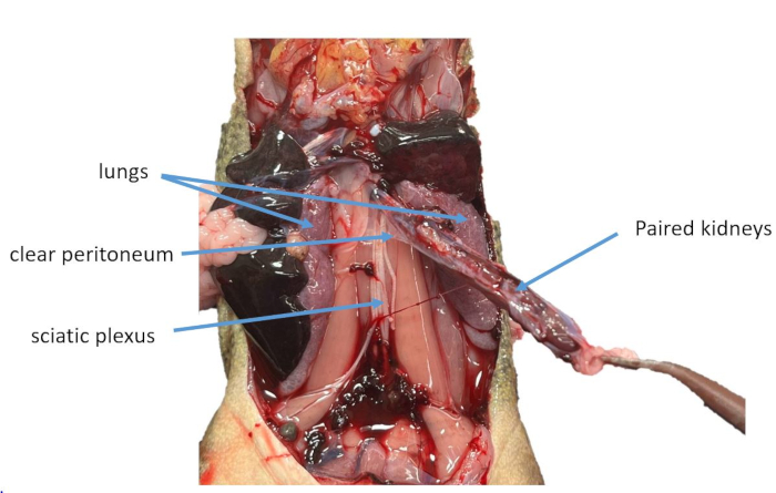
Figure 17: Kidney sampling. The paired kidneys of an unperfused X. laevis are being lifted out of the coelomic cavity. Please click here to view a larger version of this figure.
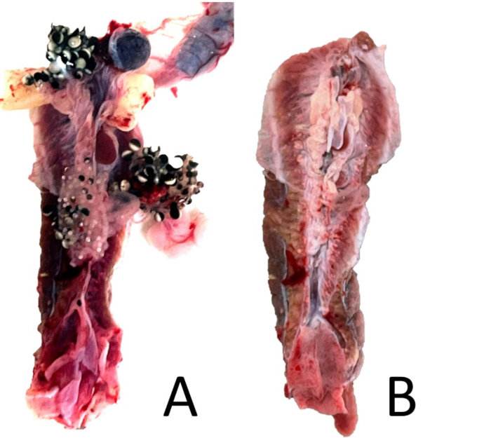
Figure 18: Kidney trimming. (A) A ventral view of an unperfused female X. laevis’s paired kidney with associated peritoneal organs attached. (B) The same kidney with associated organs removed but with some peritoneal tissue remaining. Please click here to view a larger version of this figure.
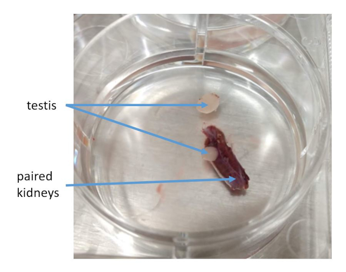
Figure 19: Testis removal. The paired kidneys of unperfused X. tropicalis with one testis removed. Please click here to view a larger version of this figure.
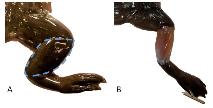
Figure 20: Skin sampling. (A) The right leg of an X. tropicalis with a dashed line indicating the area of skin to be sampled. (B) The right leg of an X. tropicalis with a skin sample removed over the tibiofibula. Please click here to view a larger version of this figure.
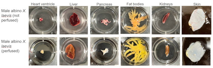
Figure 21: Representative results of organ sampling. Samples of heart ventricle, liver, pancreas, fat body, paired kidney, and skin taken from a perfused and unperfused albino X. laevis. Please click here to view a larger version of this figure.
As this protocol aims to maximize freshness, some samples may include undesired tissues. For example, the hepatopancreatic duct and some mesentery are sampled with the pancreas, and some peritoneal tissue, adrenal glands, and ureters will always be sampled with the paired kidneys. If freshness is not a concern, then more precise sampling can be achieved using modified techniques.
The appearance and location of organs are comparable between sexes and species of Xenopus. However, the color of tissues varies significantly based on whether or not the animals have been perfused. It is for this reason that images of both perfused and unperfused animals are included.
A constraint of this protocol is that speed and reproducibility are prioritized over collecting samples that best represent the entirety of the desired tissue. For example, the section of the left lobe of the liver sampled here cannot adequately represent all three lobes of liver tissue. If there are errors in sampling, options for troubleshooting are affected by the potential for variation between different sections of tissue. For example, it is not known if the right lobe of the liver, the right fat body, or a different portion of skin would be functional alternatives to the desired tissues. In these cases, discretion should be used, based on the needs of the research, before substituting sections of tissue.
Another limitation of this protocol is that if the animals being sampled have drastic anatomical defects or clinically significant health issues, the organs in the coelomic cavity may not appear as described here. Granulomas have been found in the tissues of frogs infected with Mycobacterium spp.11,12, and previous cases of ovarian hyperstimulation syndrome appear to lead to an abnormal presentation of organs13.
Though this method has been developed for laboratory Xenopus, there are significant similarities in the appearance of these organs within many non-caecilian amphibians and limbed reptiles14. The sampling portion of this protocol could easily be modified for other models, such as axolotls or the green anole.
The authors have nothing to disclose.
This work was supported by NIH's OD grant R24OD031956. We thank Samantha Jalbert, Jill Ralston, and Cora Anderson for their assistance and support as well as our editor and anonymous peer reviewers for their helpful feedback
5x Magnifying Glass with LED Light and Stand | amazon.com | B08QJ6J8P1 | light must not produce heat |
Disposable Transfer Pipets | VWR | 414004-036 | |
Dissecting Fine-Pointed Forceps | Fisher Scinetific | 08-875 | |
Dissecting scissors sharp piont, straight 6.5" | VWR | 76457-374 | |
Dissection Tray | Fisher Scinetific | 14-370-284 | styrofoam sheets are an acceptable alternative |
Euthanasia container | US Plastic | Item 2860 | alternative opaque containers acceptable |
Euthanasia container lid | US Plastic | Item 3047 | |
Iridectomy Scissors 6" | vwr | 470018-938 | iris scissors are an acceptable alternative |
MS-222: Syncaine (formerly tricaine) | Pentair AES | TRS1 | |
PBS 1x | Corning | 21-040-CV | |
Sodium Bicarbonate, Powder, USP | Fisher Scientific | 18-606-333 | |
Specimen Forceps, Serrated | VWR | 82027-442 | |
T-Pins for Dissecting | Fisher Scinetific | S99385 |