A subscription to JoVE is required to view this content. Sign in or start your free trial.

Method Article

Immunolocalization of Arabinogalactan Proteins and Pectins in Plant Tissue

1.1K views

⸱

July 8th, 2025

In This Article

Abstract

Source: Costa, M., et al. Fluorescent Immunolocalization of Arabinogalactan Proteins and Pectins in the Cell Wall of Plant Tissues. J. Vis. Exp. (2021).

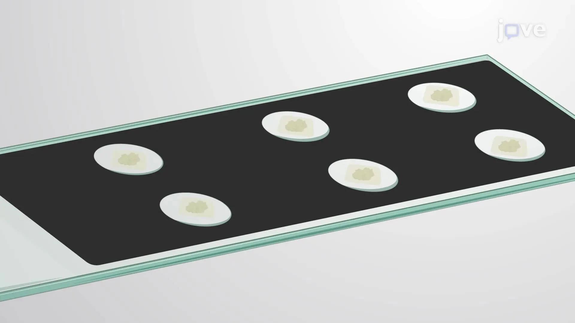
This video demonstrates an immunostaining technique to localize arabinogalactan proteins (AGPs) and pectins in plant tissues. Fixed and resin-embedded plant tissue sections are stained with AGP- and pectin-specific primary and fluorescent secondary antibodies and a cellulose-binding fluorescent dye to reveal their tissue-specific colocalization in the cell wall.
Protocol

1. Immunolocalization

NOTE: The fluorescent immunolocalization procedure relies on sequential use of two antibodies. The primary antibody is raised against a specific target antigen. The secondary antibody is raised specifically against the primary antibody and for fluorescent techniques is conjugated to a fluorophore (FITC [fluorescein isothiocyanate] in this specific protocol). The primary antibody will be used to detect the target antigen in the sample, and the secondary antibody will be used to mark the location where the primary antibody connected to the sample after washing off the excess of primary ant....

Access restricted. Please log in or start a trial to view this content.

Results

Flow cytometry sample preparation steps; includes labeling, washing, and sorting; experiment kit.
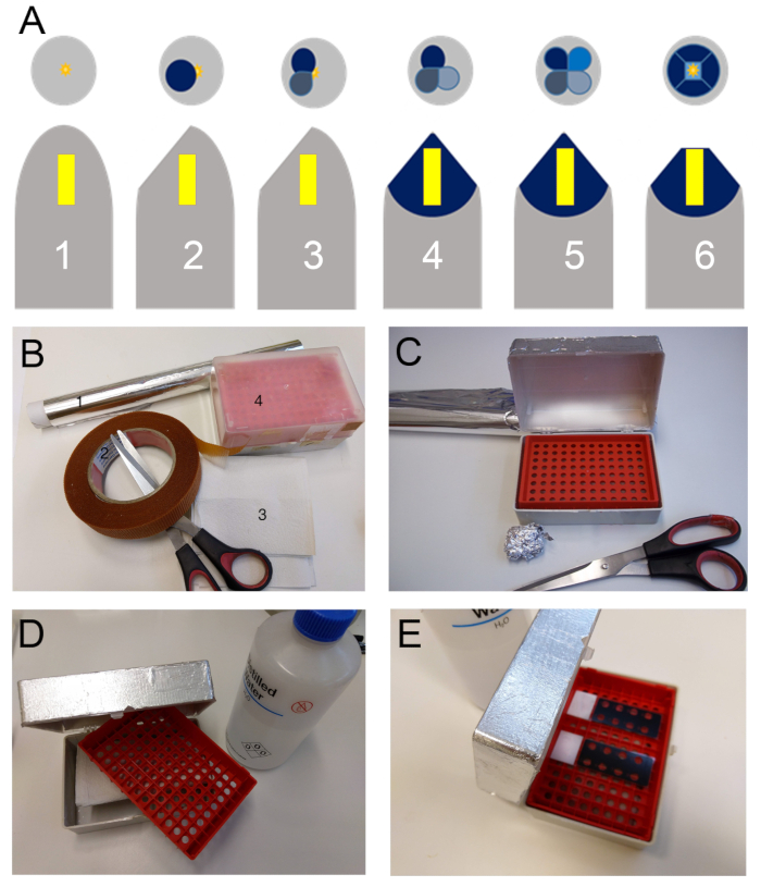
Figure 1. (A) Schematic representation of the block trimming sequence for preparing the sample (Yellow block) for sectioning with the ultramicrotome. Firstly, the gelatin capsule is removed (1). Then they are first trimmed at a 40° to 45° angle t.......

Access restricted. Please log in or start a trial to view this content.

Disclosures

No conflicts of interest declared.

Materials

List of materials used in this article
NameCompanyCatalog NumberComments
8 wells Glass reaction slidesMarinfeldMARI1216750other brands may be used
Anti-Rat IgG (wole molecule)-FITC antibody produce in GOATSigma-AldrichF6258
cover slips, 24 mm x 50 mmMarinfeldMARI0100222The cover slip should cover all the wells. Other brands may be used
ddH2Onana
Ethanol absolutenana
Fluorescent brightner 28Sigma-AldrichF-6259
Non fat dry milkNestlénaany non fat dry milk is adequate
Ovennanageneric laboratory oven
Rat generated Monoclonal Anti-BodyPlant probesnaSeveral antibodies that recognize cell wall components are available at both the Complex Carbohydrate research center (CCRC, Georgia University USA) and Plant Probes (Paul Knox Cell Wall Lab, at Leeds University UK). A short list of some commonly used MABS and where they can be purchased is presented in Supplemental Table 1
upright epifluorescence microscope with UV and FITC fluorescence filtersLeica MycrosistemsDMLb
vaccum chambernana
vaccum pumpnana
Vectashieldvecta LabsT-1000Other anti-fade may be used. Please do check for compatibility with FITC and the Fluorescente brightner 28. (Note: for a non-commercial alternative, (see Jonhson et al 198218) An antifade medium can be made by mixing 25 mg/mL of 1,4-Diazobicyclo-(2,2,2)octane (DABCO) in 9:1 (v/v) glycerol to 1xPBS. Adjust pH to 8.6 with diluted HCl.)

Explore More Articles

Pectin LocalizationImmunostaining TechniqueFluorescent AntibodiesCell Wall StainingPlant Tissue SectionsCalcofluor WhiteFluorescence MicroscopyImageJ AnalysisTissue Specific Colocalization