Many mRNAs encoding mitochondrial proteins are associated with the mitochondria outer membrane. We describe a subcellular fractionation procedure aimed at isolation of yeast mitochondria with its associated mRNAs and ribosomes. This protocol can be applied to cells grown under diverse conditions in order to reveal mechanisms of mRNA localization and localized translation near the mitochondria.
Most of mitochondrial proteins are encoded in the nucleus and need to be imported into the organelle. Import may occur while the protein is synthesized near the mitochondria. Support for this possibility is derived from recent studies, in which many mRNAs encoding mitochondrial proteins were shown to be localized to the mitochondria vicinity. Together with earlier demonstrations of ribosomes’ association with the outer membrane, these results suggest a localized translation process. Such localized translation may improve import efficiency, provide unique regulation sites and minimize cases of ectopic expression. Diverse methods have been used to characterize the factors and elements that mediate localized translation. Standard among these is subcellular fractionation by differential centrifugation. This protocol has the advantage of isolation of mRNAs, ribosomes and proteins in a single procedure. These can then be characterized by various molecular and biochemical methods. Furthermore, transcriptomics and proteomics methods can be applied to the resulting material, thereby allow genome-wide insights. The utilization of yeast as a model organism for such studies has the advantages of speed, costs and simplicity. Furthermore, the advanced genetic tools and available deletion strains facilitate verification of candidate factors.
Eukaryotic cells are organized in distinct compartments having specific functions. To accomplish its function, each compartment contains a unique set of proteins that are essential for its activity. A possible mechanism through which these proteins approach their compartment is by localized translation1,2. In this process, the protein is synthesized at its destination by ribosomes and mRNAs that are located there. Among the probable advantages of localized translation are increased efficiency of protein targeting, decreased need for protein chaperones, and enabling site-specific regulation mechanisms. Also, localized mRNAs and ribosomes can be a secluded reservoir of translation machinery in cases of cellular stress, when general translation is inhibited.
Mitochondria became in recent years a central model to study localized translation. Most mitochondria proteins are encoded in the nucleus, translated in the cytosol, and imported into the organelle. Various lines of evidence indicate that many of these proteins are produced through a local translation process. Initially, electron microscopy and biochemical fractionation studies detected ribosomes associated with mitochondria3-5. These studies where then corroborated in vivo by work on specific mRNAs, that were found to be imported only in a cotranslational manner6,7. Genome-wide studies of mRNAs association with mitochondria revealed that a significant fraction of mRNAs are localized to the mitochondria vicinity8-10. Some of these mRNAs were further characterized by in vivo fluorescence methods, such as FISH or mTAG9,11. A straight-forward interpretation of this association is that these mRNAs serve as templates for localized translation.
The mechanisms by which these mRNAs approach the mitochondria are unknown. Noncoding domains (most significantly 3’ UTRs) were shown to be involved in mRNA association to the mitochondria12. These domains are likely to serve as a binding site to RNA-binding proteins which mediate their transport. Studies in yeast revealed that a member of the PUM family of proteins (Puf3) supports mRNA association with mitochondria8,13. A plausible role for Puf3, which is based on functions of other family members, is to inhibit translation while the mRNA is en route14. Thus, mRNAs may be transported in a nontranslated status, by RNA binding proteins that interact with noncoding regions. Alternatively, a large body of work suggests that transport occurs while the protein is being synthesized. In particular, translation inhibitors were shown to affect mRNAs association8,13. Furthermore, translated features such as the AUG, mitochondrial targeting sequence (MTS) or ORF regions were shown to assist in localization8,11,15. Hsp70-family protein chaperone and protein receptor on the mitochondria outer membrane were also shown to support mRNA association, further implying that encoded-protein features are important for mRNA localization16. This is consistent with a model in which the ribosome-emerging protein serves as recognition element for targeting mRNA-ribosome-protein complex to the mitochondria17.
Localized translation near the mitochondria was studied by various methods, including electron microscopy (to visualize ribosomes)3, FISH9, green RNA (to detect specific mRNAs)11, and biochemical fractionation (to detect both RNA and ribosomes)10,18. While the former methods detect localization in vivo and may allow visualization of transport dynamics, the latter allows detection of many different mRNAs in a single experiment. Furthermore, for biochemical fractionation coding or noncoding domains do not need to be altered, therefore their specific roles can be evaluated. Biochemical fractionation has been successfully used for many years, for isolation of many different cellular compartments. Its principals and limitations are well established, and one can easily modify existing protocols for different purpose. The necessary instrumentation is standard in many labs, therefore it is usually the first method of choice for studying intracellular localization. We describe a protocol that was optimized for isolation of mRNAs while ribosomes are associated with mitochondria. This protocol is therefore optimal for studying factors involved in localized translation near mitochondria.
1. Mitochondria Purification
Weigh the pellet of cells. Roughly 0.6 g are obtained from 100 ml cells.
2. RNA Extraction
3. Preparing the RNA for Microarray Analysis
This protocol allows separation of a mitochondria-containing fraction from cytosolic components. The best way to test its success is to perform northern analysis and western analysis (Figure 1) to samples from the different isolation steps. Three parameters for the isolation quality are derived from these analyses. First, whether the RNA or proteins in the samples are intact – these will be detected as distinct bands in the analyses. Degradation events will induce the appearance of additional, shorter bands or smears. Second, comparing the signals from each step with the signal from the input (i.e. Total) provides important information regarding losses during preparation. Severe losses will be observed as a significant difference between the Total and the relative amount of signal. Lastly, and most important, the quality of separation between mitochondrial and cytosolic components can be determined. Isolation of intact mitochondria will result in signal for mitochondrially-transcribed transcript only in the mitochondria fraction and not in the cytosolic fraction (Figure 1A). Furthermore, proteins that are mitochondria-associated will appear by western blot only at the mitochondria fraction, and cytosolic proteins in the cytosolic fraction (Figure 1B).
Figure 1 demonstrates two additional outcomes that are standard for this protocol: first, association of nuclear-encoded mRNAs with mitochondria varies between genes. One can see that ACO1 mRNA, which encodes a mitochondrial protein, appears mostly in the mitochondria fraction, while the mRNA encoding the cytosolic actin protein (ACT1) is mostly in the cytosolic fraction (Figure 1C). Genome-wide analyses revealed variation among many genes8,10,21,22 and the basis for this diversity is under extensive research. Second, significant amounts of ER-related material are coisolated in the mitochondrial fraction. The mRNA encoding SEC61, which is ER-associated, is detected by northern blot (Figure 1A) and the ER-membrane protein Cue1 is detected by western blot (Figure 1B). This may be an outcome of the known physical contacts between ER and mitochondria. While further purification of mitochondria is possible, it may introduce some limitations; these are discussed in the Discussion.
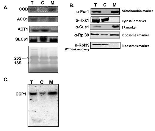
Figure 1. Quality verification of isolation. Quality is tested for samples taken in three representative steps of the procedure: Before fractionation (Total [step 1.20], T; cytosolic fraction [step 1.22], C; and mitochondrial fraction [step 1.24], M). The samples are subjected to northern analysis (A, C) or western analysis (B). A) Northern analysis for the following mRNAs: COB is an RNA that is transcribed inside the mitochondria therefore its signal should be exclusively in the mitochondria. Its appearance in the C fraction will indicate of mitochondria lysis during preparation. ACT1 is an mRNA that encodes a cytosolic protein, and therefore translated by cytosolic ribosomes and appears mostly at the C fraction. ACO1 is an mRNA that encodes a protein that is imported to the mitochondria, and appears mostly at the mitochondria fraction. SEC61 is an mRNA that encodes ER resident protein; its presence in the M fraction indicates the presence of ER components in this fraction. The bottom panel presents a methylene blue staining of the northern membrane, is which two rRNAs (18S and 25S) are detected. This panel demonstrates that significant amount of ribosomes appear in the M fraction. B) Western analysis for the following proteins: Por1 is a mitochondria outer membrane protein therefore its signal is expected only in the M fraction. Signal in the C fraction will suggest dissociation of mitochondrial components during preparation. Hxk1 is a cytosolic protein and Cue1 is an ER protein. Both report on the copurification of these fractions with the mitochondria fraction. Rpl39 is a ribosomal protein, again demonstrating the presence of ribosomes in both fractions. The panel “Rpl39 without recovery”, presents the fractionation of ribosomes when the protocol is excluded of the recovery step (step 1.14). C) Northern analysis for CCP1, an mRNA that encodes a protein that is imported to the mitochondria. The entire gel is presented to demonstrate the lack of shorter, degradation-originated bands. Please click here to view a larger version of this figure.
Technological advancements in imaging had yielded high resolution tools to study mRNA localization. Today, one can measure the movement of even a single mRNA molecule at the msec time scale23-26. Yet, traditional biochemical approaches, as the one described above, are also informative and are preferable in some cases. Biochemical isolation allows purification of a large repertoire of mRNAs and proteins, and is therefore preferable for genome-wide studies. In a single isolation, one can obtain sufficient mRNA levels to allow characterization of thousands of genes, either by DNA microarrays or RNA-seq18,27. In parallel, one can isolate proteins and perform a proteomic analysis to characterize the proteome on mitochondria28. Biochemical fractionations can be adapted to separate mitochondria from other cellular organelles, in particular the ER29. This may be advantageous in some types of cells were the high spatial overlap between ER and mitochondria obstructs microscopic localization analyses. An important advantage of biochemical fractionations is their low cost, which allows analysis of many different samples. A related advantage is their simplicity – biochemical fractionation, as the one described above, usually do not require laborious fluorescent tagging of mRNAs, as is the case in in vivo imaging studies. Finally, many pitfalls and artifacts are already diagnosed for these traditional protocols, therefore the necessary controls are well-established.
The presented protocol entails a recovery step (step 1.14) that follows the zymolyase treatment. This step is critical because the stress induced during the zymolyase treatment leads to almost immediate translational arrest and ribosomes’ dissociation from mRNAs. The actual trigger for ribosomes dissociation can any of the steps that included in this treatment, namely removal of the carbon source, exposure to high g during centrifugation, treatment with reducing agents (DTT), cell-wall removal by zymolyase or introduction of high osmotic conditions; each of these is known to induce a translational arrest. This translational arrest probably affects the association of ribosomes with mitochondria. Accordingly, we found that mRNA association with mitochondria is low after zymolyase treatment, and increases during recovery (not shown).
The mitochondrial fraction that is isolated by this protocol contains a significant amount of ER, as evident from the presence of various ER markers (e.g. Figure 1). An obvious desire is to further remove ER components and to obtain pure mitochondria, using one of several standard protocols that utilize gradients29-33. However, these protocols necessitate stripping of ribosomes of the mitochondria, either due to the stressful zymolyase treatment (Figure 1B), or the inclusion of EDTA in the buffers. Ribosomes’ stripping has significant impact on mRNA association and therefore may introduce severe biases. It is therefore unwanted here. Intriguingly, we found that these protocols are not effective in separating ER from mitochondria when ribosomes are associated with these organelles. For example, when we applied gradient separation after the recovery from the zymolyase treatment (i.e. when ribosomes reassociate with the mitochondria), both ER and mitochondria markers sedimented to the same fraction. This is probably because the associated ribosomes obscure the differences in their densities (data not shown). Thus, for ribosome association studies, alternative methods for mitochondria isolation will need to be developed.
For many studies, however, the presence of ER components is not necessarily problematic. Association of mRNAs that encode well-established mitochondrial proteins (e.g. Aconitase, ATP2, Oxa1) is likely to be with the mitochondria and not the ER component in this crude preparation. It is therefore recommended to use one of these typical mitochondrial mRNAs as readouts for impact on mitochondria. Proteins that were not verified as mitochondrial should not be used as reporters; this is important in particular when genome-wide mRNA studies are performed (e.g. microarray analysis), as mis-assigned mRNAs may introduce an error.
The major technical challenge of this protocol is minimizing mRNA degradation. While using RNAse-free reagents is always advised, one should remember that upon lysis of cells enormous amounts of RNAses are unleashed into the solution. Heparin is a very effective RNAse inhibitor and is added at significant amounts. When large extract volumes are prepared, its relatively low cost provides additional advantage compared to other RNAse inhibitors. Heparin however, also inhibits reverse transcriptase, and therefore should be removed for studies that involve such enzymatic activity (e.g. RT-PCR or DNA microarray labeling). Herein we present a removal procedure that is based on LiCl precipitation (steps 3.4-3.10). This precipitation effectively removes heparin, but apparently also significant amounts of mRNAs are lost. Alternatively, there are various spin-columns that are supposed to remove heparin from RNA samples, yet we had a limited success with these.
The authors have nothing to disclose.
We thank Drs. Erez Eliyahu, Daniel Melamed, Ophry Pines and Doron Rapaport for help and comments during the establishment of this protocol. This work is funded by the ISF (grant number 1193/09).
Yeast Extract | |||
BactoPeptone | |||
Galactose | Do not autoclave Galactose | ||
Growth medium | For mitochondrial enrichment, you should use any non- fermentable carbon source, such as Galactose-based growth medium sterilized 1% Yeast Extract, 2% BactoPeptone, 2% Galactose | ||
0.1 M TrisHCl pH9.4 | |||
30 mM TrisHCl pH7.6 | |||
Dithiothreitol (DTT) | |||
10 mM DTT Buffer | 0.1 M TrisHCl pH 9.4, 10 mM Dithiothreitol (DTT). Make fresh every time | ||
1.2 M Sorbitol | |||
4 mM KH2PO4 | |||
16 mM K2HPO4 | |||
0.2μm filter | |||
Zymolyase Buffer | 1.2 M Sorbitol, 4 mM KH2PO4 , 16 mM K2HPO4. Filter this buffer (0.2μm) and keep at room temperature for future use | ||
Zymolyase 20T | 20,000 U/gr | ||
Recovery medium | Galactose-based growth medium supplemented with 1 M Sorbitol | ||
0.1 mg/mL Cycloheximide (CHX) | |||
0.6 M Mannitol | |||
5 mM MgAc | |||
100 mM KCl | |||
0.5 mg/mL Heparin | |||
1 mM phenylmethanesulfonylfluoride (PMSF) | |||
Mannitol Buffer | 0.6 M Mannitol, 30 mM TrisHCl pH7.6, 5 mM MgAc, 100 mM KCl. Add freshly: 0.5 mg/mL Heparin, 0.1 mg/mL CHX and 1mM (PMSF). Filter this buffer and use it ice-cold | ||
8M Guanidinium-HCl | |||
100% and 70% Ethanol (EtOH) | |||
3 M Sodium Acetate pH5.2 | |||
10 M LiCl stock solution | |||
250 mM Tris HCl pH 6.8 | |||
SDS | |||
Glycerol | |||
β-mercaptoethanol | |||
Bromophenolblue | |||
4xLSB | 250 mM Tris HCl pH 6.8, 8% SDS, 40% Glycerol, 20% β-mercaptoethanol and 0.02% Bromophenol blue | ||
Dounce homogenizer of 15-ml capacity equipped with tight- fitting pestle |