This protocol aims to alleviate the limitation of poor cell engraftment for stem cell treatment of myocardial infarctions through the use of a hydrogel system and a fibrin-based glue. With this approach, cell-to-tissue contact post-infarction can be maintained, increasing the therapeutic potential of beneficial agents at the site of injury.
The murine MI model is widely recognized in the field of cardiovascular disease, and has consistently been used as a first step to test the efficacy of treatments in vivo1. The traditional, established protocol has been further fine-tuned to minimize the damage to the animal. Notably, the pectoral muscle layers are teased away rather than simply cut, and the thoracotomy is approached intercostally as opposed to breaking the ribs in a sternotomy, preserving the integrity of the ribcage. With these changes, the overall stress on the animal is decreased.
Stem cell therapies aimed to alleviate the damage caused by MIs have shown promise over the years for their pro-angiogenic and anti-apoptotic benefits. Current approaches of delivering cells to the heart surface typically involve the injection of the cells either near the damaged site, within a coronary artery, or into the peripheral blood stream2-4. While the cells have proven to home to the damaged myocardium, functionality is limited by their poor engraftment at the site of injury, resulting in diffusion into the blood stream5. This manuscript highlights a procedure that overcomes this obstacle with the use of a cell-encapsulated hydrogel patch. The patch is fabricated prior to the surgical procedure and is placed on the injured myocardium immediately following the occlusion of the left coronary artery. To adhere the patch in place, biocompatible external fibrin glue is placed directly on top of the patch, allowing for it to dry to both the patch and the heart surface. This approach provides a novel adhesion method for the application of a delicate cell-encapsulating therapeutic construct.
A myocardial infarction (MI) is defined as the interruption of blood to a region of the heart caused by the occlusion of a major coronary artery. The damage resulting from an MI is due to the remodeling of the viable heart tissue into non-functional scar tissue, which decreases the ability of the heart or, more specifically, the left ventricle, to beat properly. This results in a decrease in the volume of blood that can be delivered to the body with every heartbeat, known as the stroke volume, and the percentage of blood that is pumped out of the heart with each heartbeat, known as the ejection fraction6. These, along with other diminished functions, increases the strain on the rest of the heart to maintain adequate function. Often, this increased strain can become so severe that it causes a second heart attack, a phenomenon seen in approximately 10% of individuals7.
While medical practices have evolved to treat the immediate aftermath of an MI, no technique has been developed to halt, slow, or reverse the negative side effects of tissue remodeling. Stem cell therapies have emerged as a possible avenue for such a treatment, however, despite their promising potential, stem cells have not proven successful in the clinical setting. One theory for their shortcomings is the inability to ensure the beneficial cells remain at the site of infarction long enough to generate favorable results5. It has been shown that no more than 24% of cells that are simply injected into the site of infarction survived and remained at the damaged site 1 day post-delivery2. A possible prospect for addressing this issue of cell retention is to develop biocompatible hydrogel systems that encapsulate either cells or therapeutics, which can be delivered to the damaged site. The hydrogel of choice in this protocol is a poly(ethylene glycol) dimethacrylate due to its previous use in cell encapsulation procedures, however, any hydrogel capable of encapsulation may be used8. The delivery of the patch directly to the site of injury ensures cell-to-tissue contact over an extended period of time, increasing the length of time the cells can provide beneficial factors to the underlying myocardium.
A bottleneck to the patch approach is the difficulty of adhering the patch to the heart surface. Many groups have overcome this through a variety of techniques, the most prevalent being a simple suture to tie the construct to the heart surface9,10. This has proven successful in a number of cases in which the construct is made of a stiffer material, but fails when attempted on a hydrogel system, due to the high water concentration and delicate nature of the patch construct. To overcome this, we have utilized a fibrin glue external adhesive system that mimics the chemistry of clot formation. Fibrin glue has been used in numerous medical surgeries, including dura tears, bronchial fistulas, and corneal transplantation, highlighting the biocompatibility of the product as a wound sealant11-13. Additionally, fibrin has been used for a variety of cardiac purposes, including surgical treatment of left ventricular ruptures and coronary artery bypass surgeries, however, its use as an adhesion glue for a cardiac patch is not commonly used14-17. A simple formulation of thrombin and fibrinogen results in a biocompatible glue that can be placed directly on the outside of an external cardiac patch, providing a viable adhesion system to ensure patch to heart interaction.
Procedure is in full compatibility with IACUC protocol number 13302 and has been approved by the division of animal resources.
1. Instrument Preparation
2. Hydrogel Preparation
3. Animal Preparation and Oral intubation
4. Surgical Procedure
5. Patch Placement and Glue
6. Suturing
7. Post-surgical Treatment
8. Analysis of Heart Function and Histology
During the surgical procedure, ligation of the left coronary artery can be identified by a marked blanching downstream of the occluded artery. As a test before tying the knot, the suture can be tightened briefly to check if it is in the appropriate place. Additionally, since occlusion of the artery results in almost instantaneous decrease in the ability of the left atrium to properly beat, the left atrium will enlarge in response to a backflow of blood in the system.
M-mode echocardiography measurements taken as early as 2 days post-infarction show a cessation of left wall movement, indicative of the muscle reconstruction. Qualitative calculations made from the data show a decrease in ejection fraction and stroke volume in the infarcted hearts. At the termination of the experiment, when the hearts are collected for histological purposes, a clear dilation of the left ventricle can be seen, along with a thinning of the left ventricular wall, and the deposition of collagen that denotes scar tissue deposition in place of functioning cardiomyocytes (Figure 1).
Also seen upon histological analysis is the presence of the hydrogel construct thanks to the administration of the fibrin gel (Figure 2). For our purposes, the adhesive system must be viscous enough to allow precise placement control and minimize subsequent runoff into surrounding organs, but malleable enough not to interfere with heart function. Preliminary tests were performed to calculate the viscosity, gelation time, and stiffness of various fibrinogen/thrombin ratios, in order to determine the proper combination that suits our needs. In vivo analysis was performed to test the ability of the fibrin glue to maintain patch-to-tissue adhesion while still allowing for full heart function (data not shown). It should be noted that the fibrin glue did not harm the myocardium, as evidenced by the lack of tissue remodeling or ventricular thinning at the site (Figure 2). The cardiomyocytes remain intact despite the addition of both a cardiac patch and its accompanying glue. Additionally, viability testing confirmed that that the administration of fibrin glue to the external surface of a cell-encapsulating hydrogel patch did not affect cell survival within the patch (Figure 3).
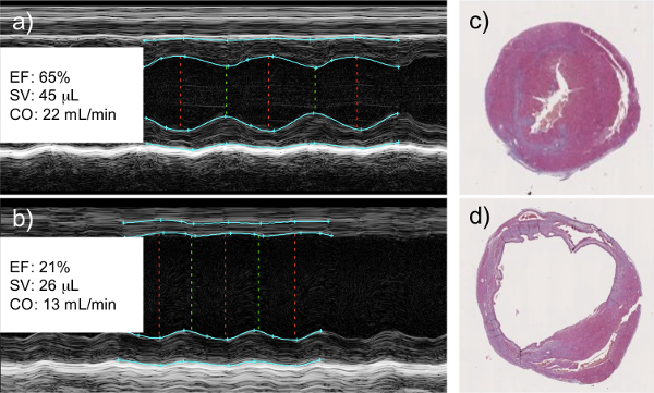
Figure 1: Left Coronary Artery Ligation Results in a Decrease in Cardiac Function as Measured by Echocardiography. M-mode echocardiography of healthy (a) and infarcted (b) hearts. Infarcted hearts show a clear lack of wall movement where viable tissue has been replaced with scar tissue. This correlates with a measureable decrease in left ventricular ejection fraction (EF), cardiac stroke volume (SV), and cardiac output (CO). Histological analysis of healthy (c) and infarcted (d) hearts shows dilation of the left ventricle and thinning of the left ventricular wall of the infarcted myocardium, both signs of tissue remodeling and scar tissue deposition. Histological images are cross sectional slices capturing the left (larger chamber) and right (smaller chamber) ventricles.
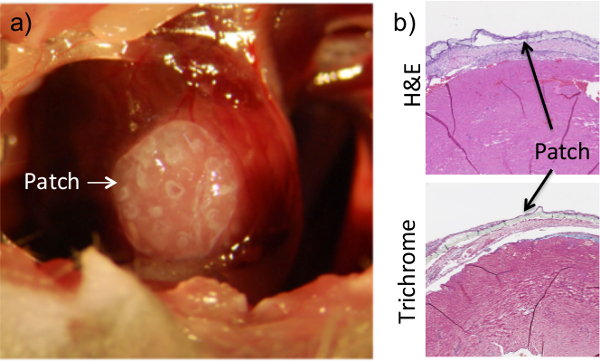
Figure 2: Application of Fibrin Flue Ensures Patch Adhesion to the Heart. The patch is placed on the surface of the heart while the chest cavity is exposed during the procedure (a). Once placed, a fibrin-based glue is added on top of the heart and allowed to dry. The patch can clearly be seen on histological sections of heart tissue that were collected at 1 week post-procedure (b). Dissociation of the patch from the heart surface in histological images is an artifact of the collection and sectioning process. Results are consistent for up to 8 weeks of adhesion.
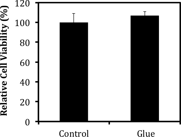
Figure 3: Fibrin-based Glue Does Not Harm Encapsulated Tissues. Cell viability was measured via MTT analysis 1 week after the administration of a fibrin-based glue using protocols described in detail in previous publications8. The viability of cells within the patch was not affected by the addition of the glue to the surface of the construct.
With this approach to the murine MI model, we have developed a system that minimizes the damage to non-myocardium areas that are associated with other murine MI techniques. These areas include damage caused by a tracheostomy, the cutting of the muscular layer, and the breakage of ribs to expose the chest cavity. We believe that these changes improve the overall surgical outcome thanks to the care taken to keep as much of the major structures, including the ribs and muscle layers, intact during the surgical procedure.
It should be noted that there are some limitations to this technique, with the largest being its time-consuming nature, resulting in only 8-10 full surgical procedures in one day. To overcome this, a second ventilator can be used, allowing for the procedure to be performed on two animals concurrently. Additionally, major vessels, such as the intercostal vessels and interior thoracic artery, should be avoided when performing the thoracotomy. If near one of these vessels, a cauterizer should be used to prevent bleeding out due to a ruptured artery.
Fibrin glue is known in both the laboratory and clinical settings thanks to its ability to quickly aid in clot formation, as well as its potential to deliver and host MSCs14-17,19. For our purposes, fibrin glue served as a biocompatible approach to adhering a water-based hydrogel construct to the surface of the heart. The use of a fibrin-based glue has proven successful in keeping the hydrogel construct at the damaged site for up to 8 weeks, with the possibility for tissue contact of a longer amount of time. The use of this adhesion system allowed control for placement of the delicate construct without the need for additional sutures that harm the myocardium as well as the construct itself. We have found that the glue itself is non-toxic to the encapsulated cells and does not negatively affect the underlying tissue. The hydrogel and fibrin-based glue system can be applied to a variety of other tissue engineering purposes that desire cell or therapeutic contact for an extended period of time. Furthermore, with the use of the SLA fabrication method, designs and structures may be incorporated within the patch to create a more complex system for cell delivery18.
One setback to the use of the fibrin-based glue is the fact that more than one researcher must be present in the surgical room during patch placement and glue addition. It is the job of the first researcher to make sure the patch remains in place and does not get lost in the chest cavity, while the second researcher mixes the solution and applies it to the patch after proper viscosity is reached. To optimize the timing of the glue delivery, it is suggested that the second researcher also tests and times the gelation of the glue prior to patch placement, since proper gelation of the glue is critical for patch adhesion.
There are a number of critical steps and error prone areas within the protocol, with the first being the induction of the myocardial infarction. It is imperative to check for signs, such as the blanching of the left ventricular wall and the enlargement of the left atrium, to ensure occlusion of the left coronary artery. This can be done prior to the addition of the stitch by simply crossing and tightening the end of the suture strings. If it is determined that the left coronary artery is not enclosed by the suture, the string can be taken out and re-applied. A second critical measure is to continue to monitor the intubation tube throughout the procedure, to ensure that it remains within the trachea and does not get pulled out mid-surgery. Finally, throughout the surgery, all attempts should be made to avoid contact with the lungs, as any contact has the potential to lead to a collapsed lung. If, during the surgery, lung collapsing occurs, simply block the outflow tube of the intubator for up to 3 sec in an attempt to re-inflate troubled regions of the lungs.
The authors have nothing to disclose.
This work was funded by US Army Grant (W81XWH-08-1-0701) and a Fellowship from Carle Foundation Hospital.
Harvard Model 687 Mouse Ventilator | Harvard Apparatus | 55-0001 | |
Inintech Biosciences LLC Dry Glass Bead Sterilizer | Fisher Scientific | NC9531961 | |
Leica MZ6 surgical microscope | Leica | ||
Cautery Kit | Gemini | GEM 5917 | |
Delicate Forceps – 0.4mm Tips Angled | Fine Science Tools | 11063-07 | |
Agricola Retractor – 3.5cm Spread | Fine Science Tools | 17005-04 | |
Spring Scissors – 2.5mm Blades Straight | Fine Science Tools | 15000-08 | |
Castroviejo Needle Holder – w/Lock Tungsten Carbide 14cm | Fine Science Tools | 12565-14 | |
Iris Scissors – Delicate Straight 10.5 cm | Fine Science Tools | 14060-10 | |
8-0 monofilament suture | Ethicon | 8730P | |
6-0 Silk suture | Ethicon | 639G | |
Thrombin | Sigma | T7009 | |
Fibrinogen | Sigma | F3879 | |
Vetbond Tissue Adhesive | 3M | 1469SB |