Using atomic force microscopy in combination with biopanning technology we created a negative and positive biopanning system to acquire antibodies against disease-specific protein variants present in any biological material, even at low concentrations. We were successful in obtaining antibodies to TDP-43 protein variants involved in Amyotrophic Lateral Sclerosis.
Because protein variants play critical roles in many diseases including TDP-43 in Amyotrophic Lateral Sclerosis (ALS), alpha-synuclein in Parkinson’s disease and beta-amyloid and tau in Alzheimer’s disease, it is critically important to develop morphology specific reagents that can selectively target these disease-specific protein variants to study the role of these variants in disease pathology and for potential diagnostic and therapeutic applications. We have developed novel atomic force microscopy (AFM) based biopanning techniques that enable isolation of reagents that selectively recognize disease-specific protein variants. There are two key phases involved in the process, the negative and positive panning phases. During the negative panning phase, phages that are reactive to off-target antigens are eliminated through multiple rounds of subtractive panning utilizing a series of carefully selected off-target antigens. A key feature in the negative panning phase is utilizing AFM imaging to monitor the process and confirm that all undesired phage particles are removed. For the positive panning phase, the target antigen of interest is fixed on a mica surface and bound phages are eluted and screened to identify phages that selectively bind the target antigen. The target protein variant does not need to be purified providing the appropriate negative panning controls have been used. Even target protein variants that are only present at very low concentrations in complex biological material can be utilized in the positive panning step. Through application of this technology, we acquired antibodies to protein variants of TDP-43 that are selectively found in human ALS brain tissue. We expect that this protocol should be applicable to generating reagents that selectively bind protein variants present in a wide variety of different biological processes and diseases.
The presence of protein variants has been implicated as a factor in the progression of many diseases including neurodegenerative diseases such as Alzheimer’s, Parkinson’s, ALS and Frontotemporal Dementia (FTD)1,2,3,4,5,6,7,8,9,10,11. Oligomeric forms of the proteins beta-amyloid and alpha-synuclein are thought to be the toxic species responsible for Alzheimer’s and Parkinson’s, respectively2,3,4,5. Aggregates of the TAR DNA-binding protein 43 (TDP-43) have been linked to ALS and FTD12,13,14. Therefore reagents such as antibodies that can selectively target the different protein variants can be powerful tools to serve as diagnostic markers and potential therapeutics. In this study, we focused on developing reagents that selectively bind variants of the TDP-43 protein implicated in ALS, however the technique outlined in this paper should be applicable to the isolation of reagents against a wide range of protein variants.
Cytoplasmic aggregation of TDP-43 has been identified as a pathological feature in ALS15,16,17,18,19. Typically TDP-43 is found in the nucleus of all cells from a normal individual, although it tends to move between the cytosol and nucleus15,17. However, in ALS aggregated forms of TDP-43 are detected in the cytoplasm of select neurons and glia with lower concentrations found in the nucleus suggesting the movement of TDP-43 from the nucleus to the cytoplasm during disease progression16,20. While aggregation of TDP-43 is found in the majority of ALS cases, it does not account for all cases since 1%-2% of total ALS cases (or 15%-20% of familial ALS cases ) are linked to mutations in the superoxide dismutase 1 (SOD1) gene15,17. Because of the important role of TDP-43 in the vast majority of ALS cases, here we focus on developing antibody based reagents that can selectively bind to TDP-43 variants that are present in human ALS brain tissue utilizing our novel AFM based biopanning techniques.
Initially we need a diverse repertoire of antibody binding domains. We combined three different phage display single chain variable domain antibody fragment (scFvs) libraries, (Tomlinson I and J and Sheets libraries21). The panning process is divided into negative and positive panning phases. Phages from the libraries are first subjected to the negative panning process during which phages reactive to multiple off-target antigens are excluded. After the completion of each round of negative panning against each off-target antigen, the process is monitored by AFM imaging to ensure that all phage binding the off-target antigens have been removed. Only after verifying by AFM imaging that all reactive phages are removed do we proceed to the next target. To isolate reagents against TDP-43 variants implicated in ALS we utilized the following negative panning antigens: 1) BSA to remove phage that bind weakly or non-specifically to proteins; 2) aggregated alpha-synuclein to remove phage that bind to generic structural elements of aggregated proteins; 3) human brain tissue homogenates to remove phage that bind to any proteins or other components present in post-mortem samples of healthy human brain tissue; 4) immunoprecipitated TDP-43 from healthy human brain to remove phage that bind to all TDP-43 forms associated with healthy human brain; and 5) immunoprecipitated TDP-43 isolated from FTD brain homogenates to remove phage that bind TDP-43 variants associated with non-ALS pathology. After removal of all phage reactive to all the off-target antigens, we then proceeded to the positive panning phase during which antibody fragments that bind the antigen of interest are isolated, in this case TDP-43 immunoprecipitated from human ALS brain tissue. These isolated antibodies may be reactive to aggregated or modified forms of TDP-43.
Conventional phage biopanning focuses mainly on the positive panning phase22,23. Usually the target of interest is immobilized, the phage library added and bound phages eluted. The phages are then amplified and added to the target again. This amplification and incubation process is usually repeated several times to increase the percentage of positive binding phage. While variations of this process have been used extensively to isolate antibody reagents against a wide range of target antigens, they generally requires large amounts of purified target antigen24,25,26,27, whereas our process requires only trace amounts of the target antigen. The protocol described here can be used to isolate reagents that selectively bind target antigens that are present at very low concentrations, without the need for purification and the panning can be performed directly against antigen present in complex tissue samples. The use of exhaustive negative panning protocols as verified by AFM ensures that clones isolated against the positive antigen should selectively bind the target even when not purified or enriched.
Kasturirangan and colleagues (2003) have carried out a similar negative and positive biopanning process to isolate antibodies reactive to oligomeric beta-amyloid using nanogram concentration of the target5. Here we expand on this process to enable the generation of reagents that selectively bind disease-specific protein variants directly from human tissue samples. In future studies we intend to further investigate not only the diagnostic value of the reagents isolated here but also assess their therapeutic relevance for treating ALS.
Overall, our novel AFM based biopanning technology should be applicable to the isolation of any disease-specific protein variant in any biological material without the need for protein purification or modification, even when the target antigen concentrations are extremely low.
1. Phage Production
2. Negative Biopanning Process
NOTE: Avoid too many freeze thaw of the phages throughout the biopanning process. Ideally, it is best to carry out as many of the negative panning steps as possible in a day. After the completion of the rounds of negative or positive panning against each target it is important to save some of the phage in case of contamination at any step.
3. Positive Panning against Immunoprecipitated TDP-43 from Individuals with ALS
4. Culturing and Phage Production of Potential Positive Clones against ALS Specific TDP-43
5. Sequencing of Potential ALS Clones
6. Screening Potential Positive Clones against ALS Specific TDP-43 Using Indirect ELISA
7. scFv Production and Screening Using Indirect ELISA
8. AFM Process
9. Immunoprecipitation of Target Antigen
10. Dot Blot Analysis
In Figure 1, the schematic demonstrates the negative panning process by which we removed phage binding off-target antigens from our library using immunotubes. We initially started with BSA since this is a common blocking agent and any phage that would react nonspecifically with this target would be problematic in future immunoassays. Next, we removed binders to aggregated alpha-synuclein to eliminate phage that are reactive with generic structures of aggregated proteins (i.e., an antibody that would be cross-reactive to aggregated alpha-synuclein, TDP-43, abeta, etc.). AFM results showed phage binding after 1 round of negative panning against the aggregated alpha-synuclein (Figure 2A) and no binding after 8 rounds (Figure 2B). We then negatively panned against healthy human brain tissue to remove phage binding the many antigens present in healthy human brain homogenate. Figure 2C showed phage binding to healthy tissue after 1 round of negative panning and no phage left binding after 10 rounds of negative panning (Figure 2D). Since the amount of healthy and FTD immunoprecipitated TDP-43 protein available for panning was low, using mica rather than immunotubes utilized less volume and therefore lowered total protein consumed (Figure 3). After 8 rounds of negative panning against healthy immunoprecipitated TDP-43, the phage was divided. Half the phage was expended in two rounds of negative panning against FTD TDP-43. The objective was to eliminate any FTD TDP-43 reactive clones (due to TDP-43’s involvement in FTD), while retaining any potential ALS TDP-43 specific clones.
For the positive panning portion (Figure 4), we also employed mica as a substrate to minimize use of material. We used the unbound phage after the FTD TDP-43 negative panning against ALS TDP-43 to acquire any ALS specific clones. We also positively panned against ALS TDP-43 twice in the event that we did not obtain clones after using the unbound phage from the negative panning against FTD TDP-43. After the entire panning process, our three elution methods yielded 154 clones from the two positive panning against ALS TDP-43 (clones that may be reactive to ALS and FTD) and 45 potential ALS specific clones (using the unbound phage from the FTD TDP-43 negative panning).
To further reduce the 45 potential ALS specific clones, the clones are sequenced and only the clones without any stop codons were considered further, except for one clone which showed up twice. This left us with 23 potential ALS specific clones. After preparing both phage and soluble scFv with these clones, they are tested in indirect ELISAs for specificity to ALS tissue. Almost all of the phages showed a preference for ALS tissue over healthy tissue (Figure 5). Similar results are obtained when comparing phage binding to ALS to FTD tissue (Figure 6). For future studies it is essential that these clones express high yields of functional scFvs, so we produced small batches of the different scFvs and carried out similar indirect ELISAs. Comparison of scFv binding to ALS tissue to both healthy (Figure 7) and FTD tissue (Figure 8) again showed selective binding to ALS tissue in almost all clones.
We used another immunoassay (dot blots) to further verify binding to ALS tissue and also to ascertain which clones are reactive in other immunological applications. Clone 2A binding to ALS tissue is shown (Figure 9). All of these clones showed promising results in either or both of the phage and scFv ELISAs. These results confirm that our AFM based biopanning process is a very powerful technique that can be used to generate reagents that selectively bind disease-specific protein variants directly from complex sources.
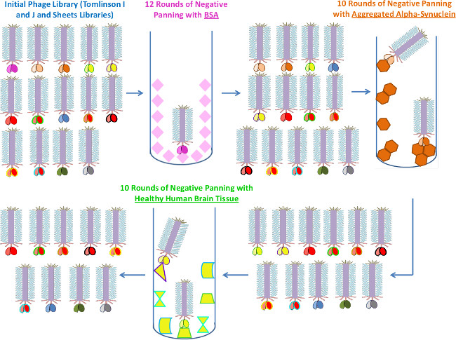
Figure 1. Negative Biopanning Process Utilizing Immunotubes. Schematic demonstrating the negative biopanning process. Phages that are reactive to proteins such as BSA, aggregated alpha-synuclein and healthy brain tissue are removed using immunotubes. Please click here to view a larger version of this figure.
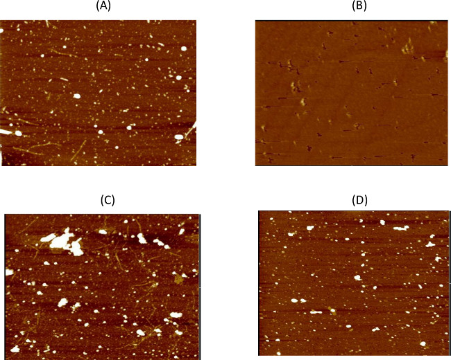
Figure 2. Confirmation of Negative Panning Results. AFM imaging is used to monitor the negative panning steps against the various targets to ensure all reactive phages are removed. Here we show some of the AFM results demonstrating the level of phage binding before and after the negative panning. (A) Phage binding can be detected to aggregated alpha-synuclein particles after the first round of negative panning, but (B) no phage are visible after 8 rounds of negative panning. (C) After the first round of negative panning against healthy tissue phage binding is evident, but (D) no binding is detected after 10 rounds of negative panning. All images are 5 μm. The large white structures on the AFM images are usually due to salts present in the buffers or residual PEG from the PEG precipitation step during phage production. Please click here to view a larger version of this figure.
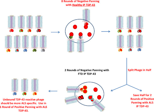
Figure 3. Negative Biopanning Process Utilizing Mica. Schematic demonstrating additional negative biopanning using mica. Due to limited sample mica surface is employed to first eliminate binders to healthy and then FTD immunoprecipitated TDP-43. Before proceeding to the two rounds of negative panning against FTD TDP-43, the phage is split in half (in the event of unsuccessful isolation of ALS TDP-43 exclusive phages during the positive panning phase). Please click here to view a larger version of this figure.
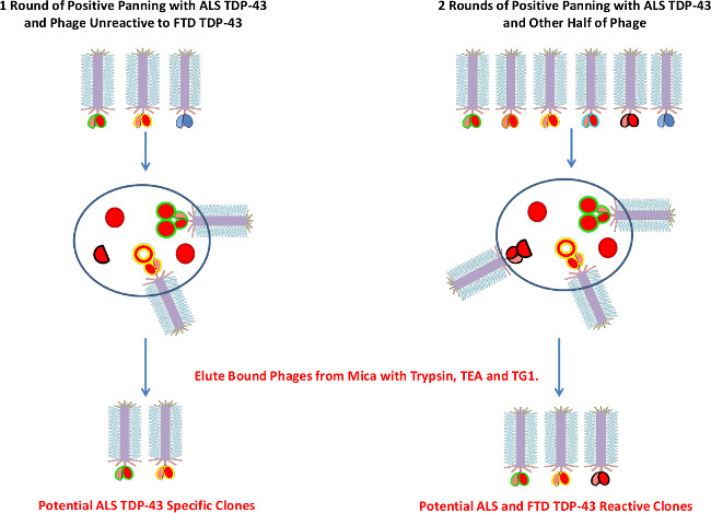
Figure 4. ALS Positive Biopanning Process. Schematic demonstrating the ALS positive biopanning process. The unbound phages after the negative panning against FTD TDP-43 are used in a round of positive panning against ALS TDP-43 to elute more ALS TDP-43 specific clones. Also, two rounds of positive panning against ALS TDP-43 are carried out using mica surface and the unbound phages after negative panning against healthy immunoprecipitated TDP-43 (the bound phages are eluted since these phages should not bind healthy TDP-43, however some may be cross-reactive with both ALS and FTD). Three elution methods are used (trypsin, TEA and TG1 cells) to ensure all bound phages are removed. Please click here to view a larger version of this figure.
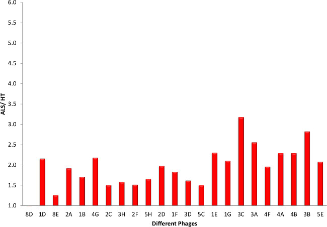
Figure 5. Phage ELISA Screening of Potential ALS Clones (ALS versus HT). Using homogenized human ALS, FTD and healthy brain tissue (HT) samples we performed indirect ELISA using phage produced from the different clones. Results are represented as the ratio to healthy tissue samples. Results showed that some clones distinguish between TDP-43 found in ALS patients and those in healthy. The ALS, FTD and HT tissues are a mix of brain samples (motor cortex) from three individuals. Please click here to view a larger version of this figure.
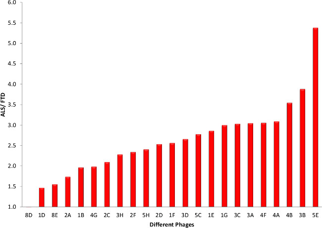
Figure 6. Phage ELISA Screening of Potential ALS Clones (ALS versus FTD). Here we show the phage ELISA results of ALS versus FTD patients. Most of the clones have a preference for ALS over FTD. Please click here to view a larger version of this figure.
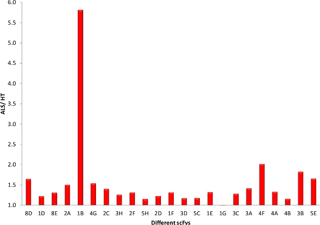
Figure 7. scFv ELISA Screening of Potential ALS Clones (ALS versus HT). Using the scFvs produced from each clone we can observe clones that have a preference for the TDP-43 from ALS patients over healthy. Please click here to view a larger version of this figure.
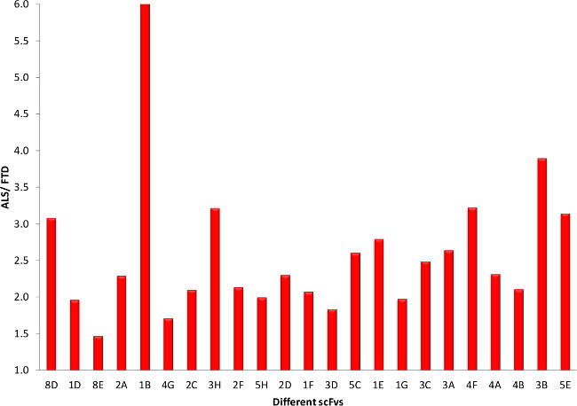
Figure 8. scFv ELISA Screening of Potential ALS Clones (ALS versus FTD). The success of our panning process is further demonstrated when comparing ALS to FTD for the different scFvs in the indirect ELISAs. Please click here to view a larger version of this figure.
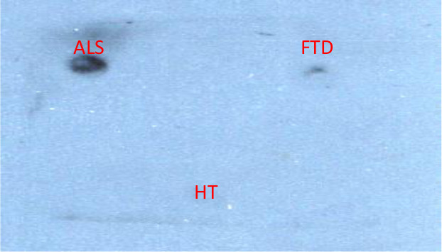
Figure 9. Dot Blot Analysis of Potential ALS Clones. Using dot blot instead of ELISA is another technique to show specificity of the clones for ALS over FTD or HT. Clone 2A is shown. Please click here to view a larger version of this figure.
Protein variants have been shown to be involved in the progression of many neurodegenerative diseases such as Alzheimer’s, Parkinson’s, ALS and FTD1,2,3,4,5,6,7,8,9,10,11. Isolation of antibodies that can selectively recognize these different protein variant targets can be effective reagents to study, diagnose and potentially treat such ailments. To generate such variant specific antibodies we have developed a novel biopanning process that utilizes atomic force microscopy to monitor the progress of each step. Previous work using a similar negative and positive biopanning combination resulted in a clone that binds oligomeric beta-amyloid5. Here we extend these studies to demonstrate that we can efficiently generate reagents that selectively bind disease-specific protein variants present in human brain tissue samples. We isolated morphology specific reagents that selectively recognize TDP-43 variants uniquely present in human ALS brain tissue13,14. Future studies necessitate characterization of the TDP-43 variants being targeted (whether aggregated or modified).
The key steps to ensuring that we can obtain phages specific to any protein variant target during the positive panning phase is to confirm that the negative panning steps have removed all potential cross-reacting phage clones. We utilized AFM imaging to verify that essentially all phage clones binding off-target antigens have been removed during each of the rounds of negative panning. Since a high percentage of the phage recovered from the positive binding are then selective for the desired target, only minimal amounts of the target antigen is needed for screening.
It is essential to perform any of the panning steps where the phages will be exposed to the external environment in a biosafety cabinet to prevent contamination by other phage. To guard against this event, it is important to save a small aliquot of phage after each panning round. If contamination is discovered, a phage aliquot from a previous round can then be used to continue the process. Freezing and thawing the phages several times may be harmful to stability and so it is beneficial to perform many of the negative panning steps continuously31. Because the panning process does not utilize phage amplification steps after each round of positive panning32 the process is less likely to isolate empty or incomplete phage sequences.
The AFM based panning process generates a number of potential protein variant specific clones after the positive panning step. To reduce the number of potential clones, we tested each clone for its ability to distinguish ALS from other diseased and healthy human brain tissue samples. The AFM based biopanning process described here represents a powerful technique that can be used to isolate reagents that selectively recognize disease-specific protein variants directly from complex human samples. The target protein variants can be present at very low concentrations, and there is no need to purify, modify or concentrate the target antigen.
The authors have nothing to disclose.
This research was supported by a grant from NIH: R21AG042066. We would like to thank Philip Schulz for his contributions in creating the screen capture videos.
Name of Reagent/ Equipment | Company | Catalog Number | Comments/Description |
Tomlinson I and J Libraries | MRC (Cambridge, England) | ||
Sheets Library | MRC (Cambridge, England) | ||
2xYT | BD Sciences | 244020 | |
Glucose | Amresco | 0188-2.5KG | |
Ampicillin | Amresco | 0339-25G | Irritant |
KM13 Helper Phage | MRC (Cambridge, England) | ||
Kanamycin | OmniPur | 5880 | Irritant |
Polyethylene Glycol 8000 | OmniPur | 6510 | Irritant |
Sodium Chloride | Macron | 7647-14-5 | |
Sodium Phosphate Dibasic | Amresco | 0404-1KG | Irritant |
Potassium Chloride | EMD | PX1405-1 | Irritant |
Potassium Phosphate Monobasic | Amresco | 0781-500G | Irritant |
TG1 Cells | MRC (Cambridge, England) | ||
Luria-Bertani Agar | EMD | 1.10283.0500 | |
Bovine Serum Albumin | Amresco | 0332-100G | |
STEN buffer | Crystalgen Inc. | 33429775 | |
Immunotubes | Thermo Scientific | 470319 | |
Mica | Spruce Pine Mica | 24365 | |
Tween 20 | EMD | ||
Trypsin | Sigma | T-0303 | Irritant |
Triethylamine | Sigma | T-0886 | Flammable |
Glycerol | Amresco | 0854-1L | Irritant |
DNA Plasmid Prep Kit | qiagen | 27106 | Irritant |
Non-Fat Milk Powder | Carnation | ||
96-Well High Binding ELISA Plate | Costar | 3590 | |
Anti-M13 HRP | GE Healthcare Life Sciences | 27-9421-01 | |
ELISA Femto Chemiluminescence Substrate Kit | Thermo Scientific | 37074 | |
Anti-TDP 43 Polyclonal Antibody | ProteinTech | 10782-2-AP | |
A/G Agarose Beads | Santa Cruz Biotechnology | sc-2003 | |
HB 2151 Cells | MRC (Cambridge, England) | ||
Isopropylthiogalactoside | Teknova | 13325 | |
9e10 HRP | Santa Cruz Biotechnology | sc-40 | |
Nitrocellulose Membrane | Biorad | 162-0115 | Flammable |
Centrifuge | Thermo Scientific | Sorvall RC 6+ | |
Nanoscope IIIa Atomic Force Microscope | Veeco | ||
AFM Probes | VistaProbes | T300R-10 |