This manuscript describes the production, characterization and potential uses of a tissue engineered 3D esophageal construct prepared from normal primary human esophageal fibroblast and squamous epithelial cells seeded within a de-cellularized porcine scaffold. The results demonstrate the formation of a mature stratified epithelium similar to the normal human esophagus.
The incidence of both esophageal adenocarcinoma and its precursor, Barrett’s Metaplasia, are rising rapidly in the western world. Furthermore esophageal adenocarcinoma generally has a poor prognosis, with little improvement in survival rates in recent years. These are difficult conditions to study and there has been a lack of suitable experimental platforms to investigate disorders of the esophageal mucosa.
A model of the human esophageal mucosa has been developed in the MacNeil laboratory which, unlike conventional 2D cell culture systems, recapitulates the cell-cell and cell-matrix interactions present in vivo and produces a mature, stratified epithelium similar to that of the normal human esophagus. Briefly, the model utilizes non-transformed normal primary human esophageal fibroblasts and epithelial cells grown within a porcine-derived acellular esophageal scaffold. Immunohistochemical characterization of this model by CK4, CK14, Ki67 and involucrin staining demonstrates appropriate recapitulation of the histology of the normal human esophageal mucosa.
This model provides a robust, biologically relevant experimental model of the human esophageal mucosa. It can easily be manipulated to investigate a number of research questions including the effectiveness of pharmacological agents and the impact of exposure to environmental factors such as alcohol, toxins, high temperature or gastro-esophageal refluxate components. The model also facilitates extended culture periods not achievable with conventional 2D cell culture, enabling, inter alia, the study of the impact of repeated exposure of a mature epithelium to the agent of interest for up to 20 days. Furthermore, a variety of cell lines, such as those derived from esophageal tumors or Barrett’s Metaplasia, can be incorporated into the model to investigate processes such as tumor invasion and drug responsiveness in a more biologically relevant environment.
The esophageal mucosa comprises a stratified, squamous epithelium above a layer of connective tissue, the lamina propria, and is one of the first sites to encounter ingested environmental stressors. Exposure to dietary toxins is implicated in the development of esophageal squamous carcinoma, while duodenogastro-esophageal reflux is a critical factor in the pathogenesis of Barrett’s Metaplasia, which is associated with increased risk of progression to esophageal adenocarcinoma. Esophageal carcinomas are the 8th most common malignant tumor in UK males and esophageal adenocarcinoma is rapidly increasing in the Western world1. Furthermore, there has been little improvement in disease prognosis, with an overall 5-year survival rate of around 15%. Consequently there is a need for experimental platforms to investigate the impact of exposure to environmental stressors on this esophageal epithelium and their potential involvement in the development of metaplasia or neoplasia.
Although immortalized or tumor cell lines allow researchers to study the response of epithelial cells to these stressors in vitro, they remain proliferative and fail to differentiate into the mature epithelial cells found on the uppermost layers of the esophageal mucosa. Furthermore, cells lines that have already undergone tumorigenesis may provide only limited information regarding the initial responses of normal cells within the epithelium to environmental factors; and this is the stage when the potential for therapeutic intervention may be highest. Finally, conventional cell culture systems fail to capture the potentially important interactions between epithelial and mesenchymal cells and between these cells and the surrounding matrix that occur within tissues in vivo.
Animal models provide a more realistic microenvironment for studying the responses of the esophageal epithelium and can incorporate the artificial induction of gastro-esophageal reflux disease2. However it can be more challenging to manipulate the environmental stressors in these models and they may not fully represent the response within the human esophagus.
Other experimental human esophageal models have been developed that utilize primary cells, immortalized cells or tumor cell lines on a collagen, or combined collagen/Matrigel, scaffold containing fibroblasts3,4. It is less labor intensive to generate these scaffolds than the acellular esophageal scaffold described in this manuscript, and these organotypic models provide a useful tool, particularly in the study of tumor invasion5,6, where tumor cell infiltration into the collagen gel can be readily observed. However these collagen gels have non-native mechanical properties and lack certain features of the original tissue, including a specific basement membrane and the appropriate surface topography. This can influence the behavior of cells resulting in, for example, poorer adhesion between the epithelium and scaffold when using a collagen gel scaffold7. As a consequence the acellular porcine esophageal scaffold was developed, with the advantage of being a more biologically realistic scaffold and thus more appropriate for use as an experimental platform. It has also been shown that it is better to incorporate primary cells into the esophageal constructs than immortalized esophageal epithelial cell lines, such as Het-1A, since these cells form a multi-layered epithelium but fail to stratify or differentiate4,7,8.
Consequently, this protocol has been adapted from a method already in use in the MacNeil laboratory for making tissue engineered skin and oral mucosa9,10 and incorporates a de-cellularized porcine esophageal scaffold combined with primary human esophageal epithelial cells and fibroblasts. This protocol produces a mature, stratified epithelium, similar to that of the normal human esophagus as demonstrated by CK4, CK14, Ki67 and involucrin staining. The resulting model provides an experimental platform to study responses to environmental stressors, and has been used effectively to investigate changes in gene expression in the esophageal epithelium in response to refluxate components11.
Human esophageal cells are obtained from patients undergoing gastric or esophageal surgery. Informed consent is obtained for the tissue to be used for research purposes, and the tissue used anonymously under the appropriate ethical approvals (SSREC 165/03, Human Research Tissue Bank Licence 12179).
1. Isolation of Human Esophageal Epithelial Cells
2. Isolation of Human Esophageal Fibroblasts
3. Preparation of the De-cellularized Esophageal Scaffold
4. Production of the Culture Media
5. Production of the Human Esophageal Mucosa Model
This manuscript describes the process required, shown in schematic form in Figure 1, to culture 3D models of the human esophageal epithelium successfully. To confirm the suitability of the model as an experimental platform histological and immunohistochemical studies have been undertaken comparing the cultured tissues with normal human esophageal squamous mucosa.
Histological assessment of the epithelium produced by the method described shows a mature, multi-layered, stratified squamous epithelium (Figure 2B) which is comparable to that observed with the normal human esophagus (Figure 2A), albeit thinner (5 to 10 layers of cells compared with 10 to 20 for the normal esophagus), with the cells becoming progressively flatter and ultimately anuclear as they migrate towards the surface.
Immunohistochemical characterization of key markers of proliferation and differentiation demonstrate that the microanatomy of the model epithelium is similar to the normal human esophageal epithelium. Comparable Ki67 expression is observed in both the native esophagus and the model epithelium, with staining restricted to a subset of cells within the basal and immediately suprabasal layers (Figures 3A and 3B). This is analogous to studies which report that less than 10% of cells generally show expression of the proliferation marker, Ki67, in the normal esophageal epithelium18. CK4 is normally expressed in stratified and columnar epithelia but is generally absent from the basal layers while CK14 is only positive in the basal layer19. In both the normal and model esophageal epithelia, CK14 is observed in all cells in the basal layer (Figures 3D and 3E) while CK4 is observed throughout the epithelium except for the two most basal layers (Figures 3G and 3H). Involucrin expression is an early marker of differentiation, expressed in the suprabasal layers of the epithelium20. Again both the normal human esophageal tissue and the model epithelium show staining that reflects this (Figures 3J and 3K).
Attempts to replace the primary esophageal epithelial cells with immortalized esophageal epithelial cells, such as Het-1A, were less successful and did not produce a valid model of the normal esophageal epithelium. A multi-layered epithelium was formed; however the proliferative marker Ki67 was detected throughout the epithelium (Figure 3C) with no expression detected for any markers of differentiation (Figures 3F, 3I and 3L), indicating that the Het-1A cells produced a hyperproliferative epithelium with no evidence of normal stratification or maturation.
However, the model has been successfully modified to incorporate tumor cells by the replacement of primary epithelial cells with either esophageal adenocarcinoma (OE33) or squamous carcinoma (OE21) cells. This demonstrates the flexibility of the model, enabling its use in investigating a range of esophageal disorders at different stages of progression through the inclusion of a number of different cell lines. It can be seen that there is a marked difference in the responses from these two cell lines. OE21 squamous carcinoma cells produces an epithelium visible on the construct as a defined yellow region (Figure 4A) with large clefts within the epithelium (Figure 4B), likely to reflect dysfunctional cell adhesion molecules. Including OE33 adenocarcinoma cells within the model results in a large amount of scaffold degradation, visible by eye as a thinning of the scaffold after 2 weeks growth at the air/liquid interface (Figure 4C) and confirmed in H+E analysis as an obvious reduction in the thickness of the scaffold in the region below the cells (Figure 4D). This is a likely to be a result of an interaction between the tumor cells and fibroblasts, since the scaffold degeneration is not observed in the absence of fibroblasts (Figures 4E and 4F). We have observed similar impacts on tumor invasion in the presence and absence of fibroblasts in equivalent melanoma models21.
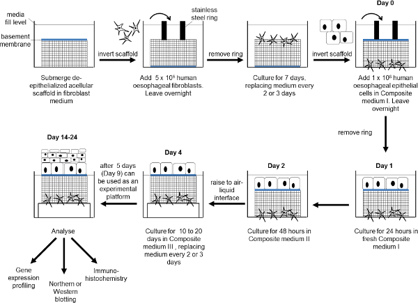
Figure 1: Schematic diagram showing the production of the esophageal mucosa model. Human esophageal fibroblasts are seeded onto the submucosal surface of the scaffold and cultured for 7 days. The scaffold is inverted, human esophageal epithelial cells added and cultured submerged for 4 days. The construct is raised to an air-liquid interface for between 10 and 20 days. At the end of the experiment the construct can undergo further analysis as required. Please click here to view a larger version of the figure.
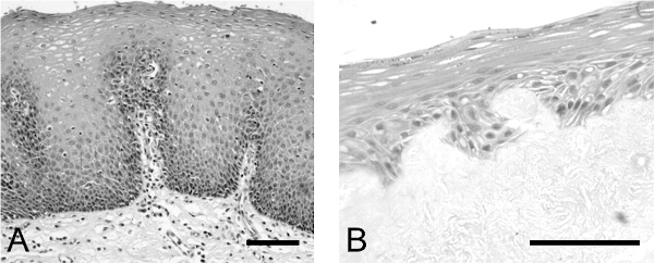
Figure 2: Comparison of epithelium produced in esophageal model with normal human esophageal epithelium. H+E analysis of (A) normal human esophageal epithelium and (B) esophageal epithelium formed in the model of the human esophageal mucosa. Scale bar is 500 µm.Please click here to view a larger version of the figure.
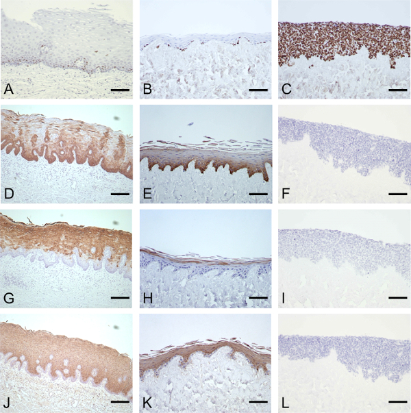
Figure 3: Immunohistochemical characterization of the esophageal epithelium produced in the model and comparison with the normal human esophagus. IHC analysis of normal esophagus (column 1) and the model esophagus produced using primary human esophageal epithelial cells (column 2) or immortalized esophageal epithelial cells (column 3). Characterization was by Ki67 (A–C), CK14 (D, E), CK4 (G, H) and involucrin staining (J, K). Scale bar is 200 µm. (This figure has been modified7.) Please click here to view a larger version of the figure.
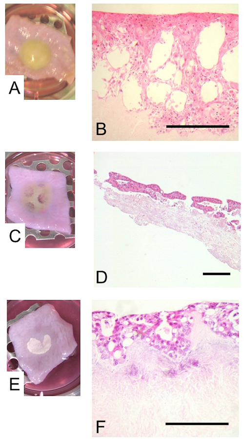
Figure 4: The inclusion of tumor cell lines within the esophageal model. The primary epithelial cells were replaced by the squamous carcinoma cell line, OE21, or the adenocarcinoma cell line, OE33. Images show the construct with the OE21 cells (A) or OE33 cells either in conjunction with (C) or in the absence of (E) fibroblasts, prior to fixing at the end of the culture period. The epithelia including the OE21 (B) or OE33 cells with (D) or without (F) fibroblasts are visualized by H+E staining. Scale bar is 500 µm. Please click here to view a larger version of the figure.
This manuscript describes the production and characterization of a biologically relevant human esophageal mucosal model suitable for use as an experimental platform to study the impact of exposure to environmental stressors upon the esophageal epithelium.
The most critical steps for the successful production of a human esophageal mucosal model are: ensuring that the majority of the epithelial cells remain proliferative and have not already begun to differentiate prior to seeding them on the scaffold; maintaining an air-liquid interface for the composite to ensure correct maturation of the epithelium; maintaining sterility throughout the extended culture period.
Due to the limited amounts of human tissue available for cell isolation it is not generally possible to obtain sufficient cells from a single tissue sample to seed freshly isolated cells directly onto the scaffold and consequently a cell expansion phase is required where the cells are grown in standard 2D cell culture conditions. It is important to ensure that cells remain proliferative during this period. This is achieved by ensuring that the cultures are never allowed to reach full confluency, using cells in the model only up to passage 4 and carefully monitoring cell morphology. Thus cells should retain their distinctive proliferative polygonal morphology and the tight “cobblestone” pavement-like appearance characteristic of proliferative epithelial cells rather than the more diffuse appearance observed in cultures which have begun to differentiate. The second critical step is controlled by ensuring that the cell culture medium covers the top surface of the stainless steel grids used to lift the cultures to air-liquid interface but the uppermost surface of the construct itself is not submerged. The third step is dependent upon strict adherence to the extended sterilization protocol when producing the de-cellularized porcine scaffold and the use of good sterile technique throughout the process.
Our results show that primary human esophageal cells are required to produce a normal, mature, stratified epithelium. If an immortalized cell line was used, albeit derived from the human esophageal epithelium, the cells remained proliferative and failed to differentiate to form a mature stratified epithelium. The resulting epithelium could not be used as a satisfactory model for the normal epithelium, demonstrating the unsuitability of the immortalized cell line Het-1A for use in the model. However other cell lines such as those derived from esophageal tumors or Barrett’s Metaplasia can be incorporated into the model either in place of or in addition to the primary epithelial cells to produce models for the study of tumor progression, invasion and the response to pharmacological agents.
Experiments can be performed using the esophageal model to simulate the exposure of the esophagus to discrete, pulsatile events during swallowing or reflux. This is achieved by repeatedly submerging the constructs in culture medium containing the compound(s) to be tested and rinsing in PBS before returning the construct to an air-liquid interface. The exposure times and frequency can be modified to reflect the process being simulated, ensuring that the epithelial layer is exposed to the environmental factor(s) while allowing the culture to continue at an air-liquid interface. This method has been used in our laboratory to investigate the impact of reflux on the normal esophageal epithelium, where the model was exposed to specific gastro-esophageal reflux components for 10 minutes twice a day for 11 days11. Alternatively the constructs can be exposed to continuous levels of environmental stressors by including these compounds in the culture medium. However, since the epithelium must be exposed to an air-liquid interface for the epithelium to mature and differentiate properly7, it is not possible to continuously submerge the constructs in the culture medium. Consequently, exposure to the stressor will only be via the submucosal surface in these experiments, which may limit the relevance of results obtained.
The technique is relatively labor intensive and consequently is better suited to the screening of relatively limited numbers of compounds. As a result, when using the model as an experimental platform, it is appropriate to first perform preliminary studies using conventional cell culture techniques to identify a limited number of conditions prior to further analysis using the more physiologically relevant esophageal model described here11. In addition to immunohistochemical analysis it has also been possible to examine changes in the epithelial gene expression profile following exposure to environmental stressors. This was achieved by manually stripping away the epithelium, extracting the RNA and converting it to cDNA to isolate the genes being expressed and analyzing by microarray to obtain gene expression profiles11. In this way it has been possible to increase the information available from the model as an experimental platform. Early changes in the epithelial gene expression profile following simulated exposure to gastro-esophageal reflux have been proposed and from these results new areas have been suggested for further investigation in the prevention of the development of Barrett’s Metaplasia, the metaplastic precursor to OAC11. The model could also be used to study protein expression using methods such as Western blot analysis15 and gene expression using Northern blot analysis16. Moreover studies of extracellular signaling molecules could be performed on the culture medium17.
The authors have nothing to disclose.
We are grateful to Mr Roger Ackroyd, Mr Andrew Wyman and Mr Chris Stoddard, Consultant Surgeons at Sheffield Teaching Hospitals NHS Foundation Trust, for their help in acquiring esophageal tissue samples and their support of our work. We thank Ashraful Haque for his help incorporating tumor cell lines into the model. We gratefully acknowledge financial support for this study by grants from the Bardhan Research and Education Trust (BRET) and Yorkshire Cancer Research (YCR).
Trypsin | BD Biosciences | 215240 | Prepare 0.1% w/v solution in PBS and filter sterilise. Warm in 37°C water bath before use |
DMEM | Labtech | LM-D1112 | Warm in 37°C water bath before use |
Ham's F12 | Labtech | LM-H1236 | Warm in 37°C water bath before use |
Foetal Calf Serum | Labtech | FB-1090 | |
Epidermal Growth Factor | R+D Systems | 236-EG-200 | Prepare 200 µg/ml stock solution in 10 mM acetic acid, 1% FCS |
Hydorcortisone | Sigma-Aldrich | H0396 | Prepare stock solution in PBS and filter sterilise before use |
Adenine | Sigma-Aldrich | A2786 | Prepare stock solution in PBS and filter sterilise before use |
Insulin | Sigma-Aldrich | I2767 | Prepare 10 mg/ml solution in 0.01M HCl, dilute 1:10 in distilled water and filter sterilise before use |
Transferrin | Sigma-Aldrich | T2036 | Prepare stock solution in distilled water and filter sterilise before use |
Triiodothyronine | Sigma-Aldrich | T2752 | Prepare stock solution in distilled water and filter sterilise before use |
Cholera toxin | Sigma-Aldrich | C8052 | Prepare stock solution in water |
L-Glutamine | Sigma-Aldrich | G7513 | |
Penicillin-Streptomycin | Sigma-Aldrich | P0781 | |
Amphotericin B | Gibco | 15290-026 | Brand name Fungizone |
PBS | Oxoid | BR0014 | Dissolve 1 tablet in 100 ml water and autoclave to sterilise |
Collagenase A | Roche | 10103578001 | |
Povidone-iodine solution | Ecolab | 10830E | Brand name Videne |
Ethanol | Sigma-Aldrich | E7023 | |
NaCl | Sigma-Aldrich | 433209 | Prepare 1M solution and autoclave to sterilise before use (121 ˚C for 15 min) |
Glycerol | Sigma-Aldrich | G2025 | Autoclave to sterilise before use (121 ˚C for 15 min) |
Chelex 100 | Sigma-Aldrich | C7901 | |
Newborn calf serum | Gibco | 26010074 | |
Progesterone | Sigma-Aldrich | P8783 | Prepare stock solution in DMEM and filter sterilise before use |
Ethanolamine | Sigma-Aldrich | E9508 | Prepare stock solution in DMEM and filter sterilise before use |
Hydrocortisone | Sigma-Aldrich | H0888 | Prepare stock solution in DMEM and filter sterilise before use use |
O-phosphorylethanolamine | Sigma-Aldrich | P0503 | Prepare stock solution in DMEM and filter sterilise before use |
ITS (insulin, transferrin, selenium) | Lonza | 17-838Z | Used for composite media preparation |
Trypsin-EDTA | Sigma-Aldrich | T3924 | Warm in 37°C water bath before use |
EDTA 0.02% solution | Sigma-Aldrich | E8008 | Warm in 37°C water bath before use |
T75 culture flask | VWR | 734-2313 | |
50 ml centrifuge tube | Fisher | 11819650 | |
15 ml universal tube | SLS | SLS7504 | |
180 ml pot | VWR | 216-2603 | |
Petri dish | SLS | 150350 | |
6 well plate | VWR | 734-2323 | |
stainless steel rings | Manufactured in house – medical grade stainless steel, internal diameter 10 mm, external diameter 20 mm | ||
steel mesh grids | Manufactured in house – sheets have 0.3 cm diameter holes, bent to produce grid 2cm (w) x2 cm (d) x 0.5 cm (h) | ||
ki67 | Novocastra | KI67-MM1-L-CE | Clone MM1 Use at 1:100 |
CK4 | Abcam | ab9004 | Clone 6B10 Use at 1:200 |
CK14 | Novocastra | LL002-L-CE | Clone LL002 Use at 1:200 |
Involucrin | Novocastra | INV | Clone SY5 Use at 1:100 |
OE21 | Sigma-Aldrich | 96062201 | |
OE33 | Sigma-Aldrich | 96070808 | |
Het-1A | ATCC-LGC | CRL-2692 | |
Mouse 3T3 fibroblasts | ATCC-LGC | CRL-1658 | previously growth arrested by irradiation (60 Gy) |