An all laser procedure is proposed in the endothelial transplant. The surgical technique is based on the use of a femtosecond laser to prepare the donor tissue. Laser welding technique is then used to secure the donor endothelium in the correct position.
The “all laser” assisted endothelial keratoplasty is a procedure that is performed with a femtosecond laser used to cut the donor tissue at an intended depth, and a near infrared diode laser to weld the corneal tissue. The proposed technique enables to reach the three main goals in endothelial keratoplasty: a precise control in the thickness of the donor tissue; its easy insertion in the recipient bed and a reduced risk of donor lenticule dislocation. The donor cornea thickness is measured in the surgery room with optical coherence tomography (OCT), in order to correctly design the donor tissue dimensions. A femtosecond laser is used to cut the donor cornea. The recipient eye is prepared by manual stripping of the descemetic membrane. The donor endothelium is inserted into a Busin-injector, the peripheral inner side is stained with a proper chromophore (a water solution of Indocyanine Green) and then it is pulled in the anterior chamber. The transplanted tissue is placed in the final and correct location and then diode laser welding is induced from outside the eyeball. The procedure has been performed on more than 15 patients evidencing an improvement in surgery performances, with a good recovery of visual acuity and a reduced donor lenticule dislocation event.
In this work we present an original approach to endothelial keratoplasty, based on the use of a femtosecond laser to prepare donor tissue and a near infrared diode laser to weld it onto the recipient bed. Intraoperative measurement of the donor cornea is necessary to correctly design the donor tissue dimensions. Endothelial keratoplasty has been proposed in the recent years to replace penetrating keratoplasty in treating endothelial disease1,2. The main advantage of this technique is a faster visual recovery, with respect to penetrating keratoplasty, reduced anesthesia during surgery, a decreased risk of graft rejection and the preservation of eye integrity. The main risk factor is postoperative donor lenticule dislocation. The standard technique is performed by inserting the donor endothelium in its final position where it is maintained by the injection of an air bubble: no sutures are used because of the mechanical, biophysical and dimensional characteristics of the endothelium. Moreover, visual acuity recovery can be limited mainly because of a mismatch between donor and recipient tissues due to a thick tissue transplanted.
Here we present a procedure in performing endothelial keratoplasty that can overcome those main problems. The donor endothelium can be assured in its final position by the use of the laser welding technique. This is a controlled and localized photothermal process: it can be induced at the donor/recipient interface. It has been studied in the last ten years and proposed in penetrating keratoplasty and in the transplant of endothelium3-5. The near infrared light (wavelength: 810 nm) emitted by a low power diode laser is delivered towards the biological tissue at the wound site. The cornea is naturally transparent to this wavelength: in order to make this tissue to absorb the laser light, it is necessary to stain it with a chromophore. The proposed dye is a sterile saturated water solution of Indocyanine Green (ICG). We demonstrated that when corneal tissue is properly stained with this ICG preparation, it shows an absorption peak at 810 nm6. Moreover, ICG is widely used in clinical diagnostics and its safety has been already demonstrated in human subjects. The stained cornea absorbs the diode laser light energy and the main resulting effect is a controlled temperature rise at the welding site. No thermal effects are induced in the unstained tissues. The temperature enhancement induces reversible thermal denaturation in the stromal collagen, with an immediate closuring of the wound walls upon cooling. This laser welding effect was firstly demonstrated in cataract surgery7,8 and penetrating keratoplasty9,10. An optimized approach that we are presenting in this paper has been studied for application in endothelial keratoplasty.
In the proposed surgery, single laser spots (lasting tens of msec) are delivered to the tissue, resulting in a photothermal effect localized within the spot dimension (a few hundreds of µm in diameter): the induced effect is a hard laser welding, consisting of a photocoagulation of the collagen confined at the donor/host interface. The result of the collagen denaturation at the welded site is a strong adhesion between the donor and host tissues, thus providing a suturing effect that is impossible to obtain with standard technique (stitches). The tissue regains his natural appearance in a short follow up (1 month) and the adhesion between donor/host tissues is improved by the welding provided in the very early stage of the healing phase.
To avoid the other main risk of the endothelial keratoplasty, i.e. the transplant of a thick donor tissue, intrasurgical optical coherence tomography (OCT) is used: a commercial device measures the thickness of donor cornea, so that a correct cut profile can be designed with the femtosec laser. The proposed “all laser” endothelial transplant thus seems to improve the clinical results of this minimally invasive surgery.
The study was conducted with the prospectively approval of the hospital’s Ethics Committee; informed consent was obtained. The study was in adherence to the tenets of the Declaration of Helsinki.
1. Donor Endothelium Preparation
2. Femtosecond Laser Preparation of Donor Endothelium
3. Recipient Eye Preparation
4. Chromophore Preparation
5. Staining of Donor Endothelium
6. Inserting the Donor Endothelium
7. Laser Welding
The “all laser” surgical procedure is proposed to perform minimally invasive corneal transplantation. The procedure is easy to perform (see Figure 1): with respect to a standard endothelial transplant only the steps of measuring the corneal thickness, staining the donor tissue and delivering the laser light are added. The achieved advantages largely compensate an increased surgical time of a few min. The use of intraoperative OCT to measure the donor cornea thickness and the femtosecond laser used to customize the donor lenticule dimensions enables the improvement of the donor/host interface adhesion (see Figure 2). In doing so, the surgery is designed following the needs and morphological characteristics of the single patient. The laser welding procedure provides an immediate closure of the donor/host interfaces4. In a standard technique, it is not possible to suture the donor tissue in any way, because of its biomechanical characteristics and location. The common postoperative risk is the donor lenticule dislocation. In our experience, donor endothelium dislocation did not occur in any of the 15 treated patients. To reach this goal it is important to deliver a complete ring of spots, covering the external diameter of the donor/recipient interface. At the beginning of the clinical trials, we performed a semicircular welding trajectory in selected patients. In one of these patients suffering from Fuch’s dystrophy with corneal deficit, a partial dislocation of the lenticule was observed (see Figure 3): interface adhesion was evident only at the welded site. For this reason, we experimented the procedure delivering a complete ring of spots, with optimized results.
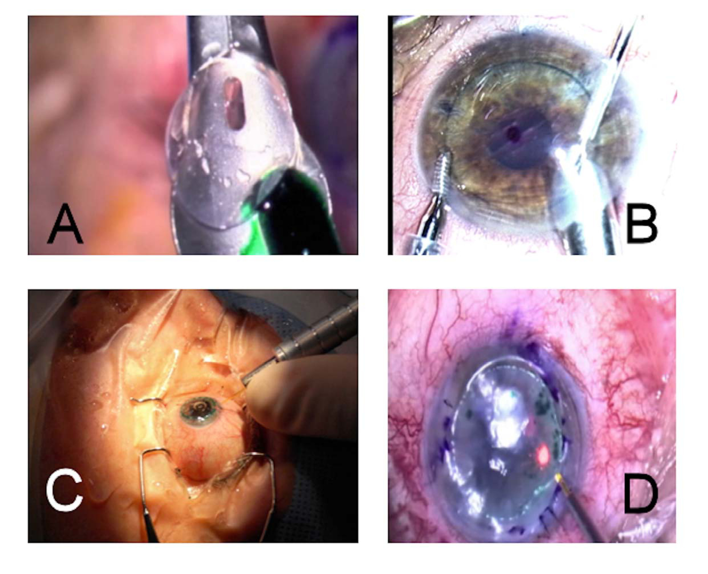
Figure 1: Endothelial Transplant. (A) The donor endothelium is put onto the injector and its inner surface is stained with a water solution of Indocyanine Green. (B) The endothelium is inserted inside the patient’s eye and positioned in its final and correct location. (C and D) Laser welding is provided from the outside, delivering single spots with a 300 µm core diameter optical fiber, mounted on a hand piece.
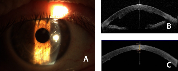
Figure 2: Postoperative results. (A) Slit lamp image of a transplanted eye, 1 week after surgery. No residual ICG is present, photothermal damage at the donor/host interface is not evident. (B) OCT image of a transplanted endothelium with the proposed “all laser” technique and without performing donor cornea thickness measurement (1 week after surgery). The transplanted endothelium is thick, with poor adhesion at the periphery. (C) OCT image of a transplanted endothelium with the proposed “all laser” technique and OCT donor cornea thickness measurement (1 week after surgery). The thickness lenticule is regular and the adhesion is good.
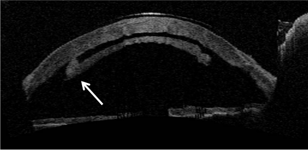
Figure 3: Laser welding efficiency. OCT image of a partially welded endothelium in a patient with Fuch’s dystrophy, 1 day after surgery. In this patient, only a portion of the endothelium was welded onto the recipient’s stroma: donor lenticule dislocation was observed the first day after surgery; this image shows the evidence that the adhesive effect was present only at the weld sites (white arrow).
The “all laser” endothelial transplant is an original approach to minimally invasive corneal transplantation.
All the procedures described within the protocol were performed in the surgery room, observing the hygienic and sterilization procedure that are common practices during surgeries, such as the use of sterilized gloves, gown, mask and cap. The ICG solution was prepared in the surgery room, soon before its application in staining the donor endothelium. The ICG powder, the water and all the tools used to prepare the staining solution were sterile and commercially available for the use in human subjects. The laser fiber optic was sterilized and it was mounted on a particular hand piece, so that it could be used under the surgical microscope.
In the proposed approach, the use of an intraoperative OCT provides a correct measurement of the donor tissue thickness. This information is used to design a personalized cut profile, with desired and reduced donor lenticule thickness, by the use of a femtosecond laser to cut the donor tissue. Laser welding procedure is provided, thus stabilizing the donor lenticule position in the recipient bed. This technique enables to reduce the donor tissue dislocation risk: to the best of our knowledge, it is the only way to suture the donor endothelium to the recipient bed.
A critical aspect of this procedure is that the ICG solution has to be prepared in the surgery room, soon before its use. This is due to the optical properties of ICG that quickly degrades. An improvement could be the realization of a ready-to-use kit. Another critical aspect is that the staining procedure introduces a difficulty in the endothelial transplant procedure, because the surgeon has to stain the inner side of the donor lenticule. Moreover, the laser welding procedure cannot be performed in donor tissue that is thinner than 50 µm.
However, the postoperative results are encouraging the dissemination of the procedure and further exploitation in other surgical fields. As it provides a method to suture thin tissues that are located in inaccessible sites, a possible application of the same procedure is in microvascular anastomosis or in the closuring of the lens capsule bag. To reach these goals, the next step in the research activities will be the standardization of the procedure, designing a platform integrating a vision system and an automated delivery system for the laser light to weld the tissue that can be adapted to different surgical scenes and target tissue.
The authors have nothing to disclose.
The authors wish to thank FORTE Project, funded by Tuscany Region (POR CReO FESR 2007-2013, Bando Unico R&S 2012), the EU FP7 ECHORD++ Experiment LA-ROSES that partially supported the research activities, and the FP7 BiophotonicPlus Project “LITE” granted by Tuscany Region.
Indocyanine Green | Pulsion Medical Systems, Germany | ICG-PULSION (http://www.pulsion.com/international-english/perfusion/icg-pulsion/) | Alternative product: IC-GREEN, Akorn Inc., Lake Forest, Illinois- US (http://www.icginjection.com/) |
Femtosecond Laser | Abbott Medical Optics, Abbott Laboratories Inc. Abbott Park, Illinois, USA | iFS150 (http://www.abbottmedicaloptics.com/products/refractive/ilasik/ifs-advanced-femtosecond-laser) | |
Optical Coherence Tomography (OCT) | Carl-Zeiss Meditec, Dublin, California- US (http://www.zeiss.com/meditec/en_de/home.html) | Visante | |
Diode Laser | E.l.En. Group s.pa., Calenzano-FI, Italy (http://www.elengroup.com/en/divisions/medical) | Mod. WELD 800 | |
Artificial Anterior Chamber | CORONET, corneal graft products. Network Medical Products Ltd. Coronet House, Kearsley Road, Ripon, North Yorkshire, HG4 2SG, UK | Artificial Anterior Chamber (A.A.C.) with large and small tissue-retaining heads. Code 51-935 (http://www.networkmedical.co.uk/ophthalmic_artificial_anterior_chamber.html) | |
Solution for tissue preservation and nutrition | AL.CHI.MI.A. Srl, Viale Austria 14, 35020 – Ponte S. Nicolò – PD ITALY |
Carry-C media for corneal deturgescence and transport at room temperature – 12 x 50 ml (http://www.alchimiasrl.com/en/organ-culture-at-31°-c-eb/carry-c-eb) |