Summary

Bedömning av neuromuskulära funktionen Använda perkutan elektrisk nervstimulering

Published: September 13, 2015
doi:

Summary

We present a protocol to assess changes in neuromuscular function. Percutaneous electrical nerve stimulation is a non-invasive method that evokes muscular responses. Electrophysiological and mechanical properties of these responses permit the evaluation of neuromuscular function from brain to muscle (supra-spinal, spinal and peripheral levels).

Abstract

Percutaneous electrical nerve stimulation is a non-invasive method commonly used to evaluate neuromuscular function from brain to muscle (supra-spinal, spinal and peripheral levels). The present protocol describes how this method can be used to stimulate the posterior tibial nerve that activates plantar flexor muscles. Percutaneous electrical nerve stimulation consists of inducing an electrical stimulus to a motor nerve to evoke a muscular response. Direct (M-wave) and/or indirect (H-reflex) electrophysiological responses can be recorded at rest using surface electromyography. Mechanical (twitch torque) responses can be quantified with a force/torque ergometer. M-wave and twitch torque reflect neuromuscular transmission and excitation-contraction coupling, whereas H-reflex provides an index of spinal excitability. EMG activity and mechanical (superimposed twitch) responses can also be recorded during maximal voluntary contractions to evaluate voluntary activation level. Percutaneous nerve stimulation provides an assessment of neuromuscular function in humans, and is highly beneficial especially for studies evaluating neuromuscular plasticity following acute (fatigue) or chronic (training/detraining) exercise.

Introduction

Perkutan elektrisk nervstimulering används ofta för att bedöma neuromuskulär funktion 1. Grundprincipen består inducera en elektrisk stimulans till en perifer motorisk nerv att framkalla en muskelsammandragning. Mekanisk (momentmätning) och elektrofysiologiska (elektromyografisk aktivitet) svar samtidigt registreras. Vridmoment, inspelad på beaktad leden, bedöms med hjälp av en ergometer. Den elektromyografisk signal (EMG) som spelats in med ytelektroder har visat sig representera aktiviteten hos muskeln 2. Denna icke-invasiv metod är inte smärtsam och lättare implementeras än intramuskulära inspelningar. Både monopolära och bipolära elektroder kan användas. Den monopolära elektrodkonfigurationen har visat sig vara mer känslig för förändringar i muskelaktivitet 3, som kan vara användbart för små muskler. Emellertid har bipolära elektroder visats vara mer effektivt för att förbättra signal-till-brus ra tio 4 och används oftast som en metod för inspelning och kvantifiera motorenhet aktivitet. Den metod som beskrivs nedan kommer att fokusera på bipolära inspelningar. EMG-aktivitet är en indikator på effektiviteten och integriteten hos neuromuskulära systemet. Användningen av perkutan nervstimulering erbjuder ytterligare insikter i neuromuskulär funktion, det vill säga förändringar på muskulös, spinal eller supra-spinal nivå (Figur 1).

Figur 1
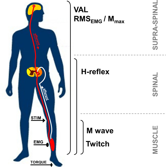
Figur 1:. Översikt över neuromuskulära mätningar Stim: nervstimulering. EMG: Elektromyografi. VAL: Frivillig aktiveringsnivå. RMS: Root Mean Square. Mmax: Maximal M-vågsamplitud.

I vila, är föreningen muskeln aktionspotentialen, även kallad M-våg på kort latens respons observerades efter stimulans artefakt, och representerar exciterbar muskelmassa genom direkt activ tion av motoriska axoner som leder till muskel (figur 2, nummer 3). M-vågsamplitud ökar med intensiteten tills den når en platå av dess maximala värde. Detta svar, som kallas M max, representerar den synkrona summering av alla motoriska enheter och / eller muskelfiberverkningspotentialer som registrerats under ytan EMG elektroderna 5. Utvecklingen av topp-till-topp-amplituden eller vågor området används för att identifiera förändringar av neuromuskulär transmission 6. Förändringar i de mekaniska svar i samband med M-vågen, det vill säga maximalt rycka vridmoment / kraft, kan bero på förändringar i muskel retbarhet och / eller inom muskelfibrerna 7. Associationen av M max amplitud och topp rycka vridmoment amplitud (Pt / M-förhållandet) ger ett index på elektromekanisk verkningsgrad muskeln 8, det vill säga mekanisk respons för en given elektrisk motor kommandot.

52974 / 52974fig2.jpg "/>
Figur 2:. Motor och reflexiva vägar aktiveras av nervstimulering Elektrisk stimulering av en blandad (motor / sensoriska) nerv (STIM) inducerar en depolarisation av både motor axon och Ia afferenta bränning. Depolarisering av la afferenter mot ryggmärgen aktiverar en alfa motoneuron, vilket i sin tur framkallar en H-reflexsvar (väg 1 + 2 + 3). Beroende på stimulusintensitet, väcker motor axon depolarisation en direkt muskelsvar: M-våg (väg 3). Vid maximal M-våg intensitet, är en antidromic ström genereras också (3 ') och kolliderar med reflex salva (2). Denna kollision helt eller delvis upphäver H-reflexen svar.

H-reflexen är en elektrofysiologisk reaktion som används för att bedöma förändringar i Ia-α motoneuron synaps 9. Denna parameter kan bedömas i vila eller under frivilliga sammandragningar. H-reflexen representerar en variant av stretchreflexen (figur 2, number 1-3). H-reflexen aktiverar motoriska enheter monosynaptically rekryterats av la afferenta vägar 10,11, och kan utsättas för perifera och centrala influenser 12. Metoden att framkalla en H-reflexen är känd för att ha en hög intraindividuella tillförlitlighet att bedöma spinal retbarhet i vila 13,14 och under isometriska kontraktioner 15.

Under en frivillig kontraktion, kan bedömas omfattningen av den frivilliga neurala disken med amplituden för EMG signalen generellt kvantifieras med användning av Root Mean Square (RMS). RMS-EMG används vanligtvis ett medel för att kvantifiera nivån av excitering av motorsystemet under frivillig kontraktion (Figur 1). På grund av den intra- och inter-individuell variabilitet 16, har RMS EMG att normaliseras med hjälp av EMG registreras under en muskelspecifikt maximal frivillig kontraktion (RMS EMGmax). Dessutom, eftersom förändringar i EMG-signalen kan be på grund av förändringar på perifer nivå, normalisering med hjälp av en perifer parameter såsom M-våg skyldig att bedöma bara den centrala delen av EMG-signal. Detta kan göras genom att dela RMS EMG av den maximala amplituden eller RMS Mmax av M-vågen. Normalisering använder RMS Mmax (dvs. RMS EMG / RMS Mmax) är att föredra eftersom det tar hänsyn till eventuella ändringar av M-våg längd 17.

Motor kommandon kan också utvärderas genom att beräkna aktiveringsnivå frivilligt (VAL). Denna metod använder rycka interpolationsteknik 18 genom att lägga en elektrisk stimulering vid M max intensitet under en maximal frivillig kontraktion. Den extra vridmoment som induceras genom att stimulera nerven jämförs med en kontrollmuskeln som produceras av samma nervstimulering i en avslappnad potentierad muskel 19. För att utvärdera maximal VAL, den ursprungliga rycka interpoförordning teknik som beskrivs av Merton 18 innebär en enda stimulus interpoleras över en frivillig kontraktion. På senare tid har användningen av parade stimulering blivit mer populärt eftersom steg framkallade vridmoment är större, lättare upptäcks, och mindre rörliga än enstaka stimuleringssvar 20. VAL ger ett index på kapaciteten hos det centrala nervsystemet till maximalt aktivera musklerna 21. För närvarande utvärderas VAL använda rycka interpolationsteknik är den mest värdefulla metoden för att bedöma graden av muskelaktivering 22. Dessutom maximala vridmomentet utvärderas med hjälp av en ergometer är den mest korrekt studerade hållfasthetsprovning parameter som gäller för användning inom forskning och kliniska miljöer 23.

Elektrisk nervstimulering kan användas i en mängd olika muskelgrupper (t.ex. armbåge flexors, handled flexors, knäextensorerna, plantar flexors). Men gör nerv tillgänglighet förteknik svårt i vissa muskler grupper. De plantar flexor muskler, särskilt triceps surae (soleus och gastrocnemii) muskler, ofta undersökts i litteraturen 24. I själva verket är dessa muskler involverade i förflyttning, motiverar sitt särskilt intresse. Avståndet mellan stimulerings webbplats och inspelning elektroder möjliggör identifiering av de olika framkallade vågor av triceps surae muskler. Den ytliga delen av den bakre tibialnerven i Poplietallymfknutor fossa och det stora antalet spindlar gör det enklare att spela in reflexsvar jämfört med andra muskler 24. Av dessa skäl fokuseras för närvarande presenteras reflex metodik på triceps surae grupp av muskler (soleus och gastrocnemius). Syftet med detta protokoll är därför att beskriva perkutan nervstimulering teknik för att undersöka neuromuskulär funktion i triceps surae.

Protocol

De experimentella förfaranden som beskrivs fått Institutionella etiskt godkännande och i enlighet med Helsingforsdeklarationen. Data samlades in från ett representativt deltagare som var medveten om de förfaranden och gav sitt skriftliga informerade samtycke. 1. Instrument Framställning Rengör huden vid elektroden plats genom rakning, och ta bort smuts med alkohol för att erhålla låg impedans (<5 kQ). Placera två AgCl ytelektroder (inspelning diameter på 1…

Representative Results

Ökande stimulusintensitet leder till en annan utveckling av respons amplituder mellan H- och M-vågor. I vila, H-reflexen når ett maximalt värde innan de helt frånvarande från EMG-signal, medan M våg ökar progressivt tills den når en platå vid maximal intensitet (se figur 4 för en grafisk bild av M-vågen och figur 6 för utvecklingen av M-vågor och H-reflexen med intensitet). För soleusmuskeln, är latensen mellan stimulans debut och M-våg ca 10 ms (figur 4A) och</…

Discussion

Perkutan nervstimulering möjliggör kvantifieringen av talrika egenskaper hos det neuromuskulära systemet, inte bara för att förstå den grundläggande kontroll av neuromotorisk funktion hos friska människor, men också för att kunna analysera akuta eller kroniska anpassningar genom utmattning eller utbildning 17. Detta är mycket fördelaktigt, särskilt för tröttande protokoll, där mätningar måste utföras så snart som möjligt efter träning slut för att undvika effekterna av snabb återhämtn…

Divulgations

The authors have nothing to disclose.

Acknowledgements

The authors have no acknowledgements.

Materials

Biodex dynamometer Biodex Medical System Inc., New York, USA www.biodex.com
MP150 Data Acquisition System Biopac Systems Inc., Goleta, USA
Acknowledge 4.1.0 software Biopac Systems Inc., Goleta, USA www.biopac.com
DS7A constant current high voltage stimulator Digitimer, Hertfordshire, UK www.digitimer.com
Silver chloride surface electrodes Control Graphique Medical, Brie-Comte-Robert, France
Computer
1 Cable for connecting the Biodex to the MP150
1 Cable for connecting the Digitimer to the MP150
1 Cable for connecting the MP150 to the computer

References

  1. Desmedt, J. E., Hainaut, K. Kinetics of myofilament activation in potentiated contraction staircase phenomenon in human skeletal muscle. Nature. 217 (5128), 529-532 (1968).
  2. Bouisset, S., Maton, B. Quantitative relationship between surface EMG and intramuscular electromyographic activity in voluntary movement. American Journal of Physical Medicine. 51 (6), 285-295 (1972).
  3. Gabriel, D. A. Effects of monopolar and bipolar electrode configurations on surface EMG spike analysis. Medical Engineering and Physics. 33 (9), 1079-1085 (2011).
  4. Merletti, R., Rainoldi, A., Farina, D. Surface electromyography for noninvasive characterization of muscle. Exercise and Sport Sciences Reviews. 29 (1), 20-25 (2001).
  5. Lepers, R. Aetiology and time course of neuromuscular fatigue during prolonged cycling exercises. Science, & Motricité. 52, 83-107 (2004).
  6. Baudry, S., Klass, M., Pasquet, B., Duchateau, J. Age related fatigability of the ankle dorsiflexor muscles during concentric and eccentric contractions. European Journal of Applied Physiology. 100 (5), 515-525 (2007).
  7. Place, N., Yamada, T., Bruton, J. D., Westerblad, H. Muscle fatigue From observations in humans to underlying mechanisms studied in intact single muscle fibres. European Journal of Applied Physiology. 110 (1), 1-15 (2010).
  8. Scaglioni, G., Narici, M. V., Maffiuletti, N. A., Pensini, M., Martin, A. Effect of ageing on the electrical and mechanical properties of human soleus motor units activated by the H reflex and M wave. The Journal of Physiology. 548 (Pt. 2), 649-661 (2003).
  9. Schieppati, M. The Hoffmann reflex a means of assessing spinal reflex excitability and its descending control in man. Progress in Neurobiology. 28 (4), 345-376 (1987).
  10. Pierrot Deseilligny, E., Burke, D. . The circuitry of the human spinal cord: its role in motor control and movement disorders. , (2005).
  11. Duclay, J., Pasquet, B., Martin, A., Duchateau, J. Specific modulation of corticospinal and spinal excitabilities during maximal voluntary isometric shortening and lengthening contractions in synergist muscles. The Journal of Physiology. 589 (Pt. 11), 2901-2916 (2011).
  12. Grosprêtre, S., Papaxanthis, C., Martin, A. Modulation of spinal excitability by a sub threshold stimulation of M1 area during muscle lengthening. Neurosciences. 263, 60-71 (2014).
  13. Mynark, R. G. Reliability of the soleus H reflex from supine to standing in young and elderly. Clinical Neurophysiology. 116 (6), 1400-1404 (2005).
  14. Palmieri, R. M., Hoffman, M. A., Ingersoll, C. D. Intersession reliability for H reflex measurements arising from the soleus peroneal and tibialis anterior musculature. The International Journal of Neuroscience. 112 (7), 841-850 (2002).
  15. Chen, Y. S., Zhou, S., Cartwright, C., Crowley, Z., Baglin, R., Wang, F. Test retest reliability of the soleus H reflex is affected by joint positions and muscle force levels. Journal of Electromyography and Kinesiology. 20 (5), 987-987 (2010).
  16. Lehman, G. J., McGill, S. M. The importance of normalization in the interpretation of surface electromyography A proof of principle. Journal of Manipulative and Physiological Therapeutics. 22 (7), 444-446 (1999).
  17. Lepers, R. Interest and limits of percutaneous nerve electrical stimulation in the evaluation of muscle fatigue. Science, & Motricité. 70 (70), 31-37 (2010).
  18. Merton, P. A. Voluntary strength and fatigue. The Journal of Physiology. 123, 553-564 (1954).
  19. Gandevia, S. C. Spinal and supraspinal factors in human muscle fatigue. Physiological Reviews. 81 (4), 1725-1789 (2001).
  20. Shield, A., Zhou, S. Assessing voluntary muscle activation with the twitch interpolation technique. Sports Medicine. 34 (4), 253-267 (2004).
  21. Rozand, V., Pageaux, B., Marcora, S. M., Papaxanthis, C., Lepers, R. Does mental exertion alter maximal muscle activation. Frontiers in Human Neuroscience. 8, 755 (2014).
  22. Place, N., Maffiuletti, N. A., Martin, A., Lepers, R. Assessment of the reliability of central and peripheral fatigue after sustained maximal voluntary contraction of the quadriceps muscle. Muscle and Nerve. 35 (4), 486-495 (2007).
  23. Kannus, P. Isokinetic evaluation of muscular performance: implications for muscle testing and rehabilitation. International Journal of Sports Medicine. 15, S11-S18 (1994).
  24. Tucker, K. J., Tuncer, M., Türker, K. S. A review of the H reflex and M wave in the human triceps surae. Human Movement Science. 24 (5-6), 667-688 (2005).
  25. Taylor, N. A., Sanders, R. H., Howick, E. I., Stanley, S. N. Static and dynamic assessment of the Biodex dynamometer. European Journal of Applied Physiology and Occupational Physiology. 62 (3), 180-188 (1991).
  26. Sale, D., Quinlan, J., Marsh, E., McComas, A. J., Belanger, A. Y. Influence of joint position on ankle plantarflexion in humans. Journal of Applied Physiology. 52 (6), 1636-1642 (1982).
  27. Cattagni, T., Martin, A., Scaglioni, G. Is spinal excitability of the triceps surae mainly affected by muscle activity or body position. Journal of Neurophysiology. 111 (12), 2525-2532 (2014).
  28. Gerilovsky, L., Tsvetinov, P., Trenkova, G. Peripheral effects on the amplitude of monopolar and bipolar H-reflex potentials from the soleus muscle. Experimental Brain Research. 76 (1), 173-181 (1989).
  29. Schieppati, M. The Hoffmann reflex a means of assessing spinal reflex excitability and its descending control in man. Progress in Neurobiology. 28 (4), 345-376 (1987).
  30. Hermens, H. J., Freriks, B., Disselhorst Klug, C., Rau, G. Development of recommendations for SEMG sensors and sensor placement procedures. Journal of Electromyography and Kinesiology. 10 (5), 361-374 (2000).
  31. Kamen, G., Sison, S. V., Du, C. C., Patten, C. Motor unit discharge behavior in older adults during maximal effort contractions. Journal of Applied Physiology. 79 (6), 1908-1913 (1995).
  32. Neyroud, D., Rüttimann, J., et al. Comparison of neuromuscular adjustments associated with sustained isometric contractions of four different muscle groups. Journal of Applied Physiology. 114, 1426-1434 (2013).
  33. Rupp, T., Girard, O., Perrey, S. Redetermination of the optimal stimulation intensity modifies resting H-reflex recovery after a sustained moderate-intensity muscle contraction. Muscle and Nerve. 41 (May), 642-650 (2010).
  34. Zehr, E. P. Considerations for use of the Hoffmann reflex in exercise studies. European Journal of Applied Physiology. 86 (6), 455-468 (2002).
  35. Gondin, J., Duclay, J., Martin, A. Soleus and gastrocnemii evoked V wave responses increase after neuromuscular electrical stimulation training. Journal of Neurophysiology. 95 (6), 3328-3335 (2006).
  36. Rochette, L., Hunter, S. K., Place, N., Lepers, R. Activation varies among the knee extensor muscles during a submaximal fatiguing contraction in the seated and supine postures. Journal of Applied Physiology. 95 (4), 1515-1522 (2003).
  37. Fuglevand, A. J., Zackowski, K. M., Huey, K. A., Enoka, R. M. Impairment of neuromuscular propagation during human fatiguing contractions at submaximal forces. The Journal of Physiology. 460, 549-572 (1993).
  38. Vandervoort, A. A., McComas, A. J. Contractile changes in opposing muscles of the human ankle joint with aging. Journal of Applied Physiology. 61 (1), 361-367 (1986).
  39. Grosprêtre, S., Martin, A. Conditioning effect of transcranial magnetic stimulation evoking motor evoked potential on V wave response. Physiological Reports. 2 (11), e12191 (2014).
  40. Allen, G. M., Gandevia, S. C., McKenzie, D. K. Reliability of measurements of muscle strength and voluntary activation using twitch interpolation. Muscle and Nerve. 18 (6), 593-600 (1995).
  41. Cooper, M. A., Herda, T. J., Walter Herda, A. A., Costa, P. B., Ryan, E. D., Cramer, J. T. The reliability of the interpolated twitch technique during submaximal and maximal isometric muscle actions. Journal of Strength and Conditioning Research. 27 (10), 2909-2913 (2013).
  42. Froyd, C., Millet, G. Y., Noakes, T. D. The development of peripheral fatigue and short term recovery during self paced high intensity exercise. The Journal of Physiology. 591 (Pt 5), 1339-1346 (2013).
  43. Pierrot Deseilligny, E., Morin, C., Bergego, C., Tankov, N. Pattern of group I fibre projections from ankle flexor and extensor muscles in man. Experimental Brain Research. 42 (3-4), 337-350 (1981).
  44. Brooke, J. D., McIlroy, W. E., et al. Modulation of H reflexes in human tibialis anterior muscle with passive movement. Brain Research. 766 (1-2), 236-239 (1997).
  45. Hultborn, H., Meunier, S., Morin, C., Pierrot Deseilligny, E. Assessing changes in presynaptic inhibition of I a fibres a study in man and the cat. The Journal of Physiology. 389, 729-756 (1987).
  46. Meunier, S., Pierrot Deseilligny, E. Cortical control of presynaptic inhibition of Ia afferents in humans. Experimental Brain Research. 119 (4), 415-426 (1998).
  47. Aymard, C., Baret, M., Katz, R., Lafitte, C., Pénicaud, A., Raoul, S. Modulation of presynaptic inhibition of la afferents during voluntary wrist flexion and extension in man. Experimental Brain Research. 137 (1), 127-131 (2001).
  48. Abbruzzese, G., Trompetto, C., Schieppati, M. The excitability of the human motor cortex increases during execution and mental imagination of sequential but not repetitive finger movements. Experimental Brain Research. 111 (3), 465-472 (1996).
  49. Garland, S. J., Klass, M., Duchateau, J. Cortical and spinal modulation of antagonist coactivation during a submaximal fatiguing contraction in humans. Journal of Neurophysiology. 99, 554-563 (2008).
  50. Rodriguez Falces, J., Place, N. Recruitment order of quadriceps motor units Femoral nerve vs direct quadriceps stimulation. European Journal of Applied Physiology. 113, 3069-3077 (2013).
  51. Rodriguez Falces, J., Maffiuletti, N. A., Place, N. Spatial distribution of motor units recruited during electrical stimulation of the quadriceps muscle versus the femoral nerve. Muscle and Nerve. 48 (November), 752-761 (2013).
  52. Bathien, N., Morin, C. Comparing variations of spinal reflexes during intensive and selective attention (author’s transl). Physiology, & Behavior. 9 (4), 533-538 (1972).
  53. Earles, D. R., Koceja, D. M., Shively, C. W. Environmental changes in soleus H reflex excitability in young and elderly subjects. The International Journal of Neuroscience. 105 (1-4), 1-13 (2000).
  54. Paquet, N., Hui Chan, C. W. Human soleus H reflex excitability is decreased by dynamic head and body tilts. Journal of Vestibular Research Equilibrium, & Orientation. 9 (5), 379-383 (1999).
  55. Miyahara, T., Hagiya, N., Ohyama, T., Nakamura, Y. Modulation of human soleus H reflex in association with voluntary clenching of the teeth. Journal of Neurophysiology. 76 (3), 2033-2041 (1996).
  56. Pinniger, G. J., Nordlund, M. M., Steele, J. R., Cresswell, a. GH reflex modulation during passive lengthening and shortening of the human triceps surae. Journal of Physiology. 534 (Pt 3), 913-923 (2001).
  57. Tallent, J., Goodall, S., Hortobágyi, T., St Clair Gibson, A., French, D. N., Howatson, G. Repeatability of corticospinal and spinal measures during lengthening and shortening contractions in the human tibialis anterior muscle). PLoS ONE. 7 (4), e35930 (2012).
  58. Grospretre, S., Martin, A. H. reflex and spinal excitability methodological considerations. Journal of Neurophysiology. 107 (6), 1649-1654 (2012).
  59. Hugon, M. Methodology of the Hoffmann reflex in man. New Developments in Electromyography and Chemical Neurophysiology. 3m, 277-293 (1973).
  60. Bigland Ritchie, B., Zijdewind, I., Thomas, C. K. Muscle fatigue induced by stimulation with and without doublets. Muscle and Nerve. 23 (9), 1348-1355 (2000).
  61. Kent Braun, J. A., Le Blanc, R. Quantitation of central activation failure during maximal voluntary contractions in humans. Muscle and Nerve. 19 (7), 861-869 (1996).
  62. Herbert, R. D., Gandevia, S. C. Twitch interpolation in human muscles mechanisms and implications for measurement of voluntary activation. Journal of Neurophysiology. 82, 2271-2283 (1999).
  63. Miller, M., Downham, D., Lexell, J. Superimposed single impulse and pulse train electrical stimulation A quantitative assessment during submaximal isometric knee extension in young healthy men. Muscle and Nerve. 22 (8), 1038-1046 (1999).
  64. Button, D. C., Behm, D. G. The effect of stimulus anticipation on the interpolated twitch technique. Journal of Sports Science and Medicine. 7 (4), 520-524 (2008).
  65. Goss, D. a., Hoffman, R. L., Clark, B. C. Utilizing Transcranial Magnetic Stimulation to Study the Human Neuromuscular System. Journal of Visualized Experiments. (59), e3387 (2012).
  66. Sartori, L., Betti, S., Castiello, U. Corticospinal excitability modulation during action observation. Journal Of Visualized Experiments: Jove. (82), 51001 (2013).
  67. Rozand, V., Lebon, F., Papaxanthis, C., Lepers, R. Does a mental training session induce neuromuscular fatigue. Medicine and Science in Sports and Exercise. 46 (10), 1981-1989 (2014).
  68. Rozand, V., Cattagni, T., Theurel, J., Martin, A., Lepers, R. Neuromuscular fatigue following isometric contractions with similar torque time integral. International Journal of Sports Medicine. 36, 35-40 (2015).
  69. Belanger, A. Y., McComas, A. J. Extent of motor unit activation during effort. Journal of Applied Physiology. 51 (5), 1131-1135 (1981).
  70. Morse, C. I., Thom, J. M., Davis, M. G., Fox, K. R., Birch, K. M., Narici, M. V. Reduced plantarflexor specific torque in the elderly is associated with a lower activation capacity. European Journal of Applied Physiology. 92 (1-2), 219-226 (2004).
  71. Dalton, B. H., McNeil, C. J., Doherty, T. J., Rice, C. L. Age related reductions in the estimated numbers of motor units are minimal in the human soleus. Muscle and Nerve. 38 (3), 1108-1115 (2008).
  72. Hunter, S. K., Todd, G., Butler, J. E., Gandevia, S. C., Taylor, J. L. Recovery from supraspinal fatigue is slowed in old adults after fatiguing maximal isometric contractions. Journal of Applied Physiology. 105 (4), 1199-1209 (2008).
  73. Jakobi, J. M., Rice, C. L. Voluntary muscle activation varies with age and muscle group. Journal of Applied Physiology. 93 (2), 457-462 (2002).
  74. Lepers, R., Millet, G. Y., Maffiuletti, N. a Effect of cycling cadence on contractile and neural properties of knee extensors. Medicine and Science in Sports and Exercise. 33 (11), 1882-1888 (2001).
  75. Duchateau, J., Hainaut, K. Isometric or dynamic training differential effects on mechanical properties of a human muscle. Journal of Applied Physiology. 56 (2), 296-301 (1984).
  76. Millet, G. Y., Martin, V., Martin, A., Vergès, S. Electrical stimulation for testing neuromuscular function From sport to pathology. European Journal of Applied Physiology. 111, 2489-2500 (2011).
  77. Cattagni, T., Scaglioni, G., Laroche, D., Van Hoecke, J., Gremeaux, V., Martin, A. Ankle muscle strength discriminates fallers from non fallers. Frontiers in Aging Neuroscience. 6, 336 (2014).
  78. Horstman, A. M., Beltman, M. J., et al. Intrinsic muscle strength and voluntary activation of both lower limbs and functional performance after stroke. Clinical Physiology and Functional Imaging. 28 (4), 251-261 (2008).
  79. Sica, R. E., Herskovits, E., Aguilera, N., Poch, G. An electrophysiological investigation of skeletal muscle in Parkinson’s disease. Journal of the Neurological Sciences. 18 (4), 411-420 (1973).
  80. Knikou, M., Mummidisetty, C. K. Locomotor Training Improves Premotoneuronal Control after Chronic Spinal Cord Injury. Journal of Neurophysiology. 111 (11), 2264-2275 (2014).
check_url/fr/52974?article_type=t

Play Video

Citer Cet Article
Rozand, V., Grosprêtre, S., Stapley, P. J., Lepers, R. Assessment of Neuromuscular Function Using Percutaneous Electrical Nerve Stimulation. J. Vis. Exp. (103), e52974, doi:10.3791/52974 (2015).

View Video