Summary

Vurdering av nevromuskulær funksjon Bruke Percutaneous elektrisk nervestimulering

Published: September 13, 2015
doi:

Summary

We present a protocol to assess changes in neuromuscular function. Percutaneous electrical nerve stimulation is a non-invasive method that evokes muscular responses. Electrophysiological and mechanical properties of these responses permit the evaluation of neuromuscular function from brain to muscle (supra-spinal, spinal and peripheral levels).

Abstract

Percutaneous electrical nerve stimulation is a non-invasive method commonly used to evaluate neuromuscular function from brain to muscle (supra-spinal, spinal and peripheral levels). The present protocol describes how this method can be used to stimulate the posterior tibial nerve that activates plantar flexor muscles. Percutaneous electrical nerve stimulation consists of inducing an electrical stimulus to a motor nerve to evoke a muscular response. Direct (M-wave) and/or indirect (H-reflex) electrophysiological responses can be recorded at rest using surface electromyography. Mechanical (twitch torque) responses can be quantified with a force/torque ergometer. M-wave and twitch torque reflect neuromuscular transmission and excitation-contraction coupling, whereas H-reflex provides an index of spinal excitability. EMG activity and mechanical (superimposed twitch) responses can also be recorded during maximal voluntary contractions to evaluate voluntary activation level. Percutaneous nerve stimulation provides an assessment of neuromuscular function in humans, and is highly beneficial especially for studies evaluating neuromuscular plasticity following acute (fatigue) or chronic (training/detraining) exercise.

Introduction

Perkutan elektrisk nervestimulering er mye brukt til å vurdere neuromuskulær funksjon 1. Grunnprinsippet består av indusere en elektrisk stimulans til en perifer motorisk nerve å fremkalle en muskelkontraksjon. Mekanisk (dreiemoment måling) og elektrofysiologiske (elektromyografi aktivitet) svar er samtidig registrert. Dreiemoment, innspilt på anses felles, er vurdert å bruke en ergometersykkel. Den elektromyografiske (EMG) signal registreres ved hjelp av overflateelektroder har vist seg å representere aktiviteten av muskelen 2. Denne ikke-invasiv metode er ikke smertefullt og lettere implementert enn intramuskulære innspillinger. Både monopolare og bipolare elektroder kan benyttes. De monopolare elektrodeutformingen har vist seg å være mer følsomme overfor forandringer i muskelaktivitet 3, som kan være nyttig for små muskler. Imidlertid har bipolare elektroder er vist å være mer effektive i å forbedre signal-til-støy-rasjo 4 og er mest brukt som en metode for opptak og kvantifisere motorenheten aktivitet. Metodikken beskrevet nedenfor vil fokusere på bipolare innspillinger. EMG-aktivitet er en indikator for effektiviteten og integriteten til det nevromuskulære system. Bruken av perkutan nervestimulering gir ytterligere innsikt i nevromuskulær funksjon, det vil si endringer på muskuløs, spinal eller supra-spinal nivå (figur 1).

Figur 1
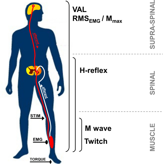
Figur 1:. Oversikt over de nevromuskulære målinger STIM: nervestimulering. EMG: Elektromyografi. VAL: Frivillig Aktivering nivå. RMS: Root Mean Square. M max: Maximal M-wave amplitude.

I hvile, er forbindelsen muskel aksjonspotensialet, også kalt M-bølge, på kort ventetid respons observert etter stimulus gjenstand, og representerer nervøs muskelmasse ved den direkte Activ asjon av motor axoner fører til muskel (Figur 2, nummer 3). M-wave amplitude øker med intensiteten til du kommer til et platå av sin maksimale verdi. Dette svaret, som kalles M max, representerer synkron summering av alle motoriske enheter og / eller muskelfiberaksjonspotensialer registrert under overflaten EMG elektroder 5. Utviklingen av topp-til-topp amplitude eller bølge område blir brukt til å identifisere endringer av neuromuskulær transmisjon 6. Endringer i de mekaniske responser forbundet med M-bølgen, dvs. topp rykk moment / kraft, kan skyldes forandringer i muskel eksitabilitet og / eller inne i muskelfibrene 7. Foreningen av M max amplitude og peak rykk dreiemoment amplitude (Pt / M ratio) gir en indeks av elektromekanisk effektiviteten av muskelen 8, dvs. mekanisk respons for en gitt elektrisk motor kommando.

52974 / 52974fig2.jpg "/>
Figur 2:. Motor og refleksive trasé aktiveres av nervestimulering Elektrisk stimulering av en blandet (motor / sensorisk) nerve (STIM) induserer en depolarisering av både motor axon og Ia afferent avfyring. Depolarisering av Ia afferente mot ryggmargen aktiverer en alfa motoneuron, som i sin tur fremkaller et H-refleks respons (bane 1 + 2 + 3). Avhengig av stimulus intensitet, fremkaller motor axon depolarization en direkte muskuløs svar: M-wave (pathway 3). Ved maksimal M-bølge intensitet, er en antidromic gjeldende også generert (3 ') og kolliderer med refleks volley (2). Denne kollisjonen helt eller delvis opphever H-refleks respons.

H-refleks er en elektrofysiologisk respons brukes til å vurdere endringer i Ia-α motoneuron synapse 9. Denne parameteren kan vurderes ved hvile eller under frivillige sammentrekninger. H-refleks representerer en variant av strekningen refleks (figur 2, number 1-3). H-refleks aktiverer motoriske enheter monosynaptically rekruttert av Ia afferente trasé 10,11, og kan bli utsatt for perifere og sentrale påvirkninger 12. Metoden for frembringer et H-refleks er kjent for å ha en høy intraindividuell pålitelighet for å vurdere spinal eksitabilitet i ro 13,14 og under isometriske kontraksjoner 15.

Under en frivillig sammentrekning, kan størrelsen av det frivillige neurale driv vurderes ved hjelp av amplituden av EMG-signalet, vanligvis kvantifisert ved hjelp av Root Mean Square (RMS). RMS EMG brukes ofte et middel for å kvantifisere nivået av magnetisering av motorsystem under frivillig kontraksjon (figur 1). På grunn av den intra- og interindividuell variasjon 16, har RMS EMG å bli normalisert ved hjelp av EMG registrert under en muskel-spesifikk maksimal frivillig kontraksjon (RMS EMGmax). I tillegg, fordi endringer i EMG-signalet kan be på grunn av endringer i perifert nivå, normalisering ved hjelp av en perifer parameter slik som M-bølgen er nødvendig for å vurdere bare den sentrale komponent av EMG-signalet. Dette kan gjøres ved å dividere RMS EMG ved maksimal amplitude eller RMS Mmax av M-bølgen. Normalisering bruker RMS Mmax (dvs. RMS EMG / RMS Mmax) er den foretrukne metoden som det tar hensyn til mulig endring av M-bølgen varighet 17.

Motoriske kommandoer kan også bli vurdert ved å beregne den frivillige aktiveringsnivå (VAL). Denne metoden bruker rykk interpolasjonsteknikk 18 ved over en elektrisk stimulering på M maks intensitet under en maksimal frivillig kontraksjon. Den ekstra dreiemoment fremkalt ved å stimulere nerve blir sammenlignet med en kontroll rykk fremstilt ved identisk nervestimulering i en avslappet forsterkes muskel 19. For å evaluere maksimal VAL, den opprinnelige rykk interposjons teknikk beskrevet av Merton 18 innebærer en enkelt stimulus interpolert over en frivillig kontraksjon. Nylig har anvendelsen av parede stimulering blitt mer populære fordi de fremkalte trinn moment er større, lettere detekteres, og mindre variabel sammenlignet med enkeltstimulerings responser 20. Den VAL gir en indeks av kapasiteten i det sentrale nervesystemet til maksimalt aktivere musklene som arbeider 21. Foreløpig VAL evaluert ved hjelp av trekning interpole teknikken er den mest verdifulle fremgangsmåte for å vurdere nivået av muskelaktivering 22. Videre maksimalt dreiemoment vurderes å bruke en ergometersykkel er det mest riktig studert styrke testing parameter som gjelder for bruk i forskning og kliniske settinger 23.

Elektrisk nervestimulering kan brukes i en rekke muskelgrupper (f.eks albue flexors, håndleddet flexors, kne extensor, plantar flexors). Men gjør nerve tilgjengelighet påTeknikken vanskelig i enkelte muskler grupper. De plantar flexor muskler, spesielt triceps surae (soleus og gastrocnemii) muskler, ofte undersøkt i litteraturen 24. Faktisk er disse musklene involvert i bevegelse, rettferdiggjør deres spesielle interesse. Avstanden mellom stimulering området og opptakselektroder muliggjør identifikasjon av de forskjellige fremkalt av bølger triceps surae de muskler. Den overfladiske del av bakre tibial nerve i popliteal fossa og det store antall spindler gjøre det lettere å ta opp refleks respons sammenlignet med andre muskler 24. Av disse grunner, fokuserer i dag presentert refleks metodikk på triceps surae gruppe av muskler (soleus og gastrocnemius). Målet med denne protokollen er derfor å beskrive perkutan nervestimulering teknikk for å undersøke nevromuskulær funksjon i triceps surae.

Protocol

De eksperimentelle prosedyrene beskrevet mottatt Institutional etisk godkjenning og er i samsvar med Helsinkideklarasjonen. Data ble samlet inn fra en representant deltaker som var klar over de prosedyrer og ga sin skriftlig informert samtykke. 1. Instrument Forberedelse Rengjør huden ved elektroden beliggenhet ved barbering, og fjerne smuss med alkohol for å oppnå lav impedans (<5 kohm). Plassere to AgCl overflateelektroder (innspilling diameter på 10 mm) ved 2/3…

Representative Results

Økende stimulus intensiteten fører til en annen utvikling av respons amplituder mellom H- og M-bølger. I hvile, når H-refleksen en maksimal verdi før de blir fullstendig fraværende fra EMG-signal, mens M bølge øker progressivt inntil nå et platå ved maksimal intensitet (se figur 4 for en grafisk fremstilling av M-bølgen og figur 6 for utviklingen av M-bølger og H-refleks med intensitet). For soleus muskelen, er det ventetid mellom stimulus utbruddet og M-bø…

Discussion

Perkutan nervestimulering muliggjør kvantifisering av mange karakteristika for det nevromuskulære system ikke bare å forstå den grunnleggende styring av neuromotor funksjon i friske mennesker, men også for å være i stand til å analysere akutte eller kroniske tilpasninger gjennom tretthet eller opplæring 17. Dette er meget fordelaktig spesielt for utmattende protokoller, hvor målinger skal utføres så snart som mulig etter trening slutt for å unngå virkningene av hurtig gjenvinning 42.

Divulgations

The authors have nothing to disclose.

Acknowledgements

The authors have no acknowledgements.

Materials

Biodex dynamometer Biodex Medical System Inc., New York, USA www.biodex.com
MP150 Data Acquisition System Biopac Systems Inc., Goleta, USA
Acknowledge 4.1.0 software Biopac Systems Inc., Goleta, USA www.biopac.com
DS7A constant current high voltage stimulator Digitimer, Hertfordshire, UK www.digitimer.com
Silver chloride surface electrodes Control Graphique Medical, Brie-Comte-Robert, France
Computer
1 Cable for connecting the Biodex to the MP150
1 Cable for connecting the Digitimer to the MP150
1 Cable for connecting the MP150 to the computer

References

  1. Desmedt, J. E., Hainaut, K. Kinetics of myofilament activation in potentiated contraction staircase phenomenon in human skeletal muscle. Nature. 217 (5128), 529-532 (1968).
  2. Bouisset, S., Maton, B. Quantitative relationship between surface EMG and intramuscular electromyographic activity in voluntary movement. American Journal of Physical Medicine. 51 (6), 285-295 (1972).
  3. Gabriel, D. A. Effects of monopolar and bipolar electrode configurations on surface EMG spike analysis. Medical Engineering and Physics. 33 (9), 1079-1085 (2011).
  4. Merletti, R., Rainoldi, A., Farina, D. Surface electromyography for noninvasive characterization of muscle. Exercise and Sport Sciences Reviews. 29 (1), 20-25 (2001).
  5. Lepers, R. Aetiology and time course of neuromuscular fatigue during prolonged cycling exercises. Science, & Motricité. 52, 83-107 (2004).
  6. Baudry, S., Klass, M., Pasquet, B., Duchateau, J. Age related fatigability of the ankle dorsiflexor muscles during concentric and eccentric contractions. European Journal of Applied Physiology. 100 (5), 515-525 (2007).
  7. Place, N., Yamada, T., Bruton, J. D., Westerblad, H. Muscle fatigue From observations in humans to underlying mechanisms studied in intact single muscle fibres. European Journal of Applied Physiology. 110 (1), 1-15 (2010).
  8. Scaglioni, G., Narici, M. V., Maffiuletti, N. A., Pensini, M., Martin, A. Effect of ageing on the electrical and mechanical properties of human soleus motor units activated by the H reflex and M wave. The Journal of Physiology. 548 (Pt. 2), 649-661 (2003).
  9. Schieppati, M. The Hoffmann reflex a means of assessing spinal reflex excitability and its descending control in man. Progress in Neurobiology. 28 (4), 345-376 (1987).
  10. Pierrot Deseilligny, E., Burke, D. . The circuitry of the human spinal cord: its role in motor control and movement disorders. , (2005).
  11. Duclay, J., Pasquet, B., Martin, A., Duchateau, J. Specific modulation of corticospinal and spinal excitabilities during maximal voluntary isometric shortening and lengthening contractions in synergist muscles. The Journal of Physiology. 589 (Pt. 11), 2901-2916 (2011).
  12. Grosprêtre, S., Papaxanthis, C., Martin, A. Modulation of spinal excitability by a sub threshold stimulation of M1 area during muscle lengthening. Neurosciences. 263, 60-71 (2014).
  13. Mynark, R. G. Reliability of the soleus H reflex from supine to standing in young and elderly. Clinical Neurophysiology. 116 (6), 1400-1404 (2005).
  14. Palmieri, R. M., Hoffman, M. A., Ingersoll, C. D. Intersession reliability for H reflex measurements arising from the soleus peroneal and tibialis anterior musculature. The International Journal of Neuroscience. 112 (7), 841-850 (2002).
  15. Chen, Y. S., Zhou, S., Cartwright, C., Crowley, Z., Baglin, R., Wang, F. Test retest reliability of the soleus H reflex is affected by joint positions and muscle force levels. Journal of Electromyography and Kinesiology. 20 (5), 987-987 (2010).
  16. Lehman, G. J., McGill, S. M. The importance of normalization in the interpretation of surface electromyography A proof of principle. Journal of Manipulative and Physiological Therapeutics. 22 (7), 444-446 (1999).
  17. Lepers, R. Interest and limits of percutaneous nerve electrical stimulation in the evaluation of muscle fatigue. Science, & Motricité. 70 (70), 31-37 (2010).
  18. Merton, P. A. Voluntary strength and fatigue. The Journal of Physiology. 123, 553-564 (1954).
  19. Gandevia, S. C. Spinal and supraspinal factors in human muscle fatigue. Physiological Reviews. 81 (4), 1725-1789 (2001).
  20. Shield, A., Zhou, S. Assessing voluntary muscle activation with the twitch interpolation technique. Sports Medicine. 34 (4), 253-267 (2004).
  21. Rozand, V., Pageaux, B., Marcora, S. M., Papaxanthis, C., Lepers, R. Does mental exertion alter maximal muscle activation. Frontiers in Human Neuroscience. 8, 755 (2014).
  22. Place, N., Maffiuletti, N. A., Martin, A., Lepers, R. Assessment of the reliability of central and peripheral fatigue after sustained maximal voluntary contraction of the quadriceps muscle. Muscle and Nerve. 35 (4), 486-495 (2007).
  23. Kannus, P. Isokinetic evaluation of muscular performance: implications for muscle testing and rehabilitation. International Journal of Sports Medicine. 15, S11-S18 (1994).
  24. Tucker, K. J., Tuncer, M., Türker, K. S. A review of the H reflex and M wave in the human triceps surae. Human Movement Science. 24 (5-6), 667-688 (2005).
  25. Taylor, N. A., Sanders, R. H., Howick, E. I., Stanley, S. N. Static and dynamic assessment of the Biodex dynamometer. European Journal of Applied Physiology and Occupational Physiology. 62 (3), 180-188 (1991).
  26. Sale, D., Quinlan, J., Marsh, E., McComas, A. J., Belanger, A. Y. Influence of joint position on ankle plantarflexion in humans. Journal of Applied Physiology. 52 (6), 1636-1642 (1982).
  27. Cattagni, T., Martin, A., Scaglioni, G. Is spinal excitability of the triceps surae mainly affected by muscle activity or body position. Journal of Neurophysiology. 111 (12), 2525-2532 (2014).
  28. Gerilovsky, L., Tsvetinov, P., Trenkova, G. Peripheral effects on the amplitude of monopolar and bipolar H-reflex potentials from the soleus muscle. Experimental Brain Research. 76 (1), 173-181 (1989).
  29. Schieppati, M. The Hoffmann reflex a means of assessing spinal reflex excitability and its descending control in man. Progress in Neurobiology. 28 (4), 345-376 (1987).
  30. Hermens, H. J., Freriks, B., Disselhorst Klug, C., Rau, G. Development of recommendations for SEMG sensors and sensor placement procedures. Journal of Electromyography and Kinesiology. 10 (5), 361-374 (2000).
  31. Kamen, G., Sison, S. V., Du, C. C., Patten, C. Motor unit discharge behavior in older adults during maximal effort contractions. Journal of Applied Physiology. 79 (6), 1908-1913 (1995).
  32. Neyroud, D., Rüttimann, J., et al. Comparison of neuromuscular adjustments associated with sustained isometric contractions of four different muscle groups. Journal of Applied Physiology. 114, 1426-1434 (2013).
  33. Rupp, T., Girard, O., Perrey, S. Redetermination of the optimal stimulation intensity modifies resting H-reflex recovery after a sustained moderate-intensity muscle contraction. Muscle and Nerve. 41 (May), 642-650 (2010).
  34. Zehr, E. P. Considerations for use of the Hoffmann reflex in exercise studies. European Journal of Applied Physiology. 86 (6), 455-468 (2002).
  35. Gondin, J., Duclay, J., Martin, A. Soleus and gastrocnemii evoked V wave responses increase after neuromuscular electrical stimulation training. Journal of Neurophysiology. 95 (6), 3328-3335 (2006).
  36. Rochette, L., Hunter, S. K., Place, N., Lepers, R. Activation varies among the knee extensor muscles during a submaximal fatiguing contraction in the seated and supine postures. Journal of Applied Physiology. 95 (4), 1515-1522 (2003).
  37. Fuglevand, A. J., Zackowski, K. M., Huey, K. A., Enoka, R. M. Impairment of neuromuscular propagation during human fatiguing contractions at submaximal forces. The Journal of Physiology. 460, 549-572 (1993).
  38. Vandervoort, A. A., McComas, A. J. Contractile changes in opposing muscles of the human ankle joint with aging. Journal of Applied Physiology. 61 (1), 361-367 (1986).
  39. Grosprêtre, S., Martin, A. Conditioning effect of transcranial magnetic stimulation evoking motor evoked potential on V wave response. Physiological Reports. 2 (11), e12191 (2014).
  40. Allen, G. M., Gandevia, S. C., McKenzie, D. K. Reliability of measurements of muscle strength and voluntary activation using twitch interpolation. Muscle and Nerve. 18 (6), 593-600 (1995).
  41. Cooper, M. A., Herda, T. J., Walter Herda, A. A., Costa, P. B., Ryan, E. D., Cramer, J. T. The reliability of the interpolated twitch technique during submaximal and maximal isometric muscle actions. Journal of Strength and Conditioning Research. 27 (10), 2909-2913 (2013).
  42. Froyd, C., Millet, G. Y., Noakes, T. D. The development of peripheral fatigue and short term recovery during self paced high intensity exercise. The Journal of Physiology. 591 (Pt 5), 1339-1346 (2013).
  43. Pierrot Deseilligny, E., Morin, C., Bergego, C., Tankov, N. Pattern of group I fibre projections from ankle flexor and extensor muscles in man. Experimental Brain Research. 42 (3-4), 337-350 (1981).
  44. Brooke, J. D., McIlroy, W. E., et al. Modulation of H reflexes in human tibialis anterior muscle with passive movement. Brain Research. 766 (1-2), 236-239 (1997).
  45. Hultborn, H., Meunier, S., Morin, C., Pierrot Deseilligny, E. Assessing changes in presynaptic inhibition of I a fibres a study in man and the cat. The Journal of Physiology. 389, 729-756 (1987).
  46. Meunier, S., Pierrot Deseilligny, E. Cortical control of presynaptic inhibition of Ia afferents in humans. Experimental Brain Research. 119 (4), 415-426 (1998).
  47. Aymard, C., Baret, M., Katz, R., Lafitte, C., Pénicaud, A., Raoul, S. Modulation of presynaptic inhibition of la afferents during voluntary wrist flexion and extension in man. Experimental Brain Research. 137 (1), 127-131 (2001).
  48. Abbruzzese, G., Trompetto, C., Schieppati, M. The excitability of the human motor cortex increases during execution and mental imagination of sequential but not repetitive finger movements. Experimental Brain Research. 111 (3), 465-472 (1996).
  49. Garland, S. J., Klass, M., Duchateau, J. Cortical and spinal modulation of antagonist coactivation during a submaximal fatiguing contraction in humans. Journal of Neurophysiology. 99, 554-563 (2008).
  50. Rodriguez Falces, J., Place, N. Recruitment order of quadriceps motor units Femoral nerve vs direct quadriceps stimulation. European Journal of Applied Physiology. 113, 3069-3077 (2013).
  51. Rodriguez Falces, J., Maffiuletti, N. A., Place, N. Spatial distribution of motor units recruited during electrical stimulation of the quadriceps muscle versus the femoral nerve. Muscle and Nerve. 48 (November), 752-761 (2013).
  52. Bathien, N., Morin, C. Comparing variations of spinal reflexes during intensive and selective attention (author’s transl). Physiology, & Behavior. 9 (4), 533-538 (1972).
  53. Earles, D. R., Koceja, D. M., Shively, C. W. Environmental changes in soleus H reflex excitability in young and elderly subjects. The International Journal of Neuroscience. 105 (1-4), 1-13 (2000).
  54. Paquet, N., Hui Chan, C. W. Human soleus H reflex excitability is decreased by dynamic head and body tilts. Journal of Vestibular Research Equilibrium, & Orientation. 9 (5), 379-383 (1999).
  55. Miyahara, T., Hagiya, N., Ohyama, T., Nakamura, Y. Modulation of human soleus H reflex in association with voluntary clenching of the teeth. Journal of Neurophysiology. 76 (3), 2033-2041 (1996).
  56. Pinniger, G. J., Nordlund, M. M., Steele, J. R., Cresswell, a. GH reflex modulation during passive lengthening and shortening of the human triceps surae. Journal of Physiology. 534 (Pt 3), 913-923 (2001).
  57. Tallent, J., Goodall, S., Hortobágyi, T., St Clair Gibson, A., French, D. N., Howatson, G. Repeatability of corticospinal and spinal measures during lengthening and shortening contractions in the human tibialis anterior muscle). PLoS ONE. 7 (4), e35930 (2012).
  58. Grospretre, S., Martin, A. H. reflex and spinal excitability methodological considerations. Journal of Neurophysiology. 107 (6), 1649-1654 (2012).
  59. Hugon, M. Methodology of the Hoffmann reflex in man. New Developments in Electromyography and Chemical Neurophysiology. 3m, 277-293 (1973).
  60. Bigland Ritchie, B., Zijdewind, I., Thomas, C. K. Muscle fatigue induced by stimulation with and without doublets. Muscle and Nerve. 23 (9), 1348-1355 (2000).
  61. Kent Braun, J. A., Le Blanc, R. Quantitation of central activation failure during maximal voluntary contractions in humans. Muscle and Nerve. 19 (7), 861-869 (1996).
  62. Herbert, R. D., Gandevia, S. C. Twitch interpolation in human muscles mechanisms and implications for measurement of voluntary activation. Journal of Neurophysiology. 82, 2271-2283 (1999).
  63. Miller, M., Downham, D., Lexell, J. Superimposed single impulse and pulse train electrical stimulation A quantitative assessment during submaximal isometric knee extension in young healthy men. Muscle and Nerve. 22 (8), 1038-1046 (1999).
  64. Button, D. C., Behm, D. G. The effect of stimulus anticipation on the interpolated twitch technique. Journal of Sports Science and Medicine. 7 (4), 520-524 (2008).
  65. Goss, D. a., Hoffman, R. L., Clark, B. C. Utilizing Transcranial Magnetic Stimulation to Study the Human Neuromuscular System. Journal of Visualized Experiments. (59), e3387 (2012).
  66. Sartori, L., Betti, S., Castiello, U. Corticospinal excitability modulation during action observation. Journal Of Visualized Experiments: Jove. (82), 51001 (2013).
  67. Rozand, V., Lebon, F., Papaxanthis, C., Lepers, R. Does a mental training session induce neuromuscular fatigue. Medicine and Science in Sports and Exercise. 46 (10), 1981-1989 (2014).
  68. Rozand, V., Cattagni, T., Theurel, J., Martin, A., Lepers, R. Neuromuscular fatigue following isometric contractions with similar torque time integral. International Journal of Sports Medicine. 36, 35-40 (2015).
  69. Belanger, A. Y., McComas, A. J. Extent of motor unit activation during effort. Journal of Applied Physiology. 51 (5), 1131-1135 (1981).
  70. Morse, C. I., Thom, J. M., Davis, M. G., Fox, K. R., Birch, K. M., Narici, M. V. Reduced plantarflexor specific torque in the elderly is associated with a lower activation capacity. European Journal of Applied Physiology. 92 (1-2), 219-226 (2004).
  71. Dalton, B. H., McNeil, C. J., Doherty, T. J., Rice, C. L. Age related reductions in the estimated numbers of motor units are minimal in the human soleus. Muscle and Nerve. 38 (3), 1108-1115 (2008).
  72. Hunter, S. K., Todd, G., Butler, J. E., Gandevia, S. C., Taylor, J. L. Recovery from supraspinal fatigue is slowed in old adults after fatiguing maximal isometric contractions. Journal of Applied Physiology. 105 (4), 1199-1209 (2008).
  73. Jakobi, J. M., Rice, C. L. Voluntary muscle activation varies with age and muscle group. Journal of Applied Physiology. 93 (2), 457-462 (2002).
  74. Lepers, R., Millet, G. Y., Maffiuletti, N. a Effect of cycling cadence on contractile and neural properties of knee extensors. Medicine and Science in Sports and Exercise. 33 (11), 1882-1888 (2001).
  75. Duchateau, J., Hainaut, K. Isometric or dynamic training differential effects on mechanical properties of a human muscle. Journal of Applied Physiology. 56 (2), 296-301 (1984).
  76. Millet, G. Y., Martin, V., Martin, A., Vergès, S. Electrical stimulation for testing neuromuscular function From sport to pathology. European Journal of Applied Physiology. 111, 2489-2500 (2011).
  77. Cattagni, T., Scaglioni, G., Laroche, D., Van Hoecke, J., Gremeaux, V., Martin, A. Ankle muscle strength discriminates fallers from non fallers. Frontiers in Aging Neuroscience. 6, 336 (2014).
  78. Horstman, A. M., Beltman, M. J., et al. Intrinsic muscle strength and voluntary activation of both lower limbs and functional performance after stroke. Clinical Physiology and Functional Imaging. 28 (4), 251-261 (2008).
  79. Sica, R. E., Herskovits, E., Aguilera, N., Poch, G. An electrophysiological investigation of skeletal muscle in Parkinson’s disease. Journal of the Neurological Sciences. 18 (4), 411-420 (1973).
  80. Knikou, M., Mummidisetty, C. K. Locomotor Training Improves Premotoneuronal Control after Chronic Spinal Cord Injury. Journal of Neurophysiology. 111 (11), 2264-2275 (2014).
check_url/fr/52974?article_type=t

Play Video

Citer Cet Article
Rozand, V., Grosprêtre, S., Stapley, P. J., Lepers, R. Assessment of Neuromuscular Function Using Percutaneous Electrical Nerve Stimulation. J. Vis. Exp. (103), e52974, doi:10.3791/52974 (2015).

View Video