RNA interference (RNAi) is an extremely valuable tool for uncovering gene function. However, the ability to target genes using RNAi during pre-adult stages is limited in the major human malaria vector Anopheles gambiae. We describe an RNAi protocol to reduce gene function via direct injection during pupal development.
RNA interference (RNAi), a naturally occurring phenomenon in eukaryotic organisms, is an extremely valuable tool that can be utilized in the laboratory for functional genomic studies. The ability to knockdown individual genes selectively via this reverse genetic technique has allowed many researchers to rapidly uncover the biological roles of numerous genes within many organisms, by evaluation of loss-of-function phenotypes. In the major human malaria vector Anopheles gambiae, the predominant method used to reduce the function of targeted genes involves injection of double-stranded (dsRNA) into the hemocoel of the adult mosquito. While this method has been successful, gene knockdown in adults excludes the functional assessment of genes that are expressed and potentially play roles during pre-adult stages, as well as genes that are expressed in limited numbers of cells in adult mosquitoes. We describe a method for the injection of Serine Protease Inhibitor 2 (SRPN2) dsRNA during the early pupal stage and validate SRPN2 protein knockdown by observing decreased target protein levels and the formation of melanotic pseudo-tumors in SRPN2 knockdown adult mosquitoes. This evident phenotype has been described previously for adult stage knockdown of SRPN2 function, and we have recapitulated this adult phenotype by SRPN2 knockdown initiated during pupal development. When used in conjunction with a dye-labeled dsRNA solution, this technique enables easy visualization by simple light microscopy of injection quality and distribution of dsRNA in the hemocoel.
Malaria is a mosquito-borne infectious disease that affects many millions of individuals every year. The World Health Organization (WHO) reports that in 2013 there were approximately 584,000 deaths due to malaria, 78 percent of which occurred in children under the age of five years1. The pathogens that cause human malaria are apicomplexan parasites within the genus Plasmodium and are transmitted between their human hosts by female Anopheles mosquitoes. Transmission occurs when the mosquito takes a blood meal from an individual who is infected, and then deposits infective parasites into an uninfected individual in a subsequent blood meal. Within the genus Anopheles, Anopheles gambiae is the species with the greatest vectorial capacity and is the most prominent malaria vector in sub-Saharan Africa1-3.
Currently, mosquito vector control by deployment of insecticides continues to be the major method employed to reduce the burden of human malaria. Although the use of insecticides since the 1960s has proven to be extremely successful, the rise of insecticide resistance has driven a need for development of novel insecticides and alternative vector control strategies4-7. During 2010, a total of 49 of 63 countries reporting to the WHO indicated the occurrence of insecticide resistance in malaria vectors1. Additionally, the IR Mapper tool, which utilizes peer-reviewed literature to assess resistance data in Afrotropical regions, reports that between 2001 and 2012 there were 46% and 27% increases in resistance to pyrethroids and dichlorodiphenyltrichloroethane (DDT), respectively7.
RNA interference (RNAi) was identified in the early 1990s as a technique that could be employed to inactivate genes in the Petunia plant8,9 and in the fungus Neurospora crassa9,10. Shortly thereafter, in 1998, RNAi was first documented in Caenorhabditis elegans as a means of reducing gene expression in an animal model by introduction of antisense or double-strand RNA (dsRNA) via injection or feeding methods9,11. Since its discovery, RNAi has revolutionized the pursuit of functional genomics by allowing researchers to utilize reverse genetics to rapidly investigate the functional roles of genes of interest via a highly selective post-transcriptional gene silencing mechanism. In some organisms, such as Drosophila melanogaster, the use of transgenic organisms that express interfering RNA constructs has been widely successful for gene knockdown (KD). Although the use of transgenes in An. gambiae for RNAi has been utilized and may prove useful for large-scale screens, the generation of transgenic mosquito strains is both labor and time intensive, generally taking two to three months to go from the identification of a gene of interest to the generation of an appropriate transgenic stock12. Currently, the primary method of gene KD in An. gambiae is by injection into the hemolymph, during the adult stage, of dsRNA specific for a given gene12,13. This process typically takes about one month to go from identification of a gene of interest to assessment of gene KD, proving to be much more rapid than transgenic methods12. Methods for larval-stage RNAi have been established recently in An. gambiae and Aedes aegypti via nanoparticle feeding14-17 or by oral delivery of microalgae-based dsRNA molecules18, offering opportunities to perform functional genomic analysis during early stages of development. In direct injection, feeding, and nanoparticle delivery methods, dsRNA is taken up autonomously by the target cell and cleaved by the enzyme Dicer into 21-25 nucleotide-long "short interfering RNAs" (siRNAs)19,20. These siRNAs are then incorporated into the RNA-induced silencing complex (RISC), from which one strand will be discarded, allowing the RNA-bound RISC complex to bind to and cleave the target mRNA and thereby reduce its level and inhibit its translation19,20.
Many intrinsic features of basic mosquito biology modulate vectorial capacity, including host preference (e.g., olfaction, gustation), mating, reproduction and immunity. Given the importance of these biological processes, it is likely that their modulation on a genetic or pharmacological level will offer new opportunities for vector control, including circumvention of insecticide resistance, and provide new tools for more broadly integrated approaches to vector management. The use of functional genomics to assess the roles of genes underlying these intrinsic biological features will enable identification of new targets and provide new insights into how we can effectively create new, more effective control strategies. We describe the development and use of a rapid injection method for initiating RNAi during the pupal stage of An. gambiae. We observe that pupal stage injection of an RNAi trigger enables observation of resultant phenotypes in early stage adults, i.e., sooner after emergence than would be observed if gene knockdown were initiated in adults post-emergence. This methods enables gene knockdown beginning during the pupal developmental interval and extending into adult stages, such that gene knockdown initiated during pupal development can persist and affect early adult hemolymph-accessible cell types, as well as cell types that are more hemolymph-accessible during metamorphosis than in the adult, such as sensory neurons found in adult appendages following emergence.
1. Synthesis and Preparation dsRNA
2. Prepare Fast Green FCF Dye (FGD) Tubes
3. Pull Injection Needles
4. Prepare Injection Station
5. Collect Pupae for Injection
6. dsRNA Injection
7. Post-injection Conditions
8. Assess Knockdown
Pupal injection for gene KD yields optimal results when injection is performed during the early pupal stage, when cuticle tanning levels are low (Figure 1A, left, and 1B). Increased tanning and hardening of cuticle, generally after 24 hours, results in increased pupal death following injection (Figure 1A, center and right). The rate of pupal development can vary depending on insectary conditions and animal density24,25; therefore, it is best to assess pigmentation visually.
During the injection process, the capillary needle is inserted into the dorsal cuticle at an angle of approximately 30° in the anterior to posterior direction (Figure 2A). Once the needle is inserted and the dsRNA + 0.01% (w/v) FGD is dispensed, the distribution of dye is evident throughout the hemolymph (Figure 2B).
Assessment of adult emergence for pupae injected with 0.01% (w/v) FGD revealed an average rate of 70% emergence, compared to 96.7% emergence of non-injected controls (Figure 3A). Of note, partial emergence from the pupal case was observed for a large number of non-surviving mosquitoes (Figure 3B). Injected animals exhibit no delays in emergence time (Figure 4A) or biased impact on either gender (Figure 4B). Additional assessment of adult survival carried out up to day 10 post-emergence reveals no evident impact on post-emergence adult survival (Figure 4C).
Validation of KD quality was assessed by the melanotic pseudo-tumor (darkly pigmented tissue cluster) phenotype associated with SRPN2 knockdown21,22 as a positive control for knockdown and the absence of phenotypes associated with dsLacZ injection as a negative control. Adult mosquitoes that emerged were scored at day 8 post-injection (day 6-7 post-emergence). Melanotic pseudo-tumors (Figure 5A and 5B) were observed through the cuticle of 93.5% of the dsSRPN2 vs. 0% of the dsLacZ adult mosquitoes (Figure 6A). Clusters of darkly melanized tissue were identified upon dissection of pigmented patches (Figure 5C). Pseudo-tumors were visible on the adult cuticle as early as day 3 post-emergence and were also present in a subset of dsSRPN2 hemolymph and gut tissues (data not shown). At 5 post-injection (early adult stage), significantly decreased SRPN2 levels in dsSRPN2, but not dsLacZ or non-injected hemolymph protein isolates was observed (Figure 6B).
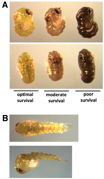
Figure 1: Developmental staging for pupal dsRNA injection. Early pupal injection of dsRNA results in optimal survival and progression into adult stage. Low levels of cuticle pigmentation (A, left, and B) can be observed within the first 0-24 hr following pupation. Tanning of the pupal cuticle preceding injection (A, center and right) results in moderate to poor survival.
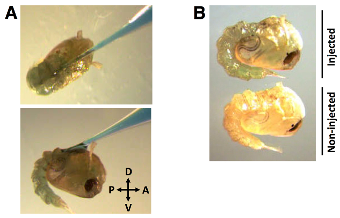
Figure 2: Injection position and distribution of dye-labeled dsRNA. (A) Capillary needle injection of dye-labeled dsRNA into the dorsal cuticle at an angle of approximately 30°, in anterior to posterior direction. (B) The dye is visibly distributed in the pupal hemolymph. dsRNA injection volume of 138 nl, labeled with 0.01% FGD (w/v).
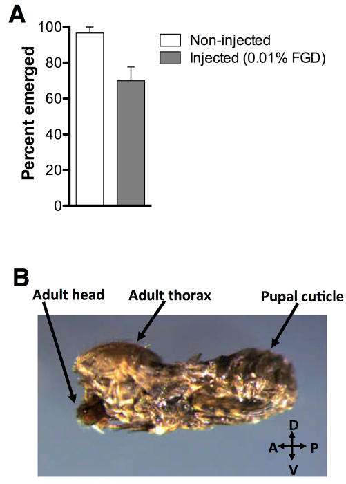
Figure 3: Post-injection adult emergence. (A) 70% of pupae injected with 0.01% FGD (w/v) successfully emerged (n = 60), compared to 96.7% of non-injected controls (n = 60). Three biological replicates were performed. (B) Partial emergence from the pupal case was observed for a large number of non-surviving mosquitoes. Error bars represent the standard error of the mean (SEM).
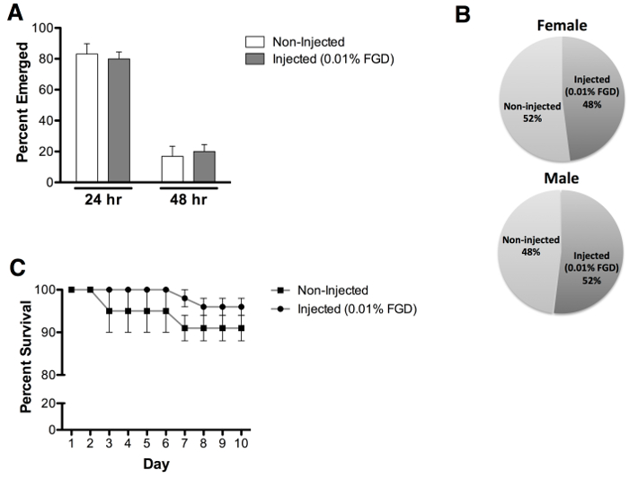
Figure 4: Emergence rate, sex assessment and adult survival. (A) Comparable emergence times were observed following pupal injection with 0.01% FGD (24 hr = 80% and 48 hr = 20%), as compared to non-injected pupae (24 hr = 83% and 48 hr = 17%). (B) Approximately equal male and female adult emergence was observed for 0.01% FGD injected pupae (female = 48% and male = 52%) and non-injected pupae (female = 52% and male = 48%). (C) Survival analysis reveals that injection with 0.01% FGD does not impact adult survival, assessed up to day 10 post-emergence. Results represent data from three independent experiments with 0.01% FGD injected (n = 60) and non-injected (n = 60) pupae (equal numbers of males and females). Error bars represent the standard error of the mean (SEM).
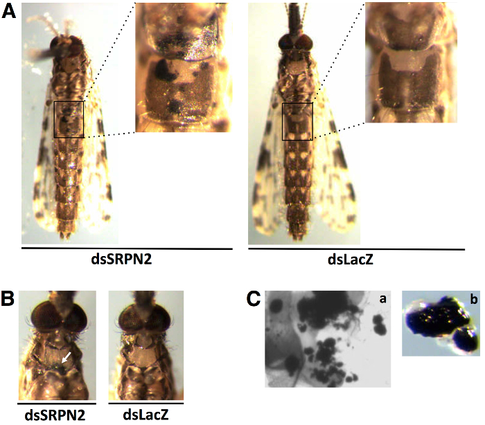
Figure 5: Pseudo-tumor positive control phenotype reflects successful knockdown. Pseudo-tumors were observed on the (A) abdominal and (B) thoracic cuticle of dsSRPN2-injected, but not dsLacZ-injected adult mosquitos at day 8 post-injection. (C) Higher magnification (400X) imaging (a) of cuticle and dissection of pigmented patches (b) reveals clusters of darkly melanized cells.
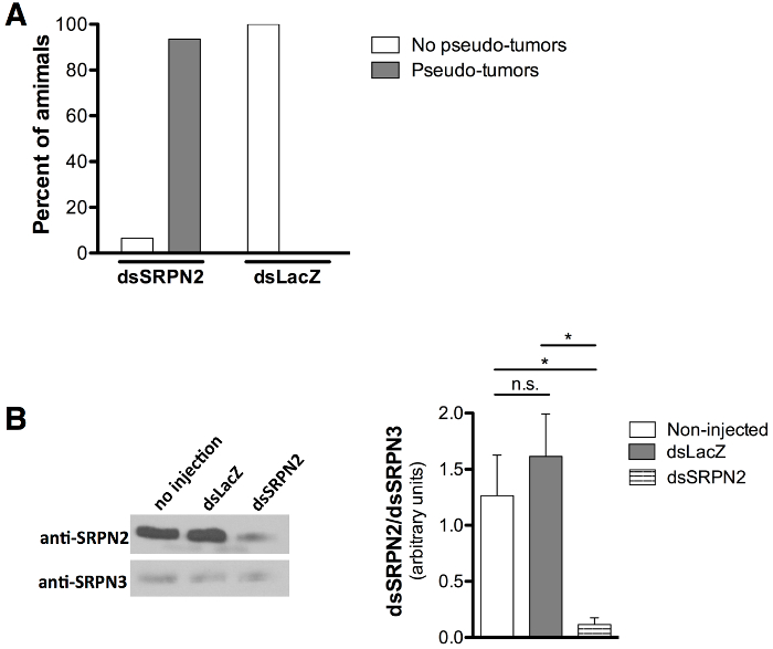
Figure 6: Quantification of pseudo-tumor formation and decreased SRPN2 protein levels. (A) Pupal stage injections result in pseudo-tumor formation in 93.5% of dsSRPN2 adults (n = 21) compared to 0% of dsLacZ controls (n = 19). Results obtained day 8 post-injection. (B) Western blot (left) shows decreased SRPN2 levels in dsSRPN2, but not dsLacZ or non-injected hemolymph protein isolates (day 5 post-injection). Results based on three independent experiments. Anti-SRPN221 and anti-SRPN326 antibody dilutions used were 1:1,000 and 1:2,000, respectively. Goat anti-rabbit IgG-HRP (Product sc-2004, Santa Cruz Biotechnology, Dallas TX) was used at 1:5,000. All protein levels were quantified (right) by band intensity (ImageJ Software, NIH, Bethesda, MD), normalized to SRPN3, and statistically compared by unpaired t test. P <0.05: *, P ≥0.05: n.s. (not significant). Error bars represent the standard error of the mean (SEM).
Current methods for inducing non-transgenic RNAi in mosquitoes involve direct injection of dsRNA into the adult hemocoel12,13 or larval feeding of RNAi trigger-coated nanoparticles14-17 or microalgae-based dsRNA molecules18. Targeting the adult mosquito, while extremely valuable, can exclude a large number of genes that function during earlier developmental periods. Knockdown initiated by larval feeding may yield inconsistent phenotypes during the adult stage due, in part, to the potential of variable protein persistence through the pupal stage. Therefore, introducing an additional method that is aimed specifically at initiating RNAi during pupal development will provide a means to more fully assess gene functions during pre-adult developmental stages, as well as enhanced abilities to assess gene function during adult stages. As with gene knockdown approach based on dsRNA injection or expression, the persistence of gene knockdown cannot be predicted. Therefore, transcript or protein levels should be assessed for gene of interest during developmental periods of interest. Although we observe a continuation of decreased protein levels at day 5 post-injection for SRPN2 in SRPN2 dsRNA-injected animals, factors such as protein turnover and half-life can differ for different targets. It is critical to ensure, as well, that the dsRNA to be used for injection is well concentrated and appears intact on an agarose gel. We recommend experimenters reassess these factors if knockdown results are not sufficient, and respective concentrations of dsRNA may need to be tested empirically for specific gene targets.
We describe a method for the initiation of RNA interference during the pupal stage of An. gambiae development. This method relies on the introduction via microinjection of dsRNA directly into the hemocoel of early pupae and allows for assessment of injection quality by the use of dye-labeled dsRNA. The ability to visualize injection quality constitutes a critical enhancement for ensuring successful knockdown and constitutes an aspect of injection-based gene knockdown that has not been considered in most previously reported dsRNA-based protocols focusing on the adult stage. By targeting the pupa at the onset of this developmental period, genes that might play a role during this critical developmental interval, or during the early stages of adulthood can be evaluated functionally. Additionally, this method may enable dsRNA delivery to cells, and establishment of RNA interference in cells that are accessible during metamorphosis, but less accessible in fully formed adult mosquitoes.
A recent microarray analysis by Harker et al. (2012) identified 560 An. gambiae transcripts that were up-regulated or down-regulated by at least 4-fold during distinct developmental stages, ranging from the embryo to adult. Of the 560 transcripts identified, a set of 309 was up-regulated during pupal development27. These findings suggest that there are many requirements for differential gene expression throughout mosquito development, including those that occur during the pupal stage, an interval during which the organism undergoes metamorphosis. In many insect species, including An. gambiae, genes involved in processes such as development (i.e., pupal cuticular and chitin-binding proteins)27-31 and immune response (i.e., Toll receptor-like proteins)27,32-34 are highly expressed during the pupal stage. Once a fully formed adult has emerged, there is continued gene expression in response to environmental and physiological changes35. Notably, during early adult development, there is an increase in the expression of developmental genes (i.e., adult cuticular and sarcoplasmic proteins)36, as well as other key genes (i.e., sperm specific protein and cytochrome P450 metabolism enzymes)27,36.
The positive control used in the development of this protocol, SRPN2, is an An. gambiae serine protease inhibitor (serpin). SRPN2 plays an important role in the negative regulation of insect melanization, a broad spectrum innate immune response in insects21,22. Knockdown of SRPN2 in adult mosquitoes results in pseudo-tumor formation21,22, a phenotype that is easily observed by use of light microscopy. Given that this distinct phenotype can be easily scored in live insects, we used SRPN2 for initial pupal stage RNAi injections. In addition, SRPN2 is expressed during all developmental stages37, thereby providing a good target for pupal stage RNAi injection and assessment of function in the early adult. We demonstrate that the method we have developed is capable of inducing similar adult melanotic pseudo-tumor formation as a consequence of dsRNA injections performed during the pupal stage of development. In developing this protocol, we have observed that injection during early pupal development (i.e., the first 24 hr after the larval-pupal molt) is critical for obtaining optimal adult emergence. In the event that poor emergence is obtained post-injection, we recommend staging larvae with greater accuracy, to obtain pupae with less extensive cuticle hardening and assure early pupal stage injection is achieved. Furthermore, minimizing damage to the cuticle results in optimal emergence rates and increasing magnification during injection can help ensure that only the intended region of the animal is punctured. If the needle should puncture through to the ventral cuticle, pooling of dye-labeled liquid will be evident on the exterior of the pupa or will saturate the filter paper. It is highly recommended to discard any pupae that have more than one cuticle puncture.
With the extensive experiences of many laboratories with the performance of adult mosquito injections, previously identified microinjection approaches can be adapted with simple protocol modifications for use in pupal RNAi experiments. Overall, the goal of this method is to provide researchers the ability to expand the timeframe during which reverse genetic analyses can be performed, further enabling research that will support the development of novel vector control strategies. Interestingly, experiments in other species, such as Rhodnius prolixus and Spodoptera frugiperda, reveal that gene silencing effects tend to be much greater when initiated during pre-adult stages38,39. During all stages of development, RNAi-mediated gene knockdown is subject to considerations regarding the rapidity and persistence of gene silencing, and the stability of proteins encoded by targeted genes. The ideal RNAi target genes tend to be those that encode a protein or RNA that has a short half-life and high turnover rate11,40.
While transgenic RNAi strategies can also be employed to address considerations regarding rapidity and persistence of RNAi during pre-adult stages, transgenic techniques have many drawbacks (e.g., time required for the generation of transgenic lines, experimental time-frames for mosquito matings to generate insects with regulated dsRNA expression, and maintenance of transgenic stocks). By contrast, our protocol affords an easier and faster method for initiating gene knockdown during pupal development and in cell types that originate and are accessible during metamorphosis but are less accessible in adults. The use of dye-labeled dsRNA suspensions allows for easy assessment of injection success and dispersal of introduced material within pupae. Our data on the onset of tumor formation in pupal-injected adults, as compared to animals injected as adults, is consistent with initiation of RNAi-mediated knockdown during the pupal stage. Using our G3 mosquito line and under our insectary conditions, we observe the adult melanotic pseudo-tumor formation as early as 10 days post-injection of adults injected three to five days post-emergence. By performing early pupal-stage injection, we observe visible melanotic pseudo-tumor formation as early as five days post-injection (i.e., three to four days post emergence). These data imply that this method enables initiation of gene knockdown during a previously under-studied developmental period (i.e., pupal development). Our dye labeling method may also prove useful for the development of new larval injection protocols, due to the translucent nature of the cuticle during all larval instars. While the control used in this study requires progression into adult stage to display a knockdown phenotype, future experiments to assess pupal stage-specific phenotypes following injection of early pupae will provide valuable insight regarding the expansion of this method into additional developmental periods such as late pupal development. In summary, this method provides a valuable RNAi protocol for pupal stage initiation of RNAi-mediated gene knockdown and expands the functional genomic tools available for use within the vector insect research community.
The authors have nothing to disclose.
We would like to thank Dr. Flaminia Catteruccia and her research group (Harvard School of Public Health, Boston, MA) for providing our laboratory with the G3 colony used in this research and for the LacZ template DNA. We thank Adam Jenkins (Boston College, Chestnut Hill, MA) for his assistance in maintaining the insectary. The Biology Department of Boston College generously funded the research associated with the development of this technique.
MEGAscript RNAi kit | Ambion | AM1626 | |
Nanoject II injector | Drummond | 3-000-204 | |
Nanoject II foot switch | Drummond | 3-000-026 | |
Borosilicate glass capillaries | Drummond | 3-000-203-G/X | |
Glass micropipette puller | Narishigne | PB-7 | |
Fast Green FCF dye | Sigma | F7258 | Can substitute with a non-toxic food dye. |
Plastic transfer pipettes | Thermo Scientific | 1371150 | ¼ inch cut from tip to create a wider opening. |
Whatman “thin” filter paper | GE Healthcare | 1001-090 | This is 9 cm diameter Grade 1, but can be altered depending on size of injection pad. |
Western blot “thick” filter paper | BioRad | 107-3931 | This is the filter paper generally used for Western blots. |
Petri dishes (60 mm x 15 mm) | Fisher Scientific | FB0875713A | |
Paint brush (size 0) | Michael’s Art | 10474940 | Brush size and brand can vary based on availability and user preference. |
Dissecting light microscope | —- | —– | This can vary based on availability and user preference. |