Summary

从背小鼠皮肤分离毛囊干细胞和表皮角质形成细胞

Published: April 29, 2016
doi:

Summary

An ideal model for studying adult stem cell biology is the mouse hair follicle. Here we present a protocol for isolating different populations of hair follicles stem cells and epidermal keratinocytes, employing enzymatic digestion of mouse dorsal skin followed by FACS analysis.

Abstract

The hair follicle (HF) is an ideal system for studying the biology and regulation of adult stem cells (SCs). This dynamic mini organ is replenished by distinct pools of SCs, which are located in the permanent portion of the HF, a region known as the bulge. These multipotent bulge SCs were initially identified as slow cycling label retaining cells; however, their isolation has been made feasible after identification of specific cell markers, such as CD34 and keratin 15 (K15). Here, we describe a robust method for isolating bulge SCs and epidermal keratinocytes from mouse HFs utilizing fluorescence activated cell-sorting (FACS) technology. Isolated hair follicle SCs (HFSCs) can be utilized in various in vivo grafting models and are a valuable in vitro model for studying the mechanisms that govern multipotency, quiescence and activation.

Introduction

成体干细胞(SCS)是用于通过替换死亡细胞维持组织稳态和在损伤修复受损的组织是至关重要的。这些雪旺由它们经受持续自我更新和分化成各种细胞谱系1-3能力来定义。最好研究的系统,这取决于他们的补给成人的SC,包括造血系统,肠和皮肤1,2,4。

在胚胎发生期间,皮肤开始作为表皮细胞的一个单层。当间质细胞填充皮肤,形成一个基本的胶原真皮5毛囊(HF)的形态开始。专业间充质细胞,后来构成真皮乳头(DP),直接组织表皮层下方,刺激上皮以形成开始向下6长出头发placodes。高度增殖基质细胞,位于在HF的底部,信封这些间充质细胞,并形成毛球,而内层开始分化成同心圆柱体,以形成毛干(HS)及周边内根鞘(IRS)2,3。

在出生后皮肤的表皮是由三个室:所述滤泡表皮(IFE),皮脂腺(SG)和HF。与此相反的IFE和SG这是在稳态的恒定状态时,HF是一个动态的微型器官而经历生长的连续周期(生长期),破坏(退化期)和其余(休止期)4,7。毛囊干细胞(HFSCs),该燃料这个永久循环,驻留在HF内的一个专门利基称为凸起4。在从DP生长期的HFSCs退出隆起,下面的启动信号,开始增殖并下降向下从而形成称为外根鞘细胞的长直线步道(ORS)8-10。基质细胞,该包围的DP在HF,快速循环的基础和迁移向上进行从而产生在HS和IRS 10( 图1)终末分化。生长期的持续时间决定了头发的长度和依赖于基质细胞6的增殖和分化的能力。当高频进入退行期,在灯泡停止过境放大基质细胞增殖,凋亡和完全消退,而向上拉动DP直到它到达的高频8,11非循环部分。在此回缩HF形成被称为上皮链,这是退行期的特征的临时结构,并且包含许多凋亡细胞。在小鼠中,退行期持续天3-4之间,并在第一毛发周期高度同步。当HF到达休止所有HFSCs变得静止。高频周期的不同阶段的特征还在于,由于到m在小鼠的皮肤的颜色变化elanin生产。从黑退行期期间生长期为深灰色期间皮肤的变化休止期6,7,12,13时为粉红色。

图1
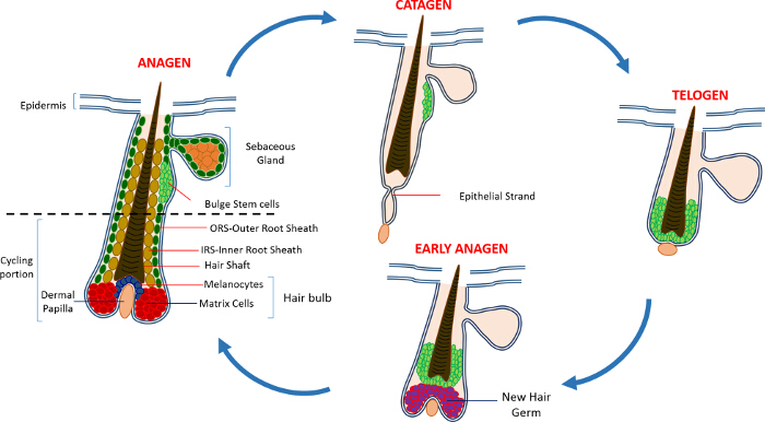
图1:毛囊周期 。在HF由永久上部,并且经历了快速增长(生长期),破坏(退行期)和相对静止阶段或休息(静止期)的连续周期不断降低重塑,单车部分。 请点击此处查看大图版本这个数字。

维持HF南海初步确定使用追踪实验,以氚化胸苷,即透露,在HF的永久居住区就在SG 14下方慢骑自行车的标签滞留细胞(LRC)的人口。在HFSC进展表征揭示少量可用于从HF利基15鉴定和分离特定的SC标记。也许HFSCs富集的最佳标记是CD34,细胞表面标记也被确定为在人16造血SC标记。在这个CD34 +人口两个不同的群体也被隔离的基础上整合α6表达式2。另一个标志物是角蛋白15(K15),其在所述凸出区域中高度表达,具有CD34的表达共定位和K15启动子被用于定位和在转基因动物15,17-19隔离HFSCs。在过去的十年中HFSCs和祖细胞的其它几个不同的种群也有报道驻留在HF 17,20-27内。

HFSCs的另一个令人兴奋的是他们的皮肤修复的贡献。在正常情况下HFSCs补充HF和不参加IFE平衡的一部分。豪版本,以应对伤人,这些细胞退出他们的SC利基,并有助于重新填充IFE 9。我们最近表明,删除小鼠促凋亡Sept4 /艺术基因显示CD34,K15和Sox9的+ HFSCs,这表明凋亡电阻的数量增加。 HFSCs从Sept4 /文理分离– / –利用背皮荧光激活细胞分选(FACS),并有在CD34 +和K15 + HFSCs的数目多于一个两倍。这些Sept4 /文理– / – HFSCs 体外扩增和不仅引起了更多的菌落,但也能够承受更苛刻的条件下与对照相比28。

作为具有HFSCs数目增加的结果,Sept4 /文理– / –小鼠显著更快响应于皮肤切除损伤愈合。引人注目的是,Sept4 /艺术– / –小鼠displayeDA大量从伤口床再生HFS和显著较小的疤痕。此外,对于XIAP(细胞凋亡的X连锁抑制 ​​剂),的文理生化靶,缺失的小鼠表现出受损的愈合28。

我们的结果及其他实验室进行工作已经表明,HFSCs作为研究成人的SC的生物学和功能的理想模型。在这里,我们描述了用于浓缩和HFSCs并基于四种标志物的表达表皮角化细胞的分离方法:整合α6;整合β1;的Sca-1(对于表皮角化细胞的标记)和CD34。也可以使用K15-GFP报告鼠标19执行K15 + HFSCs的类似的隔离。

Protocol

本研究是在严格按照指南中列出了以色列卫生部实验动物的护理和使用的建议进行的。所有动物均按照批准的机构动物护理协议技术的以色列Technion工业研究所的IL-02302-2015处理。 1.实验准备螯合胎牛血清的制备注意:上皮细胞对钙非常敏感,所以关键是要控制这些细胞对钙的曝光。为了确保细胞不分离过程螯合期间暴露于钙被用来从胎牛血清用于制备染色缓冲液<…

Representative Results

该协议详细描述了充实和两类人群的隔离:隆起种姓和表皮角质形成图2说明了协议的主要步骤。利用皮肤从背垫层8周龄小鼠的去除,我们富集使用CD34标志物,这是只在HFSCs表示隆起旺;和的Sca-1,其标签表皮角化细胞。 图3示出了皮肤上皮细胞的α6+ /β1+群体内的CD34和的Sca-1的表达的不同的模式。细胞根据整合素α6的表达第一选通<su…

Discussion

这里所描述的协议是公认的用于从成年小鼠的背部皮肤隔离HFSCs但可以同样地适用于高频结构内其他人群的隔离,基于标记2,16,23,28,29的选择。这种方法是在细胞分离的其他方法,如组织离解特别有利的,在一个特定的细胞类型,可以选择并从异质细胞群体的混合物收获。此外,这里所描述的方法是快速,可靠和可用​​于高达隔离到基于所使用的标记物的差异表达水平四个不同HFSC种群。?…

Divulgations

The authors have nothing to disclose.

Acknowledgements

This work was supported in part by NIH grant RO1GM60124 (to H.S.). H.S. is an Investigator with the Howard Hughes Medical Institute. Y.F. is supported by the Deloro Career Advancement Chair and The German Israeli Foundation (I-2381-412.13/2015). D.S. is supported by the Coleman-Cohen post-doctoral fellowship.

Materials

Isoflurane  Primal Critical Care  66794-017-10
Carbon dioxide
Electro Shaver Oster  Golden A5 Shaver from any other company could be used
70% ethanol Gadot Lab 830000051 96% ehtanol diluted with distilled water 
Dissection mat Dissection tools from any provider can be used
Forceps Dumont 11251-10 Foreceps from any other company could be used
Scissors  Dumont 14094-11 Scissors from any other company could be used
Needles/Pins
Scalpel Albion 10 Ensure that the scalpel has a blunt end
Tissue culture dish 60mm x 15mm Sigma-Aldrich CLS430166
PBS In-house PBS without Calcium and Magnesium
0.25% Trypsin/EDTA Biological Industries 03-050-1A Trypsin obtained from a different company might have a different activity and duration of the trypsin digest has to be adjusted accordingly
Pipettes 10ml Sigma-Aldrich Corning, 4488
Ice
50 ml sterie centrifuge tubes Minplast Ein-shemer 35050-43
70µM Cell strainer Fisher 22362548
40µM Cell strainer Fisher 22362549
Staining buffer
Centrifuge Eppendorf 5804 R 5805 000.017
FACS tubes with Cell strainer caps  Falcon  352235
FACS tubes  Falcon  352063
Integrin β1 eBioscience 25-0291 1:400
Integrin α6 eBioscience 15-0495 1:600
Sca I eBioscience 11-5981 1:200
CD34 eBioscience 9011-0349 1:300
DAPI Sigma-Aldrich D9542 50ng/ml
Dry Chelex BioRad 142-2842
Beaker  Pyrex
Distilled H2O 
Stir bar
NHCl BioLab 1903059
Fetal bovine serum (FBS) Beit Haemek Biological Industries 400718 FBS obtained from a different company can be used
1L glass bottle Ilmabor Boro 3.3
Bottle top filter Autofil 1102-RLS

References

  1. Wagers, A. J., Weissman, I. L. Plasticity of adult stem cells. Cell. 116, 639-648 (2004).
  2. Blanpain, C., Lowry, W. E., Geoghegan, A., Polak, L., Fuchs, E. Self-renewal, multipotency, and the existence of two cell populations within an epithelial stem cell niche. Cell. 118, 635-648 (2004).
  3. Fuchs, E. Scratching the surface of skin development. Nature. 445, 834-842 (2007).
  4. Hsu, Y. C., Fuchs, E. A family business: stem cell progeny join the niche to regulate homeostasis. Nat Rev Mol Cell Biol. 13, 103-114 (2012).
  5. Blanpain, C., Fuchs, E. Epidermal homeostasis: a balancing act of stem cells in the skin. Nat Rev Mol Cell Biol. 10, 207-217 (2009).
  6. Alonso, L., Fuchs, E. The hair cycle. J Cell Sci. 119, 391-393 (2006).
  7. Muller-Rover, S., et al. A comprehensive guide for the accurate classification of murine hair follicles in distinct hair cycle stages. J Invest Dermatol. 117, 3-15 (2001).
  8. Zhang, Y. V., Cheong, J., Ciapurin, N., McDermitt, D. J., Tumbar, T. Distinct self-renewal and differentiation phases in the niche of infrequently dividing hair follicle stem cells. Cell Stem Cell. 5, 267-278 (2009).
  9. Ito, M., et al. Stem cells in the hair follicle bulge contribute to wound repair but not to homeostasis of the epidermis. Nat Med. 11, 1351-1354 (2005).
  10. Hsu, Y. C., Pasolli, H. A., Fuchs, E. Dynamics between stem cells, niche, and progeny in the hair follicle. Cell. 144, 92-105 (2011).
  11. Fuchs, E. The tortoise and the hair: slow-cycling cells in the stem cell race. Cell. 137, 811-819 (2009).
  12. Plikus, M. V., Chuong, C. M. Complex hair cycle domain patterns and regenerative hair waves in living rodents. J Invest Dermatol. 128, 1071-1080 (2008).
  13. Ito, M., Kizawa, K., Hamada, K., Cotsarelis, G. Hair follicle stem cells in the lower bulge form the secondary germ, a biochemically distinct but functionally equivalent progenitor cell population, at the termination of catagen. Differentiation. 72, 548-557 (2004).
  14. Cotsarelis, G., Sun, T. T., Lavker, R. M. Label-retaining cells reside in the bulge area of pilosebaceous unit: implications for follicular stem cells, hair cycle, and skin carcinogenesis. Cell. 61, 1329-1337 (1990).
  15. Cotsarelis, G. Epithelial stem cells: a folliculocentric view. J Invest Dermatol. 126, 1459-1468 (2006).
  16. Trempus, C. S., et al. Enrichment for living murine keratinocytes from the hair follicle bulge with the cell surface marker CD34. J Invest Dermatol. 120, 501-511 (2003).
  17. Morris, R. J., et al. Capturing and profiling adult hair follicle stem cells. Nat Biotechnol. 22, 411-417 (2004).
  18. Lyle, S., et al. The C8/144B monoclonal antibody recognizes cytokeratin 15 and defines the location of human hair follicle stem cells. J Cell Sci. 111 (Pt 21), 3179-3188 (1998).
  19. Liu, Y., Lyle, S., Yang, Z., Cotsarelis, G. Keratin 15 promoter targets putative epithelial stem cells in the hair follicle bulge. J Invest Dermatol. 121, 963-968 (2003).
  20. Vidal, V. P., et al. Sox9 is essential for outer root sheath differentiation and the formation of the hair stem cell compartment. Curr Biol. 15, 1340-1351 (2005).
  21. Snippert, H. J., et al. Lgr6 marks stem cells in the hair follicle that generate all cell lineages of the skin. Science. 327, 1385-1389 (2010).
  22. Nijhof, J. G., et al. The cell-surface marker MTS24 identifies a novel population of follicular keratinocytes with characteristics of progenitor cells. Development. 133, 3027-3037 (2006).
  23. Jensen, U. B., et al. A distinct population of clonogenic and multipotent murine follicular keratinocytes residing in the upper isthmus. J Cell Sci. 121, 609-617 (2008).
  24. Jensen, K. B., et al. Lrig1 expression defines a distinct multipotent stem cell population in mammalian epidermis. Cell Stem Cell. 4, 427-439 (2009).
  25. Jaks, V., et al. Lgr5 marks cycling, yet long-lived, hair follicle stem cells. Nat Genet. 40, 1291-1299 (2008).
  26. Horsley, V., et al. Blimp1 defines a progenitor population that governs cellular input to the sebaceous gland. Cell. 126, 597-609 (2006).
  27. Goldstein, J., Horsley, V. Home sweet home: skin stem cell niches. Cell Mol Life Sci. 69, 2573-2582 (2012).
  28. Fuchs, Y., et al. Sept4/ARTS regulates stem cell apoptosis and skin regeneration. Science. 341, 286-289 (2013).
  29. Nowak, J. A., Fuchs, E. Isolation and culture of epithelial stem cells. Methods Mol Biol. 482, 215-232 (2009).
check_url/fr/53931?article_type=t

Play Video

Citer Cet Article
Soteriou, D., Kostic, L., Sedov, E., Yosefzon, Y., Steller, H., Fuchs, Y. Isolating Hair Follicle Stem Cells and Epidermal Keratinocytes from Dorsal Mouse Skin. J. Vis. Exp. (110), e53931, doi:10.3791/53931 (2016).

View Video