Adipose-derived stem cells (ASCs) are easily isolated and harvested from the fat of normal rats. ASC sheets can be created using cell-sheet engineering and can be transplanted into Zucker diabetic fatty rats exhibiting full-thickness skin defects with exposed bone and then covered with a bilayer of artificial skin.
Artificial skin has achieved considerable therapeutic results in clinical practice. However, artificial skin treatments for wounds in diabetic patients with impeded blood flow or with large wounds might be prolonged. Cell-based therapies have appeared as a new technique for the treatment of diabetic ulcers, and cell-sheet engineering has improved the efficacy of cell transplantation. A number of reports have suggested that adipose-derived stem cells (ASCs), a type of mesenchymal stromal cell (MSC), exhibit therapeutic potential due to their relative abundance in adipose tissue and their accessibility for collection when compared to MSCs from other tissues. Therefore, ASCs appear to be a good source of stem cells for therapeutic use. In this study, ASC sheets from the epididymal adipose fat of normal Lewis rats were successfully created using temperature-responsive culture dishes and normal culture medium containing ascorbic acid. The ASC sheets were transplanted into Zucker diabetic fatty (ZDF) rats, a rat model of type 2 diabetes and obesity, that exhibit diminished wound healing. A wound was created on the posterior cranial surface, ASC sheets were transplanted into the wound, and a bilayer artificial skin was used to cover the sheets. ZDF rats that received ASC sheets had better wound healing than ZDF rats without the transplantation of ASC sheets. This approach was limited because ASC sheets are sensitive to dry conditions, requiring the maintenance of a moist wound environment. Therefore, artificial skin was used to cover the ASC sheet to prevent drying. The allogenic transplantation of ASC sheets in combination with artificial skin might also be applicable to other intractable ulcers or burns, such as those observed with peripheral arterial disease and collagen disease, and might be administered to patients who are undernourished or are using steroids. Thus, this treatment might be the first step towards improving the therapeutic options for diabetic wound healing.
The population of diabetic patients is increasing worldwide and reached 400 million in 20151; an estimated 15 – 25% of patients with diabetes are at risk from the progression of a lower-extremity diabetic ulcer2. Lower-extremity diabetic ulcers are intractable and might require a prolonged therapeutic period with rehabilitation training after complete recovery. A long therapy period often results in a significant reduction in patient quality of life. Thus, new therapies that decrease or prevent aggravation must be developed for the treatment of diabetic wounds. To evaluate diabetic wound healing, we optimized a diabetic ulcer wound-healing model in rats, which mimics practical clinical conditions, and evaluated whether transplanting adipose-derived stem cell (ASC) sheets using cell-sheet engineering accelerated wound healing.
Mesenchymal stromal cells (MSCs) exhibit an excellent potential for accelerating wound healing because of their self-renewal capacity, their immunomodulatory effects, and their ability to differentiate into various cell lineages3. ASCs are a type of MSC derived from adipose tissue, and they exhibit several advantages over MSCs derived from other tissues, including their angiogenic potential and paracrine activity4,5. Adipose tissue is relatively abundant in the human body, and its accessibility allows for collection using minimally invasive procedures. Therefore, ASCs have been used experimentally for wound-healing applications6,7.
Previous reports have shown that the direct injection of single-cell MSC suspensions into areas around wounds can accelerate wound healing8,9. However, despite reports of the acceleration of wound healing in diabetic ulcer models following the injection of single-cell suspensions, the survival time of transplanted cells at the wound site is not clear.
In this study, we applied cell-sheet engineering using temperature-responsive culture dishes. These dishes have the temperature-responsive polymer N-isopropylacrylamide covalently bound onto their surface10. The grafted polymer layer allows for temperature-controlled cell adhesion to or detachment from the surface of the culture dish. The surface of the dish becomes hydrophobic at 37 °C, allowing cells to adhere and proliferate, whereas cells spontaneously detach from the surface when it becomes hydrophilic at temperatures below 32 °C. Cultured cells can be harvested as a contiguous cell sheet with intact cell-to-cell junctions and extracellular matrices (ECMs) simply by reducing the temperature; thus, proteolytic enzymes that damage the ECM, such as trypsin, are not required11. Therefore, cell-sheet engineering can preserve cell-to-cell connections and improve the efficacy of cell transplantation.
In addition, cell-sheet transplantation increases cell survival rates when compared to cell injection12.In this protocol, Zucker diabetic fatty (ZDF) rats were selected as a type 2 diabetes and obesity model with delayed wound healing. ZDF rats spontaneously develop obesity at approximately 4 weeks. They then develop type 2 diabetes with obesity between 8 and 12 weeks of age, at which point they exhibit hyperglycemia associated with insulin resistance, dyslipidemia, and hypertriglyceridemia13. Delayed wound healing, reduced blood flow in peripheral blood vessels, and diabetic nephropathy are also observed14,15,16. Moreover, ZDF rats might be an appropriate model for studying the healing of intractable cutaneous ulcers, such as diabetic ulcers.
The differences between humans and rodents in wound-healing mechanisms are associated with anatomical differences in the skin. Wound healing in normal rats is based on wound contraction, whereas wound healing in humans is based on re-epithelialization and granulation tissue formation. Typically, wound splinting used in rodent models helps to minimize wound contraction and allows for the gradual formation of granulation tissue17, although wounds in nondiabetic rats are almost completely closed by contraction. However, diabetic wound contraction in ZDF rats is impaired, and wound healing primarily occurs through re-epithelialization and granulation tissue formation; thus, this process is more similar to human wound healing14.
Diabetic wounds with exposed bone after debridement are often encountered clinically. Previous studies have examined 12-mm diameter full-thickness skin wounds on the backs of athymic nude mice18,19 and 10-mm diameter full-thickness skin wounds on the backs of normal mice20. To develop a clinical model for severe diabetic wounds, larger (15 x 10 mm2) full-thickness skin defects with exposed bone and without the periosteum were created, as previously described21, in rats with type 2 diabetes and obesity.
Rat ASC (rASC) sheets from the ASCs of normal Lewis rats were created through the allogenic transplantation of ASC sheets. In clinical practice, autologous transplantation is unfeasible because diabetic patients with ulcers often exhibit severe diabetic complications, such as uncontrolled high blood glucose levels and high body mass indices, and these complications cause wound-healing disorders that increase the difficulty of obtaining adipose tissue from these patients. Furthermore, ASCs from animals with diabetes exhibit altered properties and impaired function22. Therefore, the protocol presented here describes the allogenic transplantation of rASC sheets from normal rats and the application of artificial skin to diabetic rats.
The bilayer artificial skin used in this protocol prevents the spontaneous contraction of the wounds, promotes the synthesis of a new connective tissue matrix, and resembles the true dermis23. In this protocol, artificial skin is placed on an rASC sheet and fixed with nylon threads to prevent wound contraction or enlargement resulting from loose rat skin. In addition, the artificial skin provides a three-dimensional framework for the ASC sheets, maintains a moist environment for the transplanted ASC sheets and wounds, and protects the wounds from infection and external forces. Finally, a non-adhesive dressing is placed over the wound to protect it from external impact, maintain a moist wound environment, and absorb exudate.
An rASC sheet is thin, flexible, and deformable and can be adhered to moving recipient sites, such as a beating heart24. Cell-sheet engineering has been used for the reconstruction of various tissues and can generate curative effects25,26. ASC sheets that exhibit clinical therapeutic potential might accelerate the healing of many types of wounds. Moreover, the allogeneic transplantation of ASC sheets, combined with the use of artificial skin, might be applicable to the treatment of intractable ulcers or burns, such as those observed in peripheral arterial disease or collagen disease, or they may be administered to patients who are undernourished or are using steroids. This approach increases the efficiency of transplanting ASCs. The wound-healing ZDF rat model produces a severe wound condition that resembles the human wound healing process and mimics clinical conditions in a small-sized experimental animal.
All experimental protocols presented below were approved by the Animal Welfare Committee of Tokyo Women's Medical University School of Medicine and abided by all requirements of the Guidelines for Proper Conduct of Animal Experiments.
1. Preparation of Animals, Instruments, Culture Media, and Dishes
2. Isolation and Culture of rASCs
3. Creation of rASC Sheets
4. Preparation of the Full-thickness Skin Defect Wound Model and Transplantation of rASC Sheets
This protocol attempted to establish a new cell-based therapy for intractable diabetic wounds. Briefly (as illustrated in Figure 1), allogeneic rASC sheets were created from normal rats using cell-sheet engineering and were then transplanted using a bilayer of artificial skin onto a full-thickness skin defect on a diabetic rat. Light microscope images of a good example of an rASC sheet (Figure 2A) and a bad example of an rASC sheet (Figure 2B) are shown in Figure 2. When ASCs are plated onto a new culture dish, the dish should be slowly rocked back and forth and left and right in an incubator to achieve uniform rASC seeding and a uniformly-thick rASC sheet (Figure 2A). If the rASCs cannot be uniformly attached and cultured on the surface of the culture dish, the sheet cannot be collected as a contiguous ASC sheet (Figure 2B). Figure 3 shows ASC sheets that have been harvested as a contiguous cell sheet at room temperature because the ASCs were uniformly attached to the dish surface. Usually, rASC sheets can be handled with a pair of forceps. If necessary, a transfer membrane can be used to transfer a cell sheet from the culture dish to the wound site, such as if the cell sheet is brittle and fragile.
Figure 4 depicts the ZDF rats used as a diabetic wound-healing model and the transplantation of allogeneic rASC sheets combined with artificial skin. An rASC sheet is soft and flexible, adjustable in size, and capable of being extended to every corner of the wound site with a pair of forceps (Figure 4A-F). The rASC sheet-covered defect was also covered with artificial skin (15 x 10 mm2) and sutured with approximately 10 stitches using a 5-0 nylon suture (Figure 4G). To protect the wound, a moist wound environment was maintained and exudates were absorbed, a non-adhesive dressing (20 x 15 mm2) was placed over the artificial skin, and 5-0 nylon sutures were applied (Figure 4I). The non-adhesive dressing is often removed by the ZDF rats within several days of application. Therefore, the rats must be monitored after transplantation. Usually, the non-adhesive dressing is replaced every 2 days under general anesthesia. Please click here to view a larger version of this figure.
The macroscopic photographs in Figure 5 are the representative results of the transplantation of an rASC sheet. In our previous study, the average wound area in the rASC sheet transplantation group (Figure 5B) was significantly smaller than in the control group (Figure 5A). For the controls, only artificial skin was used to cover the wound, without the transplantation of an rASC sheet. These images were taken on the 14th day after the creation of the wound (n = 6 in each group)31.
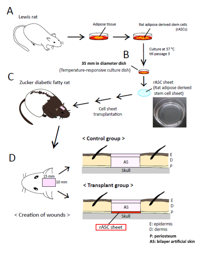
Figure 1: Schematic of the Experimental Transplantation Procedure. Schematic of the experimental transplantation procedure performed with an allogeneic rat adipose-derived stem cell (rASC) sheet and artificial skin in a rat wound-healing model of type 2 diabetes and obesity. (A) Rat adipose tissue was surgically excised from normal Lewis rats. rASCs were isolated and seeded onto a 60 cm2 culture dish and cultured at 37 °C in a 5% CO2 incubator for 7 – 8 days. (B) rASCs were subcultured every 2-3 days, and passage 3 rASCs were seeded onto a 35 mm diameter temperature-responsive culture dish. The cells were cultured in complete medium containing 16.4 µg/mL L-ascorbic acid phosphate magnesium salt n-hydrate (AA) at 37 °C in a 5% CO2 incubator for 7 – 8 days. The rASCs were harvested as a contiguous rASC sheet by reducing the temperature to 20 °C. (C) rASC sheets transplanted onto a 15 x 10 mm2 full-thickness skin defect with exposed bone on the heads of rats exhibiting diabetes and obesity (Zucker diabetic fatty (ZDF) rats) used as a wound-healing model. (D) An rASC sheet was placed on the skull directly over the defect and covered )with a 15 x 10 mm2 sheet of bilayer artificial skin, which was sutured into place with 10 nylon (5-0) sutures. Diabetes 2015;64: 2723-2734; with permission. Diabetes (c) copyright (2015) by the American Diabetes Association. Please click here to view a larger version of this figure.
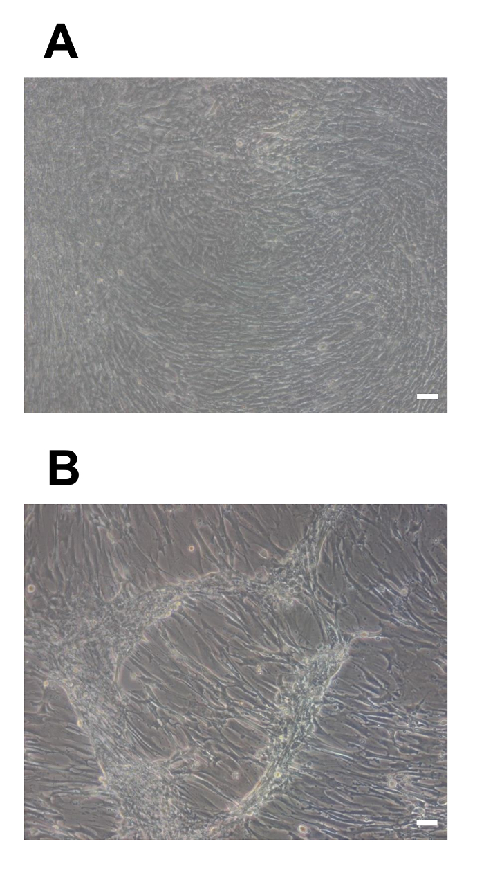
Figure 2: Light Microscope Images of ASCs. Light microscope images of ASC proliferation to the edge of culture dishes, without gaps between the ASCs. (A) An rASC sheet with a uniform thickness in all directions 7 days after the start of culturing (A). (B) An rASC sheet without uniform seeding. A contiguous rASC sheet cannot be obtained on day 7 after the start of culturing (B). Scale bars = 100 µm. Please click here to view a larger version of this figure.
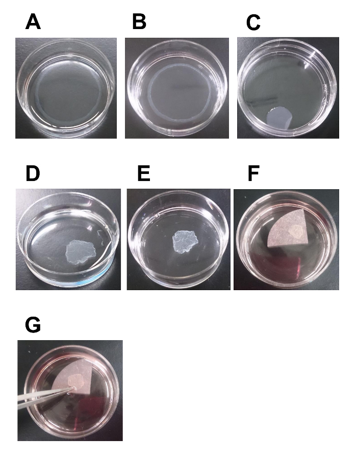
Figure 3: Time-lapse Images of rASC Sheet Statues at Room Temperature. Time-lapse images of the status of an rASC sheet at room temperature. rASCs on a 35 mm diameter temperature-responsive culture dish spontaneously and gradually detached from the dish surface at room temperature (approximately 20 °C) and were harvested as a contiguous sheet. (A) Approximately 5 min after moving the temperature-responsive culture dish to room temperature. (B) Approximately 10 min after moving the 35 mm diameter temperature-responsive culture dish to room temperature.(C) Approximately 20 – 30 min after moving the temperature-responsive culture dish to room temperature. This is a good-quality rASC sheet (C). (D-E) rASC sheet status approximately 20 – 30 min after moving the temperature-responsive culture dish to room temperature. This rASC sheet is of average quality (D). (F-G) rASC sheets are usually handled with a pair of forceps. If the cell sheet is brittle and fragile, a membrane can be used as a scaffold for transferring the cell sheet from the culture dish to the wound site. Please click here to view a larger version of this figure.
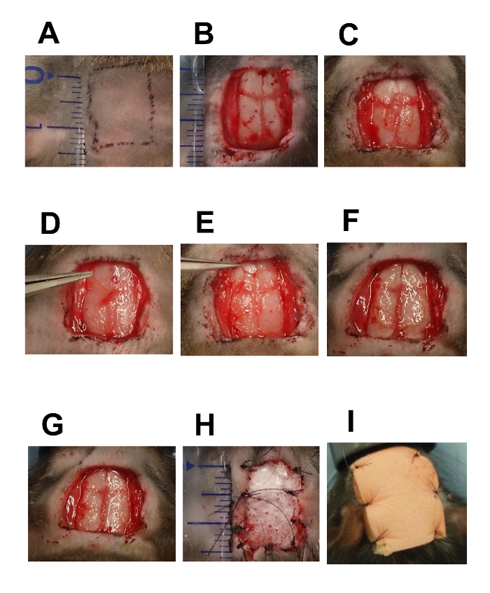
Figure 4: Time-series Images of Wound Creation and rASC Sheet Transplantation with Artificial Skin and a Non-adhesive Dressing
Time series images of wound creation and rASC sheet transplantation with artificial skin and a non-adhesive dressing. (A) The heads of ZDF rats were shaved with an electric razor. After shaving the body hair, check marks (15 x 10 mm2) were drawn using an oily hydrophobic pen. (B) A full-thickness skin defect (15 x 10 mm2) was created on the head of an anesthetized ZDF rat by removing cutaneous tissue from the epidermis to the periosteum. The skin and cutaneous tissue were excised with a scalpel, and the periosteum was removed with a periosteal raspatory. Using gauze moistened with sterile saline, pressure was applied to stop the bleeding after excision. (C) rASC sheet transplantation. An rASC sheet was placed over the defect immediately above the skull of the rat using a pair of forceps. (D-G) Adjusting rASC sheet extension to match wound size. The rASC sheet is flexible, adjustable, and can be extended to every corner of the wound site using a pair of forceps. For wider wounds, two or three flexible rASC sheets can be stacked. (H) Suturing the artificial skin covering the rASC sheet. The defect and the transplanted rASC sheet were covered with artificial skin (15 x 10 mm2), which was sutured with 10 stitches using 5-0 nylon sutures. (I) Suturing of the non-adhesive dressing (20 x 15 mm2) to the wound site covered with artificial skin. To protect the wound, non-adhesive dressing (20 x 15 mm2) was placed over the artificial skin with 5-0 nylon sutures. Diabetes 2015;64: 2723-2734; with permission. Diabetes (c) copyright (2015) by the American Diabetes Association. Please click here to view a larger version of this figure.
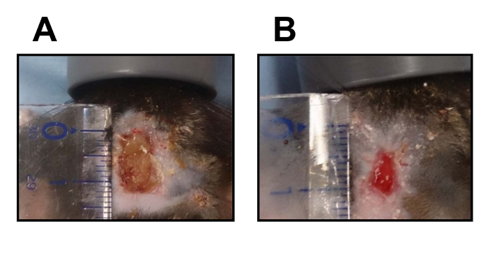
Figure 5: Macroscopic Images of Full-thickness Skin Defects. Macroscopic photographs of full-thickness skin defects without the transplantation of an rASC sheet (A) and with the transplantation of an rASC sheet (B) 14 days after creation of the wound. Please click here to view a larger version of this figure.
The most critical steps for successfully culturing an rASC sheet are as follows: 1) The temperature must be maintained at approximately 37 °C during culturing on the temperature-responsive culture dishes. During the creation of an rASC sheet, every procedure was performed on a 37 °C thermo-plate, and every reagent was warmed to 37 °C to prevent the cells from spontaneously detaching from the dish31. 2) The recipient ZDF rats must be monitored to prevent the removal of the non-adhesive dressing, which is critical for the successful transplantation of rASC sheets. If the dressing is removed, a new non-adhesive dressing must be applied to prevent the transplanted ASC sheets from detaching from the wound site.
Using this procedure, rASC sheets were generally obtained within 5 – 7 days of seeding passage 3 cells on temperature-responsive culture dishes. The culture time required to generate an rASC sheet can be adjusted according to the initial cell density and the time at which the complete culture medium containing AA is applied. If an rASC sheet detaches from the dish during cell culturing, the rASC sheet should be remade, and additional dishes should be prepared in the event of cell detachment.
The limitations of this protocol are as follows: 1) Strict temperature management must be applied to maintain an approximate temperature of 37 °C during the entire process when using temperature-responsive culture dishes. 2) After obtaining an rASC sheet, special medical devices must be used to maintain moist conditions, because the rASC sheet is sensitive to dry conditions. 3) Post-operative management, including daily observation of the condition of the recipient rat, is required.
Larger wounds with exposed bone are often observed clinically. For example, traffic accident trauma, burns, infected wounds, and damaged or necrotic wounds after debridement can develop into large wounds with exposed bone. Here, a clinical model of severe wounds was developed using a large, full-thickness skin defect with exposed bone in rats with type 2 diabetes and obesity. This model has the potential to be used as a standard model for evaluating wound healing in diabetic rats.
Artificial skin is a commercially available medical device for full-thickness skin defects after debridement, and recombinant basic fibroblast growth factor (bFGF) has been widely used for wound healing to promote angiogenesis and granulation. These two treatments have been used to achieve considerable therapeutic results, even for chronic wounds, such as diabetic wounds. It has been reported that ASCs secrete angiogeneic growth factors28, such as vascular endothelial growth factor (VEGF), hepatocyte growth factor (HGF), and bFGF, which could contribute to neovascularization29,30 and accelerate wound healing. Our previous study confirmed that ASCs continuously secrete these growth factors31. Therefore, ASC sheets, combined with artificial skin, have the potential to be used as a new therapeutic option for accelerating vascularization and wound healing31, and these sheets might be applicable to the treatment of many types of intractable ulcers or burns in human clinical settings in the future.
The authors have nothing to disclose.
The authors thank Dr. Yukiko Koga of the Department of Plastic and Reconstructive Surgery, Juntendo University School of Medicine, for providing practical advice. We also thank Mr. Hidekazu Murata of the Diabetic Center of Tokyo Women’s Medical University School of Medicine for excellent technical support. This study was supported by the Creation of Innovation Centers for Advanced Interdisciplinary Research Areas Program of the Project for Developing Innovation Systems “Cell Sheet Tissue Engineering Center (CSTEC)” from the Ministry of Education, Culture, Sports, Science and Technology (MEXT) of Japan.
α-MEM glutamax | Invitrogen | 32571-036 | Carlsbad, CA |
Fetal bovine serum (FBS) | Japan Bioserum Co Ltd. | S1650-500 | |
Penicillin/streptomycin | Life Technologies | 15140-122 | |
Collagenase A | Roche Diagnostics | 10 103 578 001 | Mannheim, Germany |
60-cm2 Primaria tissue culture dish | BD Biosciences | 353803 | Franklin Lakes, NJ |
Dulbecco's Phosphate Buffer Saline (PBS) | Life Technologies | 1490-144 | |
0.25% Trypsin-ethylenediamine tetraacetic acid (EDTA) | Life Technologies | 25200-056 | |
L-ascorbic acid phosphate magnesium salt n-hydrate | Wako | 013-19641 | |
35-mm temperature-responsive culture dish (UpcellTM) | CellSeed | NUNC-174904 | Tokyo, Japan |
Microwarm plate (MP-1000) | Kitazato Science Co., Ltd. | 1111 | |
Rodent mechanical ventilator | Stoelting | #50206 | Wood Dale, IL |
4% isoflurane | Pfizer Japan | 114-13340-3 | Tokyo, Japan |
Artificial skin (Pelnac®) | Smith & Nephew | PN-R40060 | Tokyo, Japan |
Non-adhesive dressing (Hydrosite plus®) | Smith & Nephew | 66800679 | Known as Allevyn non-adhessing® in the United State |
5-0 nylon suture | Alfresa | EP1105NB45-KF2 | |
20 CELLSTAR TUBES | greiner bio-one | 227 261 | |
15mL Centrifuge Tube | Corning Incorporated | 430791 | |
14 GOLDMAN-FOX PERIOSTEAL | Hu-Friedy | P14 | Chicago, IL |