Summary

לחקר endoplasmic reticulum ואת המיטוכונדריה אינטראקציות על ידי<em> באתרו</em> Assay קשירת הסמיכות בתאים קבועים

Published: December 10, 2016
doi:

Summary

כאן, אנו מתארים הליך לחזות ולכמת עם רגישות גבוהה האינטראקציות אנדוגני בין הרטיקולום ואת המיטוכונדריה endoplasmic בתאים קבוע. הפרוטוקול כולל אופטימיזציה assay קשירת הקרבה באתרו מיקוד אינוסיטול 1,4,5-אדנוזין קולטן / גלוקוז מוסדר חלבון 75 / ערוץ אניון תלוי מתח / cyclophilin D מורכבים על ממשק הקרום קשור המיטוכונדריה.

Abstract

Structural interactions between the endoplasmic reticular (ER) and mitochondrial membranes, in domains known as mitochondria-associated membranes (MAM), are crucial hubs for cellular signaling and cell fate. Particularly, these inter-organelle contact sites allow the transfer of calcium from the ER to mitochondria through the voltage-dependent anion channel (VDAC)/glucose-regulated protein 75 (GRP75)/inositol 1,4,5-triphosphate receptor (IP3R) calcium channeling complex. While this subcellular compartment is under intense investigation in both physiological and pathological conditions, no simple and sensitive method exists to quantify the endogenous amount of ER-mitochondria contact in cells. Similarly, MAMs are highly dynamic structures, and there is no suitable approach to follow modifications of ER-mitochondria interactions without protein overexpression. Here, we report an optimized protocol based on the use of an in situ proximity ligation assay to visualize and quantify endogenous ER-mitochondria interactions in fixed cells by using the close proximity between proteins of the outer mitochondrial membrane (VDAC1) and of the ER membrane (IP3R1) at the MAM interface. Similar in situ proximity ligation experiments can also be performed with the GRP75/IP3R1 and cyclophilin D/IP3R1 pairs of antibodies. This assay provides several advantages over other imaging procedures, as it is highly specific, sensitive, and suitable to multiple-condition testing. Therefore, the use of this in situ proximity ligation assay should be helpful to better understand the physiological regulations of ER-mitochondria interactions, as well as their role in pathological contexts.

Introduction

מיטוכונדריה reticulum endoplasmic (ER) אינה אברונים עצמאי בתא, אבל הם פועלים מבניים והן מבחינה תפקודית באתרי קשר המוגדרים ממברנות reticulum endoplasmic הקשורים המיטוכונדריה (MAM). למעשה, MAMs מתאימות באזורים שבהם קרום של ER ו המיטוכונדריה apposed מקרוב, המאפשר אינטראקציות בין חלבונים משני הצדדים. עם זאת, את הקרומים של אברונים אלה אינם מתאחים בתוך אזורים אלה, כך שהם שומרים ישויות נפרדות שלהם. MAMs לשחק תפקיד מכריע סידן (Ca 2 +) והעביר פוספוליפידים מ ER מיטוכונדריה, המשפיע חילוף אנרגיה ותא הישרדות 1-3.

העמותה בין ER ואת המיטוכונדריה היה דמיינו לראשונה בשנות ה -1970 עם מיקרוסקופ אלקטרונים. מאז, מיקרוסקופי אלקטרוני הילוכים 4,5, טומוגרפיה אלקטרון 6,7 או חיסוני לוקליזציה של fluorophore ER ואת המיטוכונדריה ספציפיs / חלבוני ניאון 8 שימשו קלאסי ללמוד אינטראקציות ER-המיטוכונדריה. כלי שימושי נוסף לניתוח MAM מבוסס על השימוש חלוק subcellular. זה מאפשר בידוד של שברים MAM ידי ultracentrifugation ההפרש מצמידים את שיפוע Percoll 9. עם זאת, המוצר הסופי מכיל שברי MAM מועשרים, ולא שברים טהורים. בסך הכל, האסטרטגיות אלה אינן רגישות במיוחד ו / או כמותית, והם לא מקובל בקלות הקרנה גדולה. לחלופין, גישות גנטיות באמצעות linkers הבין-אברון ניאון סמים מושרה צמחו, אבל הם אינם מאפשרים ניתוח של אינטראקציות אברון ברמות ביטוי אנדוגני של חלבונים 10.

בהתבסס על תגלית של Szabadkai של המתחם IP3R / GRP75 / VDAC בבית MAM 11, פיתחנו שיטה כמותית לנתח אינטראקציות ER-המיטוכונדריה. השתמשנו ב ligati הקרבה באתרועל assay כדי לזהות ולכמת אינטראקציות בין VDAC1 ו IP3R1, שני חלבונים אברון-משטח מעורב במתחם -channeling Ca 2+ על הממשק MAM בתאים קבוע 12. בקצרה, העמקנו VDAC1 על קרום המיטוכונדריה החיצוני (עכבר אנטי VDAC1 נוגדן ראשוני) ו IP3R1 על הממברנה ER (ארנב נוגדן ראשוני נגד IP3R1) (איור 1, לוח א '). לאחר מכן, על פי assay, הוספנו שני אנטי עכבר IgG נגד ארנב (עכבר בדיקות assay קשירת ארנב קרבה), אשר מצומדות כדי רחבות oligonucleotide משלימות. אם שני החלבונים הממוקדים הם במרחק מתחת ל -40 ננומטר, oligonucleotides יכול להכליא עם oligos מחבר הנוסף לאחר מכן לאפשר את היווצרות של תבנית ה- DNA מעגלי (איור 1, לוח ב '). מולקולת הדנ"א חוזר זה ligated מוגבר, יצירת מוצר דנ"א חד-גדילי המצורפת קוולנטית לאחד חלליות הקרבה (איור 1, ג פאנל) </strong>. היות והמרחק בין ER ואת המיטוכונדריה בממשק MAM נע בין 10 ננומטר ל -25 6 ננומטר, קשירת הקרבה והגברה ניתן לעשות, מה שמוביל לגילוי הבאים עקב הכלאה של בדיקות oligonucleotides אדום שכותרתו טקסס (איור 1, פאנל ד ). נקודה פלורסנט מייצגת אינטראקציות בין VDAC1 / IP3R1, ובכך מאפשר כימות של באתרו ER-המיטוכונדריה אינטראקציות בתאים בודדים.

איור 1
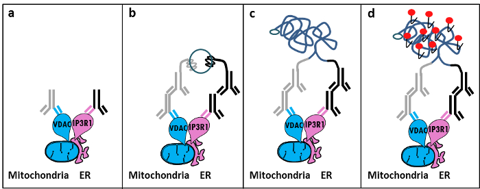
איור 1: איור סכמטי של איתור של endoplasmic אינטראקציות reticulum-המיטוכונדריה ידי ב assay קשירת Situ קרבה. א) נוגדן עכבר עיקרי המכוון נגד VDAC1 ו נוגדן ראשוני ארנב המכוון נגד IP3R1 יכול להיקשר אפיטופים שלהם בסמיכות בממשק MAM, ב) התוספת של זוג חלליות קשירת קרבהמכוון נגד עכבר IgG ארנב. בדיקות אלה יש מצורפות גדילי דנ"א שיכול ליצור תבניות עבור קשירת oligos מחבר. ג) גדיל DNA המעגלי שיקום לאחר הקשירה יכול להיות מוגבר ו ד) מדמיין ידי מיקרוסקופ כנקודת פלורסנט באמצעות oligonucleotides האדום שכותרתו טקסס. אנא לחץ כאן כדי לצפות בגרסה גדולה יותר של דמות זו.

דומה בניסויים assay קשירת הקרבה באתרו ניתן לבצע עם זוג GRP75 / IP3R1 של נוגדנים, כמו גם cyclophilin D (CypD) / נוגדנים IP3R1, בהתחשב בכך CypD הוצגה אינטראקציה עם מורכבות IP3R / GRP75 / VDAC בממשק MAM 12-14.

Protocol

1. הכנת פתרונות הכן 10 פורמלדהיד% ב PBS (מלח נמוכה) על ידי דילול 5.5 מ"ל של פורמלדהיד 37% ב 14.5 מ"ל של PBS. הכן 1 M גליצין, pH 8.0, על ידי המסת 3.8 גרם של גליצין ב 50 מ"ל של PBS; לדלל את הפתרון הזה כדי לקבל גליצין 100 מ"מ 1x PBS. <li style=";te…

Representative Results

בהתבסס על הניסיון שלנו באמצעות פרוטוקול זה, אנחנו יכולים בבטחה ממליצים על שיטה זו עבור להדמיה וכימות של אינטראקציות ER-המיטוכונדריות בתאים קבועים. נציג תמונות של באינטראקציות ER-המיטוכונדריה assay-דמיינו קשירת הקרבה באתרו הקו הסלולרי HuH7 hepatoca…

Discussion

Collectively, our studies indicate that the in situ proximity ligation assay is truly a relevant strategy to follow and quantify endogenous ER-mitochondria interactions in fixed cells, without the need for using organelle-specific fluorophores or fluorescent proteins. The specific use of VDAC1/IP3R1 antibodies has been adapted to study ER-mitochondria interactions in HuH7 cells. However, alternative isoforms of VDAC and IP3R may be used, depending on the cell type. In this case, antibodies need to be validated b…

Divulgations

The authors have nothing to disclose.

Acknowledgements

אנו מודים לכל האנשים במעבדה שלנו שתרמו לייעל ולאמת את הפרוטוקול. עבודה זו נתמכה על ידי INSERM ואת סוכנות המחקר הלאומי (ANR-09-JCJC-0116 ו ANR-11-BSV1-033-02). ET נתמכה במהלך הדוקטורט שלה על ידי מענק מחקר מהמשרד הצרפתי של השכלה גבוהות ומחקר.

Materials

Formaldehyde Sigma F-8775
Glycine Sigma G-8898
Triton Sigma T8532
35mm Glass bottom culture dishes MatTeK corporation P35G-0-14-C
Blocking solution Sigma DUO-92004 or DUO-92002 provided in the Duolink PLA probes, Sigma
VDAC1 antibody Abcam ab14734
IP3R1-H80 antibody Santa Cruz sc28614
CypD antibody Abcam ab110324
Grp75 antibody Santa Cruz sc13967
TBS 10X euromedex ET220 Dilute to obtain 1X
Tween 100X euromedex 2001-B dilute in TBS to obtain 0,01%
PLA Probes Mouse MINUS Sigma DUO-92004 Duolink, Sigma
PLA Probes Rabbit PLUS Sigma DUO-92002 Duolink, Sigma
Duolink detection reagents red Sigma DUO-92008 Duolink, Sigma
Ligation solution Sigma DUO-92008 Part of the Duolink detection reagents red, Sigma
Ligase Sigma DUO-92008 Part of the Duolink detection reagents red, Sigma
Amplification solution Sigma DUO-92008 Part of the Duolink detection reagents red, Sigma
Polymerase Sigma DUO-92008 Part of the Duolink detection reagents red, Sigma
Duolink Mounting Medium Sigma DUO80102 Duolink, Sigma
Softwares:
Blob-finder software BlobFinder is a freely distributed software that can perform calculations on cells from fluorescence microscopy images. This software can be downloaded for free from The Centre for Image Analysis at Uppsala University who have developed the software and the work was supported by the EU FP6 Project ENLIGHT and  Olink Bioscience. http://www.cb.uu.se/~amin/BlobFinder/index_files/Page430.htm
ImageJ software Can be downloaded for free from: http://rsb.info.nih.gov/ij/download.html

References

  1. Bravo-Sagua, R., et al. Organelle communication: signaling crossroads between homeostasis and disease. The international journal of biochemistry & cell biology. 50, 55-59 (2014).
  2. Giorgi, C., et al. Mitochondria-associated membranes: composition, molecular mechanisms, and physiopathological implications. Antioxidants & redox signaling. 22, 995-1019 (2015).
  3. Phillips, M. J., Voeltz, G. K. Structure and function of ER membrane contact sites with other organelles. Nature reviews. Molecular cell biology. 17, 69-82 (2016).
  4. Cosson, P., et al. The RTM resistance to potyviruses in Arabidopsis thaliana: natural variation of the RTM genes and evidence for the implication of additional genes. PLoS One. 7, 39169 (2012).
  5. Mannella, C. A. Structure and dynamics of the mitochondrial inner membrane cristae. Biochim Biophys Acta. 1763, 542-548 (2006).
  6. Csordas, G., et al. Structural and functional features and significance of the physical linkage between ER and mitochondria. The Journal of cell biology. 174, 915-921 (2006).
  7. Mannella, C. A., Buttle, K., Rath, B. K., Marko, M. Electron microscopic tomography of rat-liver mitochondria and their interaction with the endoplasmic reticulum. Biofactors. 8, 225-228 (1998).
  8. Rizzuto, R., et al. Close contacts with the endoplasmic reticulum as determinants of mitochondrial Ca2+ responses. Science. 280, 1763-1766 (1998).
  9. Wieckowski, M. R., Giorgi, C., Lebiedzinska, M., Duszynski, J., Pinton, P. Isolation of mitochondria-associated membranes and mitochondria from animal tissues and cells. Nat Protoc. 4, 1582-1590 (2009).
  10. Csordas, G., et al. Imaging interorganelle contacts and local calcium dynamics at the ER-mitochondrial interface. Mol Cell. 39, 121-132 (2010).
  11. Szabadkai, G., et al. Chaperone-mediated coupling of endoplasmic reticulum and mitochondrial Ca2+ channels. J Cell Biol. 175, 901-911 (2006).
  12. Tubbs, E., et al. Mitochondria-associated endoplasmic reticulum membrane (MAM) integrity is required for insulin signaling and is implicated in hepatic insulin resistance. Diabetes. 63, 3279-3294 (2014).
  13. Paillard, M., et al. Depressing Mitochondria-Reticulum Interactions Protects Cardiomyocytes From Lethal Hypoxia-Reoxygenation Injury. Circulation. 128, 1555-1565 (2013).
  14. Rieusset, J., et al. Disruption of calcium transfer from ER to mitochondria links alterations of mitochondria-associated ER membrane integrity to hepatic insulin resistance. Diabetologia. 59, 614-623 (2016).
  15. Allalou, A., Wahlby, C. BlobFinder, a tool for fluorescence microscopy image cytometry. Computer methods and programs in biomedicine. 94, 58-65 (2009).
  16. Theurey, P., et al. Mitochondria-associated endoplasmic reticulum membranes allow adaptation of mitochondrial metabolism to glucose availability in the liver. Journal of molecular cell biology. , (2016).
  17. de Brito, O. M., Scorrano, L. Mitofusin 2 tethers endoplasmic reticulum to mitochondria. Nature. 456, 605-610 (2008).
  18. Soderberg, O., et al. Direct observation of individual endogenous protein complexes in situ by proximity ligation. Nature methods. 3, 995-1000 (2006).
  19. De Pinto, V., Messina, A., Lane, D. J., Lawen, A. Voltage-dependent anion-selective channel (VDAC) in the plasma membrane. FEBS letters. 584, 1793-1799 (2010).
  20. Kaul, S. C., Taira, K., Pereira-Smith, O. M., Wadhwa, R. Mortalin: present and prospective. Experimental gerontology. 37, 1157-1164 (2002).
check_url/fr/54899?article_type=t

Play Video

Citer Cet Article
Tubbs, E., Rieusset, J. Study of Endoplasmic Reticulum and Mitochondria Interactions by In Situ Proximity Ligation Assay in Fixed Cells. J. Vis. Exp. (118), e54899, doi:10.3791/54899 (2016).

View Video