Summary

Struktur-funktionsstudier i mus embryonala stamceller Använda Recombinase-medierad kassett Exchange

Published: April 27, 2017
doi:

Summary

Proteins often contain multiple domains that can exert different cellular functions. Gene knock-outs (KO) do not consider this functional diversity. Here, we report a recombination-mediated cassette exchange (RMCE)-based structure-function approach in KO embryonic stem cells that allows for the molecular dissection of various functional domains or variants of a protein.

Abstract

Gene engineering in mouse embryos or embryonic stem cells (mESCs) allows for the study of the function of a given protein. Proteins are the workhorses of the cell and often consist of multiple functional domains, which can be influenced by posttranslational modifications. The depletion of the entire protein in conditional or constitutive knock-out (KO) mice does not take into account this functional diversity and regulation. An mESC line and a derived mouse model, in which a docking site for FLPe recombination-mediated cassette exchange (RMCE) was inserted within the ROSA26 (R26) locus, was previously reported. Here, we report on a structure-function approach that allows for molecular dissection of the different functionalities of a multidomain protein. To this end, RMCE-compatible mice must be crossed with KO mice and then RMCE-compatible KO mESCs must be isolated. Next, a panel of putative rescue constructs can be introduced into the R26 locus via RMCE targeting. The candidate rescue cDNAs can be easily inserted between RMCE sites of the targeting vector using recombination cloning. Next, KO mESCs are transfected with the targeting vector in combination with an FLPe recombinase expression plasmid. RMCE reactivates the promoter-less neomycin-resistance gene in the ROSA26 docking sites and allows for the selection of the correct targeting event. In this way, high targeting efficiencies close to 100% are obtained, allowing for insertion of multiple putative rescue constructs in a semi-high throughput manner. Finally, a multitude of R26-driven rescue constructs can be tested for their ability to rescue the phenotype that was observed in parental KO mESCs. We present a proof-of-principle structure-function study in p120 catenin (p120ctn) KO mESCs using endoderm differentiation in embryoid bodies (EBs) as the phenotypic readout. This approach enables the identification of important domains, putative downstream pathways, and disease-relevant point mutations that underlie KO phenotypes for a given protein.

Introduction

It is estimated that mammalian genomes contain about 20,000 protein-coding genes. Alternative splicing and posttranslational modifications further increase the protein repertoire. Proteins have a modular structure1 and often contain multiple interaction domains, which allow their recruitment into different protein complexes and their participation in multiple cellular processes2. One example is the multi-functional protein called p120ctn. p120ctn is encoded by the Ctnnd1 gene and consists of a large central armadillo repeat domain flanked by an N-terminal and a C-terminal region. The armadillo domain of p120ctn binds to a highly conserved juxtamembrane domain of classical cadherins, which are involved in cell-cell adhesion, but it also binds to the transcriptional repressor Kaiso. The N-terminal domain of p120ctn interacts with different kinases, phosphatases, small RhoGTPases, and microtubule-associated proteins3. Interestingly, as a result of alternative splicing, p120ctn isoforms can be generated from four alternative start codons4. p120ctn isoform 1A is the longest, as it is translated from the most-5' start codon and contains the full-length N-terminal segment. In p120ctn isoforms 3 and 4, this N-terminal segment is deleted partially and completely, respectively. Understanding the precise role of proteins (or protein isoforms) and their domains in different cellular functions remains a challenge.

Gene targeting in mESCs enables the study of the function of a protein through the genetic deletion of the corresponding gene and has widely contributed to the identification of developmentally important and disease-relevant genes and pathways. This breakthrough in reverse genetics was the result of advances in the fields of mESC isolation and gene targeting due to homologous recombination5. Homologous recombination is a process in which DNA fragments are exchanged between two similar or identical nucleic moieties after double-stranded (ds) DNA breaks. Normally, HR is inefficient because dsDNA breaks are infrequent. Recently, the efficiency of homology-directed gene targeting could be increased using site-specific nucleases6,7, but unfortunately, these are prone to off-target effects8. A more reliable technique to enable gene targeting is RMCE, which is based on site-specific recombination systems such as Cre/loxP or FLPe/Frt. LoxP and Frt sequence are found in bacteriophage P1 and Saccharomyces cerevisiae, respectively, and consist of 34 bp, including an asymmetric 8 bp sequence that determines the orientation of the site. On the other hand, the orientation of, for instance, two loxP sites within a DNA stretch will determine whether the floxed DNA becomes excised or inversed upon Cre-mediated recombination9. Moreover, Cre can also induce a translocation if two sites are located on different chromosomes. RMCE takes advantage of heterospecific recombination sites that do not cross-react and that are embedded in a genomic locus. In the presence of a donor plasmid that contains a DNA fragment flanked by the same heterospecific sites, the recombinase will insert this DNA fragment into the RMCE-compatible genomic locus because of double-simultaneous translocation (Figure 1). Here, only correctly RMCE-targeted clones can render drug resistance thanks to a promoter on the incoming vector that restores a "trapped," promoter-less Neomycin resistance gene (NeoR) present in the R26 genome of the docking cells (Figure 1)10,11. This results in a very high targeting efficiency, often close to 100%11,12. In conclusion, RMCE-based targeting is highly efficient and can be used for structure-functions studies; however, it requires a pre-engineered genomic locus.

Figure 1
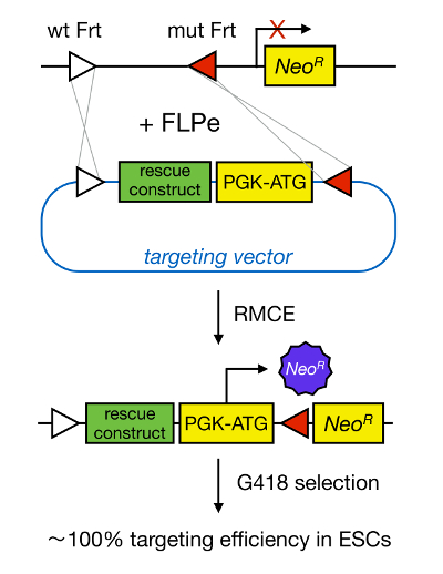
Figure 1. Schematic Representation of RMCE-mediated Targeting. RMCE allows for the exchange of DNA segments from an incoming targeting vector to a defined genomic locus if both harbor two heterospecific Frt sites (depicted by white and red triangles). In addition, the engineered genomic locus contains a promoterless and truncated neomycin-resistance (NeoR) gene. By providing a promoter and start codon in the incoming DNA fragment, only correct recombination events restore neomycin resistance, resulting in high targeting efficiencies. Please click here to view a larger version of this figure.

Genome engineering in mESCs allows for the generation of RMCE-compatible mice. In 1981, two groups succeeded in capturing pluripotent cells from the inner cell mass (ICM) of blastocysts and in maintaining them in culture13,14. mESCs are capable of self-renewal and differentiation into all types of embryonic and adult cells, including the germ-cell lineage. Therefore, gene targeting in mESCs enables reverse-genetic studies through the development of constitutive or conditional (using the Cre/LoxP system) KO mice. However, the classical way to isolate mouse ES cells is very inefficient. Several major improvements have greatly increased the success rate for deriving mESC lines, including the use of a defined serum-replacement (SR) medium15, alternating between mESC medium containing SR and fetal bovine serum (FBS)16, and the use of pharmacological compounds such as pluripotin or 2i17. Pluripotin, a small synthetic molecule, allows for the propagation of mESCs in an undifferentiated state in the absence of leukemia inhibitory factor (LIF) and mouse embryonic fibroblasts (MEFs)18. Finally, it has been shown that mESCs can be isolated with a very high efficiency (close to 100%) when an SR/FBS medium alternation protocol is combined with LIF and pluripotin19,20. These protocols enable the efficient isolation of RMCE-compatible KO mESCs that can subsequently be used for structure-function studies.

This paper describes a method that enables one to identify the key domains or residues within a protein that are responsible for specific cellular processes. To this end, a pipeline of advanced technologies that enable efficient mESC isolation, targeting vector assembly, and mESC targeting was created. As such, large panels with protein isoforms, domain mutants, and downstream effectors can be introduced in KO mESCs and can be evaluated for their ability to rescue the in vitro KO phenotype.

Protocol

All experiments on mice were conducted according to institutional, national, and European animal regulations. 1. Isolation of RMCE-compatible KO mESCs Breed heterozygous KO mice with RMCE-compatible mice, such as ROSALUC mice10 or ROSA26-iPSC mice21. Both RMCE-compatible mice were maintained on a mixed 129/C57BL6/Swiss background. NOTE: Crossing with heterozygous KO mice is advised to overcome embryonic lethality in homozygous KO mice. Us…

Representative Results

The procedure to isolate RMCE-compatible KO mESC lines is depicted in Figure 2. Two consecutive breedings are required to obtain RMCE-compatible KO blastocysts. First, heterozygous KO mice are crossed with RMCE-compatible mice to obtain RMCE-compatible, heterozygous KO mice. These mice are then used for timed matings with other heterozygous KO mice to obtain 3.5-dpc, RMCE-compatible, homozygous KO blastocysts. The chance of obtaining such an embryo is one in eight, as pre…

Discussion

Our mESC isolation method is user-friendly and does not require advanced skills or equipment, such as microsurgery of blastocysts. Thus, this technology is accessible to a large proportion of the scientific community. Anyone with basic cell culture experience can propagate ICM outgrowths and establish mESCs lines. However, the flushing and handling of blastocysts requires some practice. A mouth pipette is used to transfer blastocysts and consists of a micropipette, a micropipette holder, tubing, and an aspirator mouthpie…

Divulgations

The authors have nothing to disclose.

Acknowledgements

We thank Jinke D'Hont, Frederique Van Rockeghem, Natalie Farla, Kelly Lemeire, and Riet De Rycke for their excellent technical support. We also thank Eef Parthoens, Evelien Van Hamme, and Amanda Goncalves from the Bioimaging Core Facility of the Inflammation Research Center for their expert assistance. We acknowledge members of our research group for valuable discussions. This work was supported by the Belgian Science Policy (Belspo Interuniversity Attraction Poles – Award IAP VII-07 DevRepair; https://devrepair.be), by the Queen Elisabeth Medical Foundation, Belgium (GSKE 2008-2010; http://www.fmre-gske.be), and by the Concerted Research Actions (GOA 01G01908) of Ghent University, Belgium (http://www.ugent.be/en/ghentuniv). SG is a postdoctoral fellow of the Flanders Research Funds (FWO-V).

Materials

ROSALUC Mice made in house frozen sperm available upon request
R26-iPSC mice made in house frozen sperm available upon request
Vessel probe Fine Science Tools 10160-13 to check for copulation plugs
M2 medium Sigma-Aldrich M7167 make aliquots and store at -20°C
Fine forceps (Dumont #5 Standard tip Student forceps) Fine Science Tools 11251-10 spray with 70% EtOH before use (do not autoclave)
23G needles Fine-ject 8697
1-ml syringes Soft-ject 6680
60-mm bacterial grade plates (for flushing)  Gosselin BB60-01
Mouth pipette made in house see discussion
Mouse embryonic fibroblasts (MEFs, TgN (DR4)1 Jae strain) ATTC SCRC-1045
TgN (DR4)1 Jae mice The Jackson Laboratory 3208
Mitomycin C  Sigma-Aldrich M0503
Phosphate buffered saline (PBS) without calcium or magnesium Gibco 14190-094
Tg(DR4)1Jae/J mice JAX 3208 mice that contain four drug-selectable genes and DR4 MEFS confers resistance to neomycin, puromycin, hygromycin and 6-thioguanine
0.1% Gelatin Sigma-Aldrich G1393 Dissolve 0.5 g in 500 ml distilled water, autoclave and store at 4°C.
Trypsin (0.25%)  Gibco 25200-056
2 μM pluripotin Cayman Chemical 10009557
pRMCE-DV1 BCCM/LMBP collection  LMBP 08870 public available from the BCCM/LMBP collection (http://bccm.belspo.be) 
cre-excised pRMCE-DV1 BCCM/LMBP collection  LMBP 08195 public available from the BCCM/LMBP collection (http://bccm.belspo.be) 
pCAG-FlpE-IRES-Puro-pA  Addgene 20733
heat-shock competent DH5α bacteria  made in house
Gateway pDONR221 vector Thermo Fisher 12536-017 contains kanamycin-resistance gene
BP clonase II mix Thermo Fisher 11789-020
LR clonase II mix Thermo Fisher 11791-020 
Luria Broth (LB) 
Ampicillin 
Applied Biosystems 3730XL DNA Analyzer Thermo Fisher 3730XL
G418 Thermo Fisher 11811-023
Lipofectamine 2000 transfection reagent Thermo Fisher 11668027
Lipofectamine LTX transfection reagent Thermo Fisher 15338100
Effectene transfection reagent Qiagen 301425
GATEWAY pENTR 1A vector Thermo Fisher A10462 recombination-compatible vector
mouse monoclonal anti-p120ctn antibody BD Transduction Laboratories 610134
mouse monoclonal anti-Ecadherin antibody BD Transduction Laboratories 610181
General equipment:
Sterile dissection tools fine scissors and forceps for dissecting the uterus
Sterile pipettes: 5 ml, 10 ml and 25 ml
15-ml and 50-ml conical centrifuge tubes
96-well culture plates V-shaped bottom and flat bottom) 
Culture dishes: 24 wells, 12 wells and 6 wells
Multichannel pipettes (to pipette 30, 50, 100 and 200 μl) 
Sterile multichannel reservoirs
Access to a laminar air flow
Access to an incubator at 37°C with 5% CO2
Access to an inverted microscope
Access to a bench-top centrifuge
Access to a stereo microscope with transmitted-light 
Culture media:
MEF Medium: stored at 4°C; warm 30 min at 37°C before use
 Dulbecco’s modified Eagle’s medium (DMEM) Gibco 41965-062
10% fetal bovine serum (FBS) Sigma-Aldrich F-7524
L-glutamine (2 mM) Gibco 25030-024 
Sodium pyruvate  (0.4 mM) Gibco 11360-039 
penicillin (100 U/ml) Gibco 15140-122 
streptomycin (100 µg/ml) Gibco 15140-122 
SR-based mESC medium: stored at 4°C; warm 30 min at 37°C before use
DMEM/F12 Gibco 31330-038 mixed in a 1:1 ratio
15% knock-out serum replacement (SR ) Gibco 10828–028
L-glutamine (2 mM) Gibco 25030-024 
0.1 mM non-essential amino acids  Gibco 11140-050
penicillin (100 U/ml) Gibco 15140-122 
streptomycin (100 µg/ml) Gibco 15140-122 
β-mercaptoethanol (0.1 mM)  Sigma-Aldrich M 3148 
2,000 U/ml recombinant mouse LIF  (IRC/VIB Protein Service facility)
FBS-based mESC medium (similar to SR-based mESC medium): stored at 4°C; warm 30 min at 37°C before use
Knockout DMEM Gibco 10829-018 
15% FBS Hyclone SH30070.03E
Differention Medium stored at 4°C; warm 30 min at 37°C before use
Iscove's Modified Dulbecco's Medium (IMDM) Gibco 21980-032 
15% FBS Hyclone SH30070.03E
5% CD Hybridoma Medium(1x) liquid   Gibco 11279-023 
 2 mM L-glutamine Gibco 25030-024 
0.4 mM 1-thioglycerol Sigma-Aldrich M-6145
50 μg/ml ascorbic acid Sigma-Aldrich A-4544 
penicillin (100 U/ml) Gibco 15140-122 
streptomycin (100 µg/ml) Gibco 15140-122 
2i
1 μM Erk inhibitor PD0325901  Axon Medchem Axon 1408
3 μM Gsk3 inhibitor CHIR99021 Axon Medchem Axon 1386

References

  1. Gul, I. S., Hulpiau, P., Saeys, Y., van Roy, F. Metazoan evolution of the armadillo repeat superfamily. Cell Mol Life Sci. , (2016).
  2. Valenta, T., Hausmann, G., Basler, K. The many faces and functions of beta-catenin. EMBO J. 31, 2714-2736 (2012).
  3. Pieters, T., van Hengel, J., van Roy, F. Functions of p120ctn in development and disease. Front Biosci. 17, 760-783 (2012).
  4. Keirsebilck, A., et al. Molecular cloning of the human p120ctn catenin gene (CTNND1): Expression of multiple alternatively spliced isoforms. Genomics. 50, 129-146 (1998).
  5. Capecchi, M. R. Gene targeting in mice: functional analysis of the mammalian genome for the twenty-first century. Nat Rev Genet. 6, 507-512 (2005).
  6. Wang, H., et al. One-step generation of mice carrying mutations in multiple genes by CRISPR/Cas-mediated genome engineering. Cell. 153, 910-918 (2013).
  7. Yang, H., et al. One-step generation of mice carrying reporter and conditional alleles by CRISPR/Cas-mediated genome engineering. Cell. 154, 1370-1379 (2013).
  8. Fu, Y., et al. High-frequency off-target mutagenesis induced by CRISPR-Cas nucleases in human cells. Nat Biotechnol. 31, 822-826 (2013).
  9. Branda, C. S., Dymecki, S. M. Talking about a revolution: The impact of site-specific recombinases on genetic analyses in mice. Dev Cell. 6, 7-28 (2004).
  10. Sandhu, U., et al. Strict control of transgene expression in a mouse model for sensitive biological applications based on RMCE compatible ES cells. Nucleic Acids Res. 39, 1 (2010).
  11. Haenebalcke, L., et al. Efficient ROSA26-based conditional and/or inducible transgenesis using RMCE-compatible F1 hybrid mouse embryonic stem cells. Stem Cell Rev. 9, 774-785 (2013).
  12. Pieters, T., et al. p120 Catenin-Mediated Stabilization of E-Cadherin Is Essential for Primitive Endoderm Specification. PLoS Genet. 12, e1006243 (2016).
  13. Evans, M. J., Kaufman, M. H. Establishment in culture of pluripotential cells from mouse embryos. Nature. 292, 154-156 (1981).
  14. Martin, G. R. Isolation of a pluripotent cell line from early mouse embryos cultured in medium conditioned by teratocarcinoma stem cells. Proc Natl Acad Sci U S A. 78, 7634-7638 (1981).
  15. Cheng, J., Dutra, A., Takesono, A., Garrett-Beal, L., Schwartzberg, P. L. Improved generation of C57BL/6J mouse embryonic stem cells in a defined serum-free media. Genesis. 39, 100-104 (2004).
  16. Bryja, V., et al. An efficient method for the derivation of mouse embryonic stem cells. Stem Cells. 24, 844-849 (2006).
  17. Ying, Q. L., et al. The ground state of embryonic stem cell self-renewal. Nature. 453, 519-523 (2008).
  18. Chen, S., et al. Self-renewal of embryonic stem cells by a small molecule. Proc Natl Acad Sci U S A. 103, 17266-17271 (2006).
  19. Pieters, T., et al. Efficient and User-Friendly Pluripotin-based Derivation of Mouse Embryonic Stem Cells. Stem Cell Rev. 8, (2012).
  20. Yang, W., et al. Pluripotin combined with leukemia inhibitory factor greatly promotes the derivation of embryonic stem cell lines from refractory strains. Stem Cells. 27, 383-389 (2009).
  21. Haenebalcke, L., et al. The ROSA26-iPSC Mouse: A Conditional, Inducible, and Exchangeable Resource for Studying Cellular (De)Differentiation. Cell Rep. 3, (2013).
  22. Tucker, K. L., Wang, Y., Dausman, J., Jaenisch, R. A transgenic mouse strain expressing four drug-selectable marker genes. Nucleic Acids Res. 25, 3745-3746 (1997).
  23. Nyabi, O., et al. Efficient mouse transgenesis using Gateway-compatible ROSA26 locus targeting vectors and F1 hybrid ES cells. Nucleic Acids Res. 37, 55 (2009).
  24. Katzen, F. Gateway((R)) recombinational cloning: a biological operating system. Expert Opin Drug Discov. 2, 571-589 (2007).
  25. Schaft, J., Ashery-Padan, R., vander Hoeven, F., Gruss, P., Stewart, A. F. Efficient FLP recombination in mouse ES cells and oocytes. Genesis. 31, 6-10 (2001).
  26. Spencer, H. L., et al. E-cadherin inhibits cell surface localization of the pro-migratory 5T4 oncofetal antigen in mouse embryonic stem cells. Mol Biol Cell. 18, 2838-2851 (2007).
  27. Stryjewska, A., et al. Zeb2 Regulates Cell Fate at the Exit from Epiblast State in Mouse Embryonic Stem Cells. Stem Cells. , (2016).
  28. Nagy, A., Gertsenstein, M., Vintersten, K., Behringer, R. . Manipulating the mouse embryo: A laboratory manual. , (2003).
  29. van den Berghe, V., et al. Directed migration of cortical interneurons depends on the cell-autonomous action of Sip1. Neuron. 77, 70-82 (2013).
  30. Li, J., et al. The EMT transcription factor Zeb2 controls adult murine hematopoietic differentiation by regulating cytokine signaling. Blood. , (2016).
check_url/fr/55575?article_type=t

Play Video

Citer Cet Article
Pieters, T., Haenebalcke, L., Bruneel, K., Vandamme, N., Hochepied, T., van Hengel, J., Wirth, D., Berx, G., Haigh, J. J., van Roy, F., Goossens, S. Structure-function Studies in Mouse Embryonic Stem Cells Using Recombinase-mediated Cassette Exchange. J. Vis. Exp. (122), e55575, doi:10.3791/55575 (2017).

View Video