This work describes a fluorescence microscopy-based method for the study of platelet adhesion, spreading, and secretion under flow. This versatile platform enables the investigation of platelet function for mechanistic research on thrombosis and hemostasis.
Blood platelets are essential players in hemostasis, the formation of thrombi to seal vascular breaches. They are also involved in thrombosis, the formation of thrombi that occlude the vasculature and injure organs, with life-threatening consequences. This motivates scientific research on platelet function and the development of methods to track cell-biological processes as they occur under flow conditions.
A variety of flow models are available for the study of platelet adhesion and aggregation, two key phenomena in platelet biology. This work describes a method to study real-time platelet degranulation under flow during activation. The method makes use of a flow chamber coupled to a syringe-pump setup that is placed under a wide-field, inverted, LED-based fluorescence microscope. The setup described here allows for the simultaneous excitation of multiple fluorophores that are delivered by fluorescently labeled antibodies or fluorescent dyes. After live-cell imaging experiments, the cover glasses can be further processed and analyzed using static microscopy (i.e., confocal microscopy or scanning electron microscopy).
Platelets are anucleate cells that circulate in the blood stream. Their main function is to seal vascular breaches at sites of injury and to prevent blood loss. At these sites of injury, subendothelial collagen fibers become exposed and are subsequently covered by the multimeric protein, von Willebrand factor (VWF). VWF interacts with the platelets in circulation in a mechanism that depends upon the glycoprotein Ibα-IX-V complex on the cell surface1, slowing down the speed of the platelets. This is particularly important at high shear rates. The platelets subsequently undergo morphological changes while receiving activating impulses from collagen. This leads to irreversible spreading and eventually to platelet aggregation. Both processes depend upon the secretion of granule contents to facilitate platelet-platelet crosstalk. Amongst others, platelet α-granules contain fibrinogen and VWF to assist platelet adhesion and to bridge platelets together in an integrin-dependent manner. The platelet-dense granules contain inorganic compounds2, including calcium and adenosine diphosphate (ADP), which help to reinforce platelet activation. Furthermore, platelets contain mediators of (allergic) inflammation3, complement-controlling proteins4, and angiogenesis factors5,6, raising the questions of whether and how these contents are differentially released under varying conditions.
Since the 1980s, the study of platelet function in flow models has been valuable to the investigation of thrombotic mechanisms7. Since then, much technical progress has been made, and flow models that include fibrin formation are currently developed to assay the hemostatic potential of therapeutic platelet concentrates ex vivo8 or to investigate the influence of disturbances in shear rates on thrombus morphology9. The differences in the molecular and cell-biological mechanisms that drive stable adhesion and physiological thrombus formation (hemostasis) versus pathological thrombus formation (thrombosis) may be very subtle and motivate the development of flow models that allow for the real-time visualization of these subcellular processes.
An example of a process for which such a setup would be valuable is the (re)distribution of intracellular polyphosphate and the recruitment of clotting factors to uncover the time-dependent impact that this has on fibrin ultrastructure10. Studies are often limited to end-point analyses. The main aim of the described method is to enable the real-time visual investigation of dynamic subcellular processes that take place during platelet activation under flow.
The local Medical Ethical Committee of the University Medical Center Utrecht approved the drawing of blood for ex vivo research purposes, including those of this study.
1. Solution Preparation
2. Cover Glass Preparation
3. Preparation of Platelet-rich Plasma (PRP)
4. Preparation of Washed Platelets
5. Flow Chamber Assembly
NOTE: These experiments make use of an in-house developed flow chamber12. A variety of commercially produced flow chambers can be used, as long as they can hold a cover glass, which preferentially is removable for further analyses. The flow chamber used for the described experiments is made from poly(methyl methacrylate) to precisely fit a microscope insert stage. It contains an inlet and outlet (Figure 1A–C; 1, 2), as well as a vacuum connection (Figure 1A and C; 3). A customized silicone sheet (Figure 1A and D; 4) is placed on top. Two cut-outs form vacuum channels (Figure 1A and D; 5) to attach the cover glass (Figure 1A; 6) tightly to the chamber. A central cut-out forms the flow channel (Figure 1A and D; 7; 2.0 mm width and 0.125 mm height). The inlet and outlet are connected to the flow channel, and the vacuum is connected to the vacuum channel.
6. Pre-staining of the Platelets and Preparation of the Antibodies
7. Perfusion
8. Sample Preparation for Confocal Fluorescence Microscopy
Figure 1 shows images of the flow chamber and experimental setup; the position and dimensions of the silicon sheet; and tubing connections. Figure 2 provides details on the dimensions of the flow chamber. Figure 3 and Movie 1 show a time-series of images of platelet adhesion and spreading on immobilized VWF. CD63 is a transmembrane protein that is inserted into the membrane of intracellular dense granules of resting platelets15. Its time-dependent mobilization onto the platelet surface is shown in green. Similarly, P-selectin (Psel) is a transmembrane protein inserted in the membrane of intracellular α-granules of resting platelets. Its time-dependent mobilization onto the platelet surface is shown in orange. Figure 3B shows a representative image at a 45° angle, acquired by confocal fluorescence microscopy after a flow experiment. Movie 2 shows a tilt series of the same experiment. Note that CD63 is mainly located at the central granulomere, while P-selectin is distributed over the entire cell body and appears concentrated at the edges. Figure 4A and Movie 3 show the time-dependent mobilization of CD63 onto the platelet surface (green). In these experiments, platelets (which do not have nuclear DNA) were pre-incubated with DAPI (blue), which stains polyphosphate in dense granules16. Remarkably, despite clear signs of dense granule release (single-cell analyses are shown in Figure 4B and Movie 4), polyphosphate is retained within or on the outside of these platelets. Figure 5 and Movie 5 show time-dependent mobilization (or recruitment) of VWF, a thrombogenic multimeric protein17 stored in α-granules, to the platelet surface (green). Similar to polyphosphate, VWF remains associated with the surface of the activated platelets.
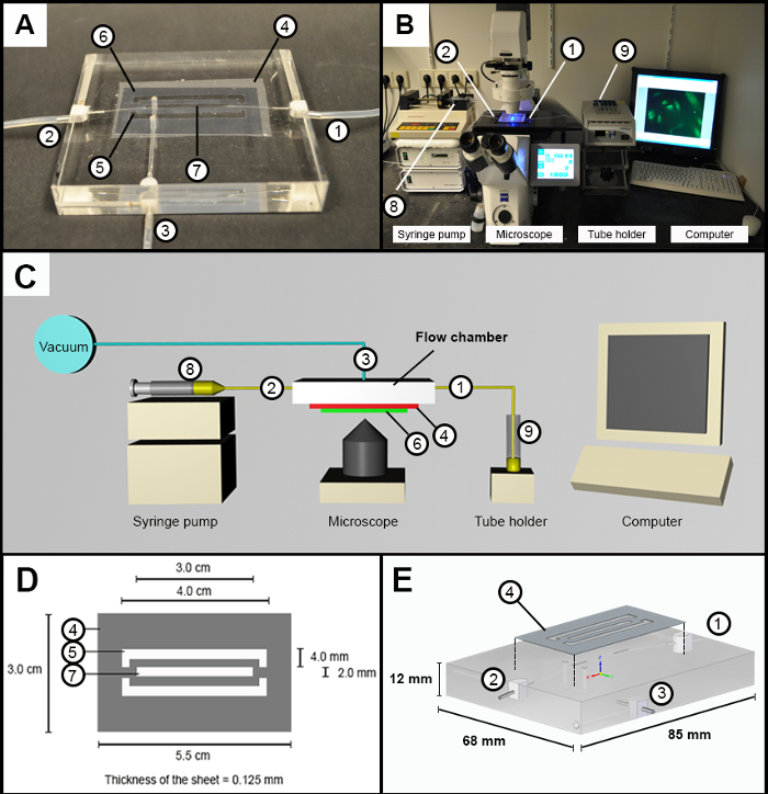
Figure 1: Flow Chamber and Experimental Setup. (A) Image of flow chamber design with an inlet and outlet (1 and 2), a vacuum connection (3) on which the silicone sheet (4) is placed. Two outer cutouts (5) in the silicon sheet form the vacuum channels to attach the cover glass (6). A central cutout (7) forms the flow channel (B) Image of the experimental setup. Inlet and outlet (1 and 2), syringe pump (8), and sample holder (9). (C) Schematic overview of the experimental setup; the numbers are identical to those in A and B. (D) Schematic overview with the dimension of the customized cut silicone sheet. (E) General schematic overview of the poly(methyl methacrylate) flow chamber. Please click here to view a larger version of this figure.
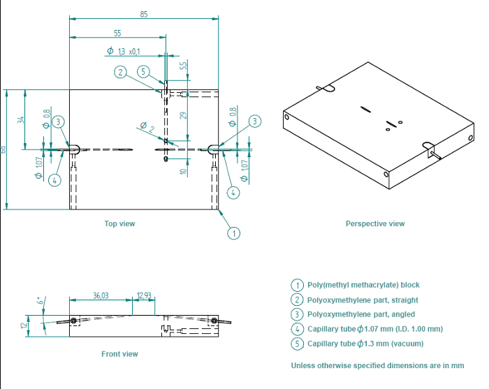
Figure 2: Detailed Blueprint of Flow Chamber. The flow chamber consists of a poly(methyl methacrylate) block (1), one straight polyoxymethylene insert for the vacuum inlet (2), and two angled polyoxymethylene inserts for the sample inlet and outlet (3). The angled polyoxymethylene inserts contain metal capillary tubes, with diameters of 1.07 mm. The straight insert contains a capillary tube with a diameter of 1.3 mm. Please click here to view a larger version of this figure.
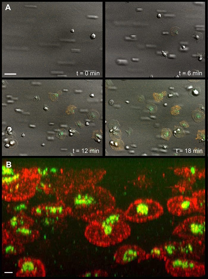
Figure 3: Platelet Spreading and Degranulation on Immobilized von Willebrand Factor. (A) Time-dependent mobilization of CD63 and P-selectin onto the surface of spreading and degranulating platelets. The white scale bar (upper left) indicates 10 µm. (B) Representative confocal fluorescence microscopy image at a 45° angle. The white scale bar (lower left) indicates 2 µm. Green, CD63; Orange, P-selectin. Please click here to view a larger version of this figure.
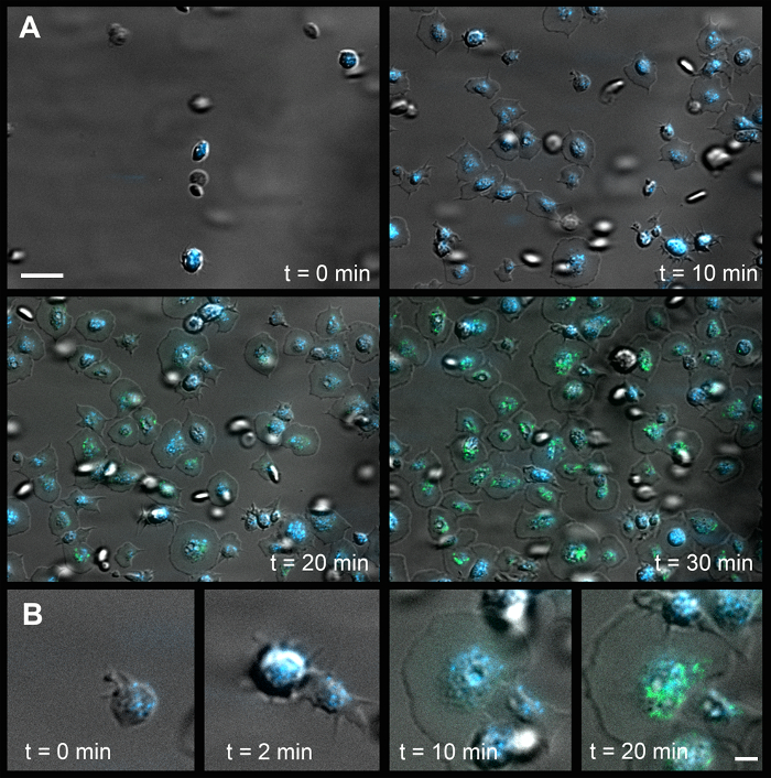
Figure 4: Platelet Spreading, Degranulation, and Mobility of Dense Granules on Immobilized Fibrinogen. (A) Time-dependent mobilization of CD63 and polyphosphate onto the platelet surface. The white scale bar (upper left) indicates 10 µm. (B) Time series of a single platelet (t = 0 represents the first adhesion to surface). The white scale bar (lower right) indicates 2 µm. Green, CD63; Blue, DAPI. Please click here to view a larger version of this figure.
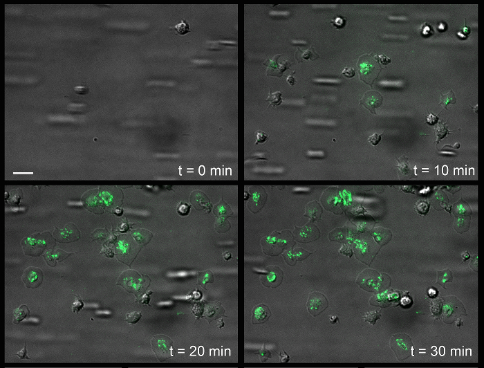
Figure 5: Surface Association of von Willebrand Factor onto the Platelet Surface of Immobilized Fibrinogen. Time-dependent mobilization (or recruitment) of VWF to the platelet surface. The white scale bar (upper left) indicates 5 µm. Green, VWF. Please click here to view a larger version of this figure.
Setting | Value |
Exposure time FITC (Alexa488) | 200 ms / 300 ms |
LED 488 intensity FITC (Alexa488) | 50% |
Exposure time DIC | 30 ms |
Voltage halogen lamp | 4.5 Volt |
Exposure time TRITC (Alexa 546) | 400 ms |
LED 555 intensity FITC (Alexa546) | 50% |
Exposure time DAPI | 500 ms |
LED 365 intensity | 50% |
Filterset FITC | Filterset 10 |
Filterset TRITC | Filterset 20 |
Filterset DAPI | Filterset 01 |
Objective | Alpha Plan-Fluar 100x/1.45 Oil |
Frame rate, recording time | 1 frame / 10 s, 30 min |
File compression movies | MOV (h.264), AVI(uncompressed) |
File compression pictures | JPEG, TIF |
Table 1: Microscope settings. Settings used for the representative experiments.
Target | Antibody / Dye | Concentration |
CD63 | Anti-CD63-biotin *) | 325 ng/mL |
P-selectin | Anti-CD62P-biotin **) | 90 ng/mL |
DNA | DAPI | 10 µg/mL |
VWF | Anti-human VWF- FITC | 5 µg/mL |
*) anti-CD63-biotin antibody is mixed with streptavidin-Alexa488 in a molar ratio of 1:1 before use | ||
**) anti-CD62P-biotin antibody is mixed with streptavidin-Alexa546 in a molar ratio of 1:1 before use |
Table 2: Antibodies and Dyes. Recommended antibody and dye concentrations for the shown representative experiments.
Reagent category | Example compounds |
Platelet agonists | Thrombin, ADP, PAR-1 or -4 activating peptides, U46619, Thromboxane A2, ADP |
Enzyme inhibitors | Hirudin, PPACK, heparinoids (indirect), corn trypsin inhibitor, soy bean trypsin inhibitor |
Receptor antagonists | anti-Glyprotein 1bα, anti-Glycoprotein VI; clopidogrel, (cyclic) RGD-containing peptides |
Activation inhibitors | Aspirin pretreatment, fixation |
Table 3: Potentially useful reagents.
Supplemental Movies: Movie 1: Time-series of images of platelet (see Figure 1), Movie 2: tilt series of Figure 3B, Movie 3: Time-dependent mobilization of CD63 onto the platelet surface (see Figure 4), Movie 4: Time series of single-cell analyses (see Figure 4B), Movie 5: Time-dependent mobilization of VWF (see Figure 5).
Please click here to download Movie 1.
Please click here to download Movie 2.
Please click here to download Movie 3.
Please click here to download Movie 4.
Please click here to download Movie 5.
Worldwide, thrombosis is a leading cause of death and morbidity, and platelets play a central role in its development. This work describes a method for the live-cell imaging of platelet degranulation under flow. It is generally assumed that, when platelets become activated, all granular contents are directly released into solution. The accompanying results suggest that this is not necessarily the case. During adhesion and degranulation, platelets retain a significant amount of polyphosphate (Figure 4). Additionally, the multimeric protein VWF is associated with the platelet surface after degranulation (Figure 5). This indicates that the molecular mechanisms that control platelet granule degranulation are subtler than generally assumed.
To enable the live-cell imaging of platelet degranulation under flow, the experimental conditions were selected to keep platelet aggregation to a minimum. Platelet counts of 150,000 platelets/µL were used. This is at the lower end of the normal range (150,000-450,000 platelets/µL). Furthermore, platelet adhesion and spreading were studied on immobilized VWF or fibrinogen, which led to mild platelet activation (in contrast to, for example, collagen). In studies with washed platelets, increased shear rates move these cells to the center of the flow. This interferes with adhesion and subsequent spreading and complicates the investigation of degranulation.
In physiological hemostasis, platelet margination is important. This process is driven by erythrocytes, explaining why low anemia is sometimes accompanied by a bleeding diathesis. A limitation of the presented technique is that the visualization of platelet degranulation (with the presented materials and reagents) is disturbed by red blood cells. Platelet function is influenced by shear stress, which is a function of the viscosity of the fluid (µ), as well as of the shear rate. Hence, it is recommended to study platelet function in flow models in solutions with comparable viscosities (i.e., washed platelets, reconstituted blood, PRP, or whole blood).
There are several practical factors to take into account when performing these experiments. First, fluorescence signals are strongly influenced by the focus area. If the microscope is not in focus, the signal is easily blurred or even lost. Prior to the arrival of platelets into the flow channel, it is best to focus on the channel itself (begin at the edge of the silicon sheet/flow channel wall). When platelets start to adhere, focus on this adhesion event. A second point of concern is the bleaching of certain fluorophores (e.g., FITC). Repeated excitation will lead to a loss of signal, disturbing the investigation. These obstacles can be overcome by using more stable fluorophores, reducing the intensity of the exciting LED light, shortening the exposure times, and/or lowering the frame rate.
The model described here is very useful for performing a detailed study of platelet responses to a wide variety of immobilized proteins and at different flow rates. Potentially, this method can also be used to study their interaction with activated endothelial cells18. The technical advances that allow for the flexible designs of the special flow chambers are currently opening avenues for biocompatibility studies19. The live-cell imaging of platelet reactivity under flow will reveal valuable insights on the complexity of the platelet adhesion response. Ultimately, these experiences should lead to the development experimental flow models that allow for the combined investigation of platelet function and influence on the coagulation system.
The authors have nothing to disclose.
CM acknowledges financial support from the International Patient Organization for C1-Inhibitor Deficiencies (HAEi), Stichting Vrienden van Het UMC Utrecht, and the Landsteiner Foundation for Blood Transfusion Research (LSBR).
4-(2-hydroxyethyl)-1-piperazineethanesulfonic acid (HEPES) | VWR | 441476L | |
Na2HPO4 | Sigma | S-0876 | |
NaCl | Sigma | 31434 | |
KCl | Sigma | 31248 | |
MgSO4 | Merck KGaA | 1.05886 | |
D-glucose | Merck KGaA | 1.04074 | |
Prostacyclin | Cayman Chemical | 18220 | |
Tri-sodium citrate | Merck KGaA | 1.06448 | |
Citric acid | Merck KGaA | 1.00244 | |
Cover glasses | Menzel-Gläser | BBAD02400500#A | 24x50mm, No. 1 = 0.13-0.16 mm thickness. |
Chromosulfuric acid (2% CrO3) | Riedel de Haen | 07404 | CAS [65272-70-0]. |
Von Willebrand factor (VWF) | in-house purified | ||
Fibrinogen | Enzyme Research Laboratories | FIB3L | |
4 well dish, non-treated | Thermo Scientific | 267061 | |
Human Serum Albumin Fraction V | Haem Technologies Inc. | 823022 | |
Blood collection tubes, 9 ml, 9NC Coagulation Sodium Citrate 3.2% | Greiner Bio-One | 455322 | |
Cell analyser | Abbott Diagnostics | CELL-DYN hematology analyzer | |
Paraformaldehyde | Sigma | 30525-89-4 | |
Syringe pump | Harvard Apparatus, Holliston, MA | Harvard apparatus 22 | |
10 mL syringe with 14.5 mm diameter | BD biosciences | 305959 | Luer-Lok syringe |
Anti-CD63-biotin | Abcam | AB134331 | |
Anti-CD62P-biotin | R&D Systems | Dy137 | |
4’,6-Diamidino-2-phenylindole dihydrochloride (DAPI) | Polysciences Inc. | 9224 | |
Streptavidin, Alexa Fluor 488 conjugate | Thermo Scientific | S11223 | |
Immersion oil | Zeiss | 444963-0000-000 | |
Detergent solution | Unilever, Biotex | ||
Glycine | Sigma | 56-40-6 | |
Polyvinyl alcohol | Sigma | 9002-89-5 | Mowiol 40-88. |
Tris hydrochloride | Sigma | 1185-53-1 | |
1,4-Diazabicyclo[2.2.2]octane (DABCO) | Sigma | 280-57-9 | |
Sheep Anti-hVWF pAb | Abcam | AB9378 | |
Alexa fluor 488-NHS | Thermo Scientific | A20000 | |
Glycerol | Sigma-Aldrich | 15523-1L-R | |
Parafinn film | Bemis | PM-996 | 4 in. x 125 ft. Roll. |
Silicone sheet non-reinforced | Nagor | NA 500-1 | 200mmx150mmx0.125mm. |
Customized cut silicone sheet with perfusion and vacuum channels | in-house made | Made of Silicone sheet non-reinforced (Nagor, NA 500-1) | |
1.5 mL tubes | Eppendorf AG | T9661-1000AE | |
Fluorescent microscope | Zeiss Observer Z1 | Equiped with LED excitation lights. | |
Microscope software | Zeiss ZEN 2 | blue edition | |
18 G needle (18 G x 1 1/2") | BD biosciences | 305196 | |
NaCl | Riedel de Haen | 31248375 | |
Tris | Roche | 10708976 | |
Plastic pasteur pipet | VWR | 612-1681 | 7 ml non sterile, graduated up to 3ml. |
Silicone tubing | VWR | 228-0656 | Inner diamete. x Outer diameter x Wall thickness = 1.02 x 2.16 x 0.57 mm. |
Microscope slides | Thermo Scientific | ABAA000001##12E | 76 x 26 x 1 mm, ground edges 45°, frosted end. |