We present a protocol to isolate adipose tissue-derived microvascular fragments that represent promising vascularization units. They can be rapidly isolated, do not require in vitro processing and, thus, may be used for one-step prevascularization in different fields of tissue engineering.
A functional microvascular network is of pivotal importance for the survival and integration of engineered tissue constructs. For this purpose, several angiogenic and prevascularization strategies have been established. However, most cell-based approaches include time-consuming in vitro steps for the formation of a microvascular network. Hence, they are not suitable for intraoperative one-step procedures. Adipose tissue-derived microvascular fragments (ad-MVF) represent promising vascularization units. They can be easily isolated from fat tissue and exhibit a functional microvessel morphology. Moreover, they rapidly reassemble into new microvascular networks after in vivo implantation. In addition, ad-MVF have been shown to induce lymphangiogenesis. Finally, they are a rich source of mesenchymal stem cells, which may further contribute to their high vascularization potential. In previous studies we have demonstrated the remarkable vascularization capacity of ad-MVF in engineered bone and skin substitutes. In the present study, we report on a standardized protocol for the enzymatic isolation of ad-MVF from murine fat tissue.
Tissue engineering focuses on the fabrication of tissue and organ substitutes that maintain, restore or augment the function of inoperable in vivo counterparts1,2. The fate of engineered tissue constructs crucially depends on an adequate vascularization3. Microvascular networks within these constructs should be hierarchically organized with arterioles, capillaries, and venules to allow efficient blood perfusion after inosculation to the recipient's vasculature4. The generation of such networks is among the key challenges in tissue engineering. For this purpose, a broad spectrum of experimental vascularization strategies has been introduced over the last two decades5,6.
Angiogenic approaches stimulate the ingrowth of recipient microvessels into engineered tissues by means of structural or physicochemical scaffold modification, such as the incorporation of growth factors7. However, for the vascularization of large three-dimensional constructs, angiogenesis-dependent strategies are markedly limited by slow growth rates of developing microvessels8.
In contrast, the concept of prevascularization aims for the generation of functional microvascular networks within tissue constructs prior to their implantation9. Conventional prevascularization involves the co-culture of vessel-forming cells, such as endothelial cells, mural cells or stem cells10, within scaffolds. After microvascular network formation, the prevascularized constructs can then be implanted into tissue defects. Noteworthy, this prevascularization approach is difficult to apply in a clinical setting, because it is based on complex and time-consuming in vitro procedures, which are restricted by major regulatory hurdles9. Accordingly, there is still a need for the development of novel prevascularization strategies that are more suitable for a broad clinical application.
Such a prevascularization strategy may be the application of adipose tissue-derived microvascular fragments (ad-MVF). ad-MVF represent potent vascularization units that can be harvested in large amounts from the fat tissue of rats11,12 and mice13. They consist of arteriolar, capillary, and venular vessel segments, which exhibit a physiological microvessel morphology with a lumen and stabilizing perivascular cells14,15. This unique feature allows the immediate implantation of ad-MVF-seeded scaffolds into tissue defects without precultivation. There, the ad-MVF rapidly reassemble into functional microvascular networks. Furthermore, ad-MVF represent a rich source of mesenchymal stem cells16, which may additionally contribute to their striking regenerative capacity. Accordingly, ad-MVF are increasingly used in different fields of tissue engineering14,15,17,18,19,20,21.
The isolation of ad-MVF has originally been established in rats11,12. Herein, we describe a protocol, which allows the standardized isolation of murine ad-MVF from epididymal fat pads. This may provide further insights into molecular mechanisms underlying ad-MVF function by using transgenic mouse models.
All procedures were performed according to the National Institute of Health guidelines for the use of experimental animals and followed institutional guidelines (Landesamt für Soziales, Gesundheit und Verbraucherschutz, Abt. Lebensmittel- und Veterinärwesen, Zentralstelle, Saarbrücken, Germany).
1. Preparation of Surgical Instruments
2. Animals and Anesthesia
3. Harvesting of Epididymal Fat Pads
4. Isolation of ad-MVF
In the present study we performed six ad-MVF isolation procedures with fat tissue from 7- to 12-month-old male wild-type C57BL/6 mice (mean body weight: 35 ± 1 g). Figure 1 illustrates the harvesting of murine epididymal fat pads with subsequent mechanical and enzymatic ad-MVF isolation. The time required for the harvesting of fat was 30 min and for the isolation of ad-MVF was 120 min. In total, the procedure took 150 min.
We harvested 1.2 ± 0.1 mL of adipose tissue per donor animal. This amount of fat allowed the isolation of 42,000 ± 2,000 ad-MVF per mL. The mean length of the isolated ad-MVF was 42 ± 1 µm (Figure 2A). The duration of enzymatic digestion was determined by means of microscopic control as shown in Figure 2B. ad-MVF exhibited a typical microvessel morphology with hierarchical vessel segments (Figure 2C).
We additionally characterized the ad-MVF by means of flow cytometry. For this purpose, ad-MVF were further digested in cell detachment solution for 30 min into single cells15. The flow cytometric analyses revealed that ad-MVF contain 26 ± 2% CD31-positive endothelial cells, 17 ± 2% α-smooth muscle actin (SMA)-positive perivascular cells, and 9 ± 1% cells positive for the mesenchymal stem cell marker CD117 (Figure 3A-C). After seeding on a dermal skin substitute, which was fixed and sectioned immediately for histological analyses, larger ad-MVF were mainly localized on the implant's surface (Figure 3D). However, capillary vessel segments could also be detected within its pores. Immunohistochemical staining of seeded ad-MVF further revealed physiological microvessel configuration with arteriolar, venular and capillary-like fragments (Figure 3E).
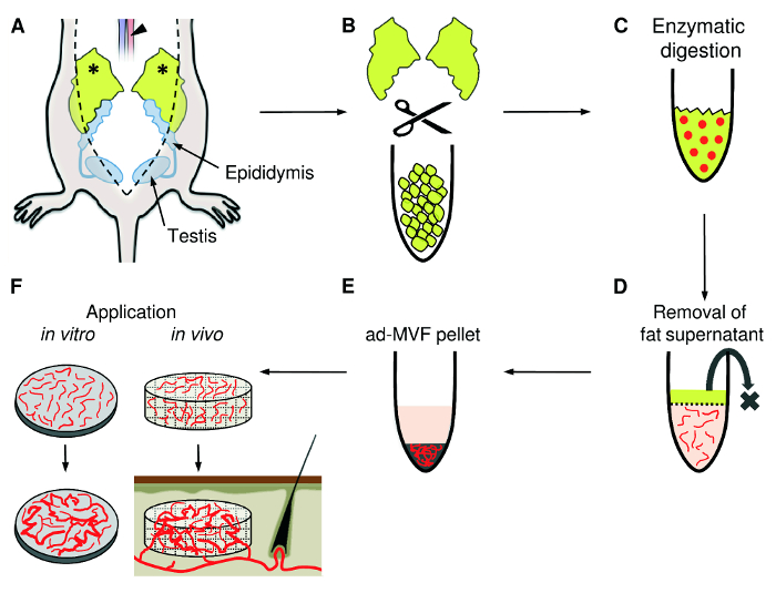
Figure 1: ad-MVF Isolation. A) Following midline laparotomy, the epididymal fat pads (asterisks) are identified. The adipose tissue is harvested with a small preparation scissors and the animal is sacrificed by incision of the abdominal aorta (arrowhead). B) Mechanical mincing of the fat pads with fine scissors until the tissue suspension appears homogeneous. C) For enzymatic digestion, collagenase is added to the tissue suspension. D) The cell-vessel suspension is incubated and the supernatant fat layer is discarded. E) After centrifugation, the pellet containing ad-MVF is resuspended and can be used for different applications, such as the in vitro analysis of microvascular network formation or the seeding of ad-MVF on scaffolds for in vivo implantation into tissue defects (F). Please click here to view a larger version of this figure.
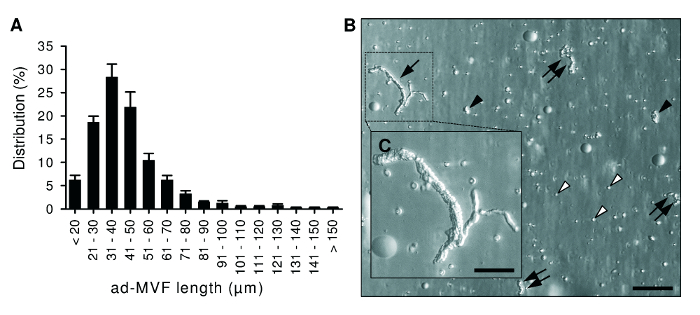
Figure 2: Morphological Characteristics of ad-MVF. A) Length distribution of freshly isolated ad-MVF. Mean ± standard error of the mean (n = 6). B) Microscopic image of a cell-vessel suspension smear with large (arrow), medium-sized (double arrows) and small (black arrowheads) ad-MVF as well as single cells (white arrowheads). Scale bar = 110 µm. Higher magnification reveals that ad-MVF exhibit a mature microvessel morphology with hierarchical microvessel segments (C, inset of B). Scale bar: C = 50 µm. Please click here to view a larger version of this figure.
Figure 3: Flow Cytometric, Histological and Immunohistochemical Characteristics of ad-MVF. A-C) Flow cytometric analyses of freshly isolated ad-MVF illustrating CD31-positive endothelial cell (A), α-SMA-positive perivascular cell (B) and CD117-positive mesenchymal stem cell (C) fractions. Appropriate isotype-identical controls were used to adjust threshold levels. Mean ± standard error of the mean (n = 6). D) Hematoxylin and eosin-stained section of a seeded dermal skin substitute. A few capillary vessel segments are located in the implant's pores (arrowheads). Larger microvessels with venular (double arrow) or arteriolar (arrow) morphology are localized on its surface. Scale bar = 40 µm. Broken line = implant border. E) Characterization of ad-MVF by means of immunohistochemical detection of the endothelial cell marker CD31 (red) and the perivascular cell marker α-SMA (green). Cell nuclei were stained with a DNA-binding fluorescent dye (blue). Arteriolar ad-MVF (arrow) are characterized by a thicker α-SMA-positive cell layer when compared to venular fragments with a larger lumen (double arrow). Arrowheads = capillary ad-MVF. Scale bar = 25 µm. Please click here to view a larger version of this figure.
In this study we present a well-established protocol for the isolation of ad-MVF. Obtaining ad-MVF from murine adipose tissue is a straightforward procedure with a few critical steps. Mice exhibit different subcutaneous and intraabdominal fat deposits. As previously described for rats, the most suitable fat source for the isolation of ad-MVF are the epididymal fat pads due to their size, homogeneous structure and minimal contamination with larger blood vessels11,12. In contrast, subcutaneous fat deposits in mice are much smaller and more adherent to the surrounding tissue. However, the excision of epididymal fat pads bears the risk of accidental injury of the epididymis and spermatic cords, resulting in contamination of the harvested tissue with sperms. Therefore, the identification of epididymis, testis and spermatic cords is of major importance in this procedure. Moreover, the fat pads should be rapidly processed after harvesting to minimize ex vivo tissue damage. Finally, it should be considered that there are major age-dependent variations in the vascularization of murine epididymal fat pads23. In our experience, the use of 6- to 12-month-old male donor mice provides sufficient amounts of ad-MVF for in vitro and in vivo studies. However, it may also be possible to isolate ad-MVF from younger mice, which exhibit smaller but highly vascularized fat pads24.
The enzymatic digestion of the harvested epididymal fat pads crucially determines the final quality of isolated ad-MVF. Too short exposition to collagenase leads to incomplete separation of ad-MVF from surrounding adipocytes. On the other hand, prolonged digestion results in the destruction of ad-MVF to a single cell suspension, the so-called stromal vascular fraction (SVF). The SVF is a heterogeneous cell population containing mesenchymal stem cells, endothelial cells, pericytes, macrophages and many other cell types with regenerative potential through paracrine and differentiation mechanisms25. The use of ad-MVF exhibits considerable advantages when compared to SVF-based approaches. The fully functional microvessel morphology of ad-MVF allows their immediate implantation for in vivo experiments without time-consuming and complex in vitro cultivation. Moreover, the isolation of the SVF usually requires the digestion of the tissue with collagenase up to 90 min26. In contrast, tissue degradation should not exceed 10 min for the isolation of ad-MVF. This supports the idea of intraoperative one-step applications of ad-MVF9. For this purpose, ad-MVF may be automatically isolated from autologous lipoaspirates and transferred back into a tissue defect of the patient without leaving the operation theatre. This is a realistic goal considering the fact that the automated isolation of SVF from lipoaspirates is already a clinically established procedure in plastic surgery27,28,29.
Recently, we have demonstrated a strong effect of ad-MVF on the in vivo vascularization of bone14 and skin15 substitutes. Of interest, we found that it is beneficial not to remove single cells from the ad-MVF isolate, because they may provide a more physiological environment that contributes to the high vascularization capacity of ad-MVF13. Therefore, we only used a 500-µm filter during the isolation of ad-MVF for the removal of larger non-digested fat clots. Moreover, we found that ad-MVF induce lymphangiogenesis in implanted skin substitutes15. Hence, ad-MVF may also be promising building blocks for lymphatic tissue engineering and lymphedema therapy.
For the successful translation of these experimental findings into clinical practice, the next logical step is to clarify whether human adipose tissue is equally suited for the isolation of ad-MVF as rodent fat. In this context, a critical issue is the amount of fat required to isolate large amounts of ad-MVF. The herein described protocol resulted in approximately 40,000 ad-MVF per mL fat tissue. Accordingly, 3 mL fat tissue would be necessary to prevascularize 1 cm2 of dermal skin substitute15. Hence, this approach may not be feasible for the treatment of larger wounds or severely ill patients due to a limited amount of available fat tissue for ad-MVF isolation.
Beside a broad spectrum of potential in vivo applications in tissue engineering and regenerative medicine, ad-MVF are also suitable for in vitro modeling of angiogenic processes. For instance, rat ad-MVF have been cultured in three-dimensional collagen hydrogels to study the formation of new microvascular networks30. Similar approaches have been used to evaluate sophisticated imaging techniques for the visualization of neovessel growth13 or interstitial matrix remodeling during angiogenesis31.
Taken together, these findings indicate that ad-MVF are not only promising vascularization and lymphangiogenic units for future therapeutic strategies, but also for basic research in the field of angiogenesis and vascular physiology.
The authors have nothing to disclose.
We are grateful for the excellent technical assistance of Janine Becker, Caroline Bickelmann and Ruth Nickels. This study was funded by a grant of the Deutsche Forschungsgemeinschaft (DFG – German Research Foundation) – LA 2682/7-1.
1.5-mL conical microcentrifuge tube | VWR, Kelsterbach, Germany | 700-5239 | |
100-µL precision pipette | Eppendorf, Hamburg, Germany | 4920000059 | |
10-mL measuring pipette | Costar, Corning Inc., New York, USA | 4488 | |
14-mL PP tubes | Greiner bio-one, Frickenhausen, Germany | 187261 | |
1-mL precision pipette | Eppendorf, Hamburg, Germany | 4920000083 | |
500-µm filter (pluriStrainer 500 µm) | HISS Diagnostics, Freiburg, Germany | 43-50500-03 | |
50-mL conical centrifuge tube | Greiner bio-one, Frickenhausen, Germany | 227261 | |
50-mL Erlenmeyer flask | VWR, Kelsterbach, Germany | 214-0211 | |
96-well plate | Greiner bio-one, Frickenhausen, Germany | 65518 | |
cell detachment solution (Accutase) | eBioscience, San Diego, CA USA | 00-4555-56 | |
C57BL/6 mice | Charles River, Cologne, Germany | 027 | |
C57BL/6-Tg(CAG-EGFP)1Osb/J mice | The Jackson Laboratory, Bar Harbor, USA | 003291 | |
CD117-FITC | BD Biosciences, Heidelberg, Germany | 553373 | |
CD31-PE | BD Biosciences, Heidelberg, Germany | 553354 | |
Collagenase NB4G | Serva Electrophoresis GmbH, Heidelberg, Germany | 17465.02 | Lot tested by manufacturer for enzymatic activity |
Dissection scissors | Braun Aesculap AG &CoKG, Melsungen, Germany | BC 601 | |
DNA-binding dye (Bisbenzimide H33342) | Sigma-Aldrich, Taufkirchen, Germany | B2261 | |
Dulbecco's modified Eagle medium (DMEM) | PAN Biotech, Rickenbach, Germany | P04-03600 | |
Fetal calf serum (FCS) | Biochrom GmbH, Berlin, Germany | S0615 | |
Fine forceps | S&T AG, Neuhausen, Switzerland | FRS-15 RM-8 | |
Fine scissors | World Precision Instrumets, Sarasota, FL, USA | 503261 | |
Dermal skin substitute (Integra) | Integra Life Sciences, Sain Priest, France | 62021 | |
Ketamine | Serumwerk Bernburg AG, Bernburg, Germany | 7005294 | |
M-IgG2akAL488 | eBioscience, San Diego, CA USA | 53-4724-80 | |
Octeniderm (disinfecting solution) | Schülke & Mayer, Norderstedt, Germany | 118211 | |
Penicillin/Streptomycin | Biochrom, Berlin, Germany | A2213 | |
Petri dish | Greiner bio-one, Frickenhausen, Germany | 664160 | |
Phosphate-buffered saline (PBS) | Lonza Group, Basel, Switzerland | 17-516F | |
pluriStrainer 20-µm (20 µm filter) | HISS Diagnostics, Freiburg, Germany | 43-50020-03 | |
Rat-IgG2akFITC | BD Biosciences, Heidelberg, Germany | 553988 | |
Rat-IgG2akPE | BD Biosciences, Heidelberg, Germany | 553930 | |
Small preparation scissors | S&T AG, Neuhausen, Switzerland | SDC-15 R-8S | |
Surgical forceps | Braun Aesculap AG &CoKG, Melsungen, Germany | BD510R | |
Tape (Heftpflaster Seide) 1.25 cm | Fink & Walter GmbH, Mechweiler, Germany | 1671801 | |
Xylazine | Bayer Vital GmbH, Leverkusen, Germany | 1320422 | |
α-SMA-AL488 | eBioscience, San Diego, CA USA | 53-9760-82 | Intracellular labeling additionally requires Cytofix/Cytoperm (BD Biosciences, Heidelberg, Germany; #554722) |