Summary

3 차원 조직 설계 정렬 사이토 네트워크 발달 메커니즘을 정리 하 고 신경 재생을 촉진 하

Published: January 10, 2018
doi:

Summary

우리 쇼케이스 경도 정렬된 astrocytic somata 소설 소재 넣음 내 프로세스의 자기 조립, 3 차원 번들의 개발. 이러한 “생활 건설 기계”, 아직 테스트-베드 neurodevelopmental 메커니즘을 공부 하거나 감독 신경 마이그레이션 및 axonal neuroregeneration를 용이 하 게 하 역할 수 센티미터 길이, 확장 미크론 단위 직경을 전시 설계 길입니다.

Abstract

Neurotrauma 및 신경 퇴행 성 질환 자주 손실된 신경 대체 axonal 경로 재생성 하 중앙 신경 시스템 (CNS)의 제한 된 용량으로 인해 신경학 상 적자 지속 될. 그러나, 신 시스템 개발, 신경 마이그레이션 및 axonal 확장 하는 동안 자주 발생 “건설 기계 생활”로 다른 세포에 의해 형성 하는 경로 따라 합니다. 이러한 메커니즘을 에뮬레이션 circumvents CNS의 금지 환경 전략을 디자인 하 고 추구,이 원고 제공 조직 설계를 조작 하는 프로토콜 사이토 기반 “건설 기계 생활”. 이러한 구조를 만들려면, 우리는 바이 폴라 경도 정렬 somata 및 프로세스의 고밀도 3 차원 번들으로 조립 자체 이다 유도 하는 소설 소재 넣음 구조 고용. 첫째, 빈 히드로 마이크로-열 조립 했다, 그리고 내부 루멘 기질-콜라겐 코팅 했다. 해리 대뇌 피 질 이다 다음의 중요 한 내부 직경, 및 원통형 마이크로 열의 루멘으로 배달 되었다 < 350 µ m, 자발적으로 자기 정렬 및 밀도 번들으로 구성 된 긴 섬유 같은 케이블을 생산 하는 계약을 체결 사이토 프로세스와 콜라겐 소 측정의 97% 생존 세포와 거의 독점적으로 했다 이다 표현 하는 중간 필 라 멘 트 단백질 폐해 fibrillary 산 성 단백질 (GFAP), vimentin의 조합 및 nestin의 구성. 이 사이토 네트워크 신경 첨부 파일에 대 한 관대 한 기질을 제공 하는 것을 발견 했다 고 neurite 연장 정렬 정렬. 또한, 이러한 구조에 맞춤 히드로 넣음, CNS 이식 적합 하에서 추출 될 때 무결성 유지 관리 합니다. 이러한 미리 형성한 구조는 구조적으로 자연스럽 게 발생의 주요 cytoarchitectural 요소를 에뮬레이션 “건설 기계 생활” glial 기반 에 비보. 따라서, 이러한 설계 생활 건설 기계 테스트-베드 neurodevelopmental 메커니즘에서 생체 외에서 연구 또는 신경 마이그레이션 및 CNS 변성 vivo에서 다음 axonal 길 여 neuroregeneration 촉진 역할 수 있습니다. .

Introduction

중앙 신경 조직 (CNS) 중화 손실 및/또는 신경의 외상 성 뇌 손상 (TBI), 같은 조건이 동반 axonal 경로 장애 제한 용량이 뇌졸중, 척수 상해 (SCI), 및 신경 퇴행 성 질병1 ,2,3,,45. CNS에 신생 방해 손실된 신경6,7의 복원, 두뇌에 있는 영역의 제한 된 수에 제한 됩니다. 또한, CNS에서 손실된 axonal 통로의 재생 감독된 지침의 부족, 파생물 억제제, 및 신경 조직2,8에 손상 다음 반응 astrogliosis의 존재 충분 하지 않습니다. 9,10. 이다는 일반적으로 이온 항상성, 신경 전달 물질 클리어런스, 시 냅 스 형성, 및11를 커플링 하는 혈관과 신경 지원에 다양 한 기능을가지고. 그럼에도 불구 하 고, 신경 조직에 가벼운 손상, 다음 이다 수 있습니다 받을 분자, 구조적, 기능적 변화 그들은11hypertrophic 상태 전환. 심한 neurotrauma에 대응, 이러한 변경 결과 흉터의 형성에 포함 된 마이그레이션 반응성 이다는 파열된 혈액-뇌 장벽 (BBB) microglia에서 유출 하는 백혈구를 포함 하는 병 변 코어 penumbra oligodendrocytes, 그리고 섬유 아 세포11,,1213. 이러한 반응성 이다 달성 filamentous, 비 조직 프로세스의 형태 그리고 중간 필 라 멘 트 단백질 및 신경 재생12방해 콘 proteoglycans (CSPGs)의 증가 식 전시. Glial 흉터 처음 BBB 무결성을 복원 하 고 건강 한 티슈를 에워싸는 것 염증 반응의 전송을 방지 하는 데 도움이, 비록 그것은 축 삭 재생12,14에 대 한 물리적 및 생화학 장벽 역 ,,1516. 예를 들어, glial 흉터 발생 axons 주먹코 dystrophic 성장 콘을 표시 하 고 성장12저하. 또한, 부상 후 astrocytic 프로세스의 해체는 축 삭17회생의 확장을 방해 한다. 이러한 억제 특성의 결과 종종 영구 신체적, 신경 장애 환자 겪는 심각한 neurotrauma, TBI 및 문화를 포함 한 후에 각 성

외부 직면 CNS 기능 재생에 축 삭 재생성 하는 본질적인 능력을가지고 표시 되었습니다. 예를 들어, glial 흉터 문의 dystrophic 성장 콘의 동적 특성 제안 이러한 엔딩 확장12하 그들의 능력을 유지 합니다. 따라서, 그것은 있다고 믿고 axonal 다시 성장에 주요 방해 후 부상 CNS와 감소 glial 흉터 및 흉터에서 재생 다리 것 제공을 통해 더 환경을 제공 하는 금지 환경 유리. 실제로, 이전 학문은 설명 했다 CNS 뉴런 축 삭 재생12,18에 대 한 더 많은 유리한 환경을 제시, 교량으로 주변 신경 이식 술을 사용 하 여 병 변을 통해 축 삭 연장 가능 했다 19. 여러 가지 다른 전략이 흔적 재생 능력을 악용 하 추구 되어 있다. 예를 들어 다양 한 부상 모델에서 세포 성장 신호 통로의 조작 axonal 재생 및 glial 흉터 감소10,,2021에 결과 있다. 또한, 연구 치료 chondroitinase CSPGs에 설탕 체인의 대부분을 앞, ABC CSPGs 반응 이다22분 비의 억제 효과 줄이는 나타났습니다. 도 불구 하 고 고무적인 결과, 이러한 방식을 벗어난 재생12, 잠재적으로 발생할 수 있습니다 하 고 또한 신경의 손실에 대 한 계정을 하지 않습니다 성장 콘의 지도 감독을 제공 하지 않습니다. 셀 기반 접근 시도 glial 흉터의 효과 극복 하 고 보충 손실된 세포, 특히 신경에 이용 되어 있다. 다른 CNS 병 변 축 삭 재생23,24, 을 추진 하 고 다시 상해 지역에 신경 조상 세포를 이식 하는 동안 일부 그룹 뉴런으로 반응 이다를 dedifferentiated는 그러나 25., 줄기 세포 이식 혼자 낮은 생존 율, 불 쌍 한 통합 및 손상 된 조직을5겸손 보존에 의해 제한 됩니다. 또한, 이러한 세포 기반 전략 제어 방식 특히 장거리 axonal 책자를 복원 실패. 따라서, 다른 접근에와 함께 바이오는 다양 한 신경에 대 한 배달 차량으로 탐험 되 고 조상 세포 및 성장 요인26. 소재 기반 접근 디자인 컨트롤 구문 특정 물리적, haptotaxic 모방 생산의 높은 학위와 대상 호스트 조직27의 3 차원 (3D) microenvironment chemotaxic 단서 제시 28,29,30,31,32,,3334. 이러한 환경 신호 복제는 네이티브 같은 형태학, 확산, 마이그레이션, 및 다른 neurobiological 특성29중 신호 이식된 세포에 대 한 최고입니다. 이러한 유리한 속성 시드 전통적인 셀 소재 건설 기계 넘어 발전은 동시에 감독된 장거리 axonal 재생을 촉진 하 고 손실 된 신경 세포를 대체 하는 데 필요한.

“생활 건설 기계”, 다른 네이티브 neuroanatomy를 에뮬레이트하는 미리 형성한 cytoarchitecture와 살아있는 신경 세포의 존재로 인해 다른 셀 기반 접근을 설계 유망한 다른 접근은 신경 조직 기반 및 타겟된 교체, 개조, 및 신경 회로4,35의 재생을 촉진 하기 위하여 발달 기계 장치. 건설 기계 생활의 디자인에 대 한 고려 사항 포함 고기 그리고 신경 세포, 기계/물리 속성의 소스 및 생 화 확 적인 신호 어떤 동반 바이오35의 구성에 의해 결정. 제조 후 체 외에, 이러한 생활 건설 기계 이식 수 비보에 현재 세포 접착 분자 및 혈 및 科 신호 신경 세포 이동과 축 삭 가지 상태에 따라 적극적으로 규제를 하 고 재생의 진행35를처리합니다. Glial 세포는 이후 다양 한 개발 메커니즘에 vivo에서이 세포 중재 건설 기계 생활 설계 된 cytoarchitecture에 대 한 기준으로 사용할 수 있습니다. 두뇌 개발, 새로운 신경 세포 기저 프로세스 개발 외피 격판덮개로 심 실 영역에서 방사형 명과 감독된 마이그레이션36,37건설 기계 생활으로 확장에 의존 합니다. 또한, 콘은 도표 glial 세포, elicited 매력적이 고 구 충 제 신호를 감지 하 여 스스로 방향을 표시 하 고 소위 “개척” 축 삭 성장 확장 제안 하 고 폐해 미리 패턴을 따라 확장 하 여 올바른 목표 도달 35,,3839투어 따라서, glial 세포는 축 삭, 나중으로 축 삭을 기반으로 봉사 하는 개척의 지도에 필요한 “건설 기계 생활” “추종자” 축 삭의 투영을 직접. 또한, 명과 중재 성장 메커니즘 표시 되었습니다 유지 postnatally, neuroblasts 따라 rostral 철새 스트림 (RMS) 帯 (SVZ), 성체 뇌에서 신생의 몇 가지 남아 있는 분야 중 하나에서 탐색 하는 후 각 전구 (OB)40. RMS에서이 neuroblasts 경도 정렬 astrocytic 프로세스, 직접 셀 유착을 통해 이루어진 고 성 요인37, 지역화 glial 튜브 (그림 1A-1), 내 마이그레이션 41. 마지막으로, 포유류 원인에 CNS 손상 중단 astrocytic 프로세스 배열 axonal 재생17를 물리적으로 방해 glial 흉터를 형성 하는 동안 많은 비 포유류 시스템 부족 해로운 glial 흉터의 형성. 오히려, 비 포유류 종의 glial 세포 유지 더 조직, 부상된 지역17,,4243를 통해 가이드로 사용 되는 패턴을 정렬 합니다. 예를 들어, 비 포유류 과학 모델에서 축 삭 glial 다리 건너 기판 axonal 재생과 기능 회복 (촉진으로 조직 된 폐해 건설 기계에 대 한 중요 한 역할을 제안 하는 병 변와 가까운 협회에서 성장에 표시 됩니다. 그림 1A -2) 42 , 44 , 45. 신경 해부학 기능 및 위에서 설명한 발달/재생 메커니즘의 재현 부 동시에 미 숙 신경 마이그레이션 구동할 수 조작된 폐해-기반 생활 장비의 새로운 클래스를 얻을 수 있습니다 및 axonal 그렇지 않으면 비 허용 환경, 그로 인하여 잠재적으로 신경의 효과 완화를 통해 길 및 축 삭으로 변성 CNS 상해 및 질병과 관련 된.

연구 그룹은 이전 여러 종류의 재건을 위한 건설 기계 생활 설계 및 CNS와 말 초 신 경계 (PNS) 마이크로 조직 통해 axonal 책자의 재생 신경 네트워크 (마이크로-TENNs) 및 조직 설계 신경 이식 (TENGs), 각각27,,4647,48를 설계. 두 전략 biomimicry에 근본적으로 근거한 다. 마이크로-TENNs는 해부학 영감 구조 구조적 및 기능적으로 연결 하는 뇌의 뚜렷한 신경 인구 axonal 책자를 대체 하도록 설계 되었습니다. TENGs 축 삭 촉진 axonal 중생, 타겟된 호스트 axonal 재생35,,4648를 달성 하기 위해 “선구자” 축 삭을 따라 “추종자” 축 삭 성장에 의해 exemplified의 개발 메커니즘을 악용 합니다. 우리는 최근 생활 발판의 다양성에 대문자로 기술 개발을 통해 제시 하는 마이크로 TENNs로 비슷한 넣음 스키마를 사용 하 고 명과 기반 메커니즘에서 영감을 추구. 여기, 우리는 히드로 마이크로 열49의 collagenous 루멘에 걸친 정렬된 astrocytic 번들으로 구성 된 구조를 개발 했다. 이러한 astrocytic 생활 건설 기계 만들 해당 직경의 하는 외경 (OD) 및 내경 (ID)와 함께 빈 원통형 히드로 액체 agarose는 모 세관 튜브-침술 바늘 어셈블리를 작성 하 여 개발 되는 튜브와 바늘, 각각. Agarose 겔 화와 히드로 마이크로 열 모 세관 튜브에서 추출, 빈 내부는 유형 나 사이토 접착을 위한 관대 한 환경을 제공 하는 콜라겐 코팅 및 정렬 번들 형성 (그림 1B -1)입니다. 이후에, 루멘은 대뇌 피 질 이다 출생 후 쥐 새끼 (그림 1B-2)에서 절연 된 시드. 2 차원 (2D) 정렬 기법 (ECM) 단백질 패턴, 전기 분야, micropatterned 홈 및 세포 외 매트릭스의 응용 프로그램에 의존 하는 생활 발판에서 사이토 맞춤 반대로 기술에 의존 자기 조립 제어 변수 같은 기판 곡률 (열 ID), 셀 밀도 및 콜라겐 농도50,,5152에 따르면. 이다 계약, 콜라겐을 리 모델링 하 고 바이 폴라, 경도 정렬 형태에서 관찰 vivo에서 (그림 1B-3) 자연 건설 기계와 유사한 취득. 실제로, 우리는 적극적으로 추구 하 고이 케이블 같은 구조를 사용 하 여 미 성숙한 신경 세포 이주 특히 손상 된 CNS의 불리 한 환경을 통해 axonal 재생 촉진의 대상된 지도 대 한 실제 기판으로 포유류 glial 흉터 (그림 1C). 이 문서는 astrocytic 마이크로-열에 대 한 자세한 제작 방법을 제시, 단계 예상된 cytoarchitecture의 현재 한계에 대 한 포괄적인 토론 명암과 면역 형광 이미지 및의 미래 방향에 기술입니다.

Figure 1
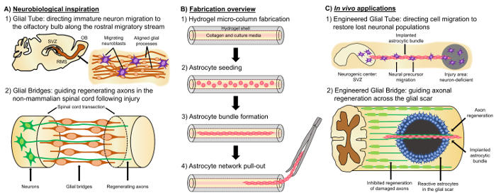
그림 1: 영감, 제조 프로토콜 및 정렬된 Astrocytic 네트워크에 대 한 제안 된 응용 프로그램. (A) Neurobiological 영감: neurogenic 帯 (SVZ)에서 발생 하는 (1) Neuroblasts 후 각 전구 (OB); 향해 감독 마이그레이션 rostral 철새 스트림 (RMS)에 경도 정렬된 glial 관 활용 (2) 비-포유류 양서류, 물고기 등 신경 조직의 병 변 (예: transected 척수)의 끝을 연결 하 고의 지도 위한 비 계 역할 glial 다리의 형성으로 인해 일부 손상 후 재생을 유지할 수 있는 축 삭 재생 (B) 제조 개요: ECM, 코팅 루멘과 미크론 크기, 빈 히드로 마이크로 열의 건설 (1) (2) 기본 외피 이다 출생 후 쥐 새끼, 경도 중심의 (3) 자기 조립에서 고립의 시드 문화와 미래 이식 연구 소재 넣음에서 번들의 (4) 추출에서의 번들 (C) vivo에서 응용 프로그램: (1) 이러한 생활 건설 기계 신경 결핍 지역; 다시 neurogenic 센터에서 감독된 신경 마이그레이션에 대 한 설계 glial 튜브 역할을 수 있습니다 (2) 축 삭 지도 개척의 발달 메커니즘 및 비 포유류에서 glial 교량의 재생 메커니즘의 재현 부 비 허가 걸쳐 축 삭 재생을 직접 수 용량을 가진이 astrocytic 건설 기계를 부여 수 있습니다. 포유류 glial 흉터의 환경입니다. 이 그림의 더 큰 버전을 보려면 여기를 클릭 하십시오.

Protocol

모든 절차는 기관 동물 관리 및 사용 위원회 펜실베니아 대학의 마이클 J. Crescenz 노 병 사변 의료 센터에 의해 승인 되었고 자비 롭에 NIH 공중 보건 서비스 정책에 명시 된 지침을 준수 관리 및 실험실 동물 (2015)의 사용 합니다. 1. 개발의 Agarose 히드로 마이크로-열 Agarose의 3 g의 무게와 Dulbecco의 버퍼링 하는 인산 염 (DPBS) 100 mL를 포함 하는 메 마른 비 커에 전송 agarose 3% ?…

Representative Results

처음에, 단계 대조 현미경 검사 법 사이토 접착 및 번들 형성의 진행과 시간의 함수로 cytoarchitecture의 전반적인 안정성을 모니터링 하는 데 사용 되었다. 도금 후 1 시간에서 이다 둥근 형태 (그림 2A)와 마이크로 열의 루멘을 통해 발견 되었다. 5 h 이다 시작 프로세스를 확장 하 고 8 h 여 계약 셀 완전 한 프로세스 베어링 형태를 전시 케이블 마이크로 ?…

Discussion

PNS 더 지원 환경에 비해, CNS neurotrauma 및 neurodegeneration의 해로운 결과 처리에 특히 제한 됩니다. 포유류 CNS에 심각한 모욕 후 glial 흉터 형성, 분열된 반응 이다 축 삭 발전이 억제 proteoglycans14분 비의 조밀한 meshwork에 둘러싸인 거리 및 염증 세포의 핵심 구성. 이 흉터는 축 삭 엔딩12의 재생에 대 한 물리적 및 생화학 방해 역할을 합니다. 또한, 성인 포유류 CNS 복원 …

Divulgations

The authors have nothing to disclose.

Acknowledgements

재정 지원 건강의 국가 학회 [U01-NS094340 (컬) & F31-NS090746 (Katiyar)], 마이클 제이 폭스 재단 [치료 파이프라인 프로그램 #9998 (컬)], 펜 의학 신경 과학 센터 조종사 상 (컬 린)에 의해 제공 되었다 국립 과학 재단 [대학원 연구 장학 DGE-1321851 (Struzyna)], 재향 군인의 부서 담당 [RR & D 장점 검토 #B1097-난 (컬)], 미국 육군 의료 연구 및 물자 명령 [#W81XWH-13-207004 (컬) & W81XWH-15-1-0466 (컬)].

Materials

Acupuncture needle (300 µm diameter) Lhasa Medical HS.30×40 The diameter may be varied according to the desired size for the micro-column lumen.
Petri dish Fisher 08772B
Dulbecco's phosphate buffered saline (DPBS) Invitrogen 14200075
Polystyrene disposable serological pipet Fisher 13-678-11D
Agarose Sigma A9539-50G
Microliter glass capillary tube (701 µm) Fisher 21-170J The diameter may be varied according to the desired size for the micro-column shell.
Microcap bulb dispenser Fisher 21-170J Bulb comes with the microcap tubes.
Hot plate Fisher SP88857200
Magnetic bar Fisher 1451352
Micropipette Sigma Z683884-1EA
25 mm gauge needle Fisher 14-826-49
Microscalpel Roboz Surgical RS-6270
Scissors Fine Science Tools 14081-09
Forceps World Precision Instruments 501985
Hot bead sterilizer Sigma Z378550-1EA
Stereoscope Nikon SMZ800N
Micro-spatula Fisher S50821
Rat tail type I collagen Corning 354236 Maintain at 4 ºC and remove only when needed. Use ice to preserve its temperature when in use.
Microcentrifuge tube Fisher 02-681-256
Sodium hydroxide (NaOH) Fisher SS2661
Hydrochloric acid (HCl) Fisher SA48-1
Litmus paper Fisher 09-876-18
Hank's balanced salt solution (HBSS) Invitrogen 14170112 Store at 4 ºC.
0.25% Trypsin-EDTA Invitrogen 25200056 Store at -20 ºC and warm at 37 ºC before use.
Bovine pancreatic deoxyribonuclease (Dnase) I Sigma 10104159001 Store at -20 ºC and warm at 37 ºC before use.
Dulbecco's Modified Eagle Medium (DMEM) with Ham's F-12 Nutrient Mixture Gibco 11330-032 Store at 4 ºC.
Fetal bovine serum (FBS) Atlanta Biologicals S11195 Store at -20ºC.
Postnatal day 0 or day 1 Sprague Dawley rat pups Charles River Strain 001
Neurobasal embryonic neuron basal medium Invitrogen 21103049 Store at 4ºC and warm at 37 ºC before use.
B-27 serum free supplement Invitrogen 12587010 Store at -20 ºC and warm at 37 ºC before use.
L-glutamine Invitrogen 35050061 Store at -20 ºC and warm at 37 ºC before use.
G5 astrocytic supplement Invitrogen 17503012
Sprague Dawley embryonic day 18 rats Charles River Strain 001
Pasteur pipette Fisher 22-042816
15 mL centrifuge tube EMESCO 1194-352099
Vortex Fisher 02-215-414
Centrifuge Fisher 05-413-115
Hemocytometer Fisher 02-671-6
Incubator Fisher 13 998 076
Formaldehyde 40% Fisher F77P-4 Formaldehyde is a toxic compound known to be carcinogenic, and must be disposed of in a separate container.
Glass cover slip Fisher 12-548-5M
Nail polish Electron Microscopy Sciences (EMS) 72180
Fluoromont mounting medium Southern Biotech 0100-01
Poly-L-lysine Sigma P4707
Phosphate buffered saline Fisher BP3994
Triton X-100 Sigma T8787
Normal horse serum Gibco 16050-122
Rabbit anti-glial acidic fibrillary protein (GFAP) primary antibody Millipore AB5804 Store at -20ºC.
Mouse anti-beta-tubulin III primary antibody Sigma T8578 Store at -20ºC.
Rabbit anti-collagen I primary antibody Abcam ab34710 Store at -20ºC.
Rabbit anti-vimentin Millipore AB3400 Store at -20ºC.
Mouse anti-nestin Millipore AB5326 Store at -20ºC.
Donkey anti-mouse 568 secondary antibody Invitrogen A10037 Store at 4ºC.
Donkey anti-rabbit 568 secondary antibody Invitrogen A10042 Store at 4ºC.
Donkey anti-rabbit 488 secondary antibody Invitrogen A21206 Store at 4ºC.
Hoechst 33342, Trihydrochloride Invitrogen H3570 Store at 4ºC. Hoechst is a known mutagen that should be treated as a carcinogen. Therefore, it must be disposed of in a separate container.
Calcein AM Sigma C1359 4 mM in anhydrous DMSO
Ethidium homodimer-1 Life Technologies E1169 2 mM in DMSO/H2O 1:4 (v/v)
Dimethyl sulfoxane (DMSO) Sigma 276855
A1RSI Laser Scanning Confocal Microscope Nikon ————– Used for taking the confocal reconstructions of immunolabeled constructs.
Eclipse Ti-S Microscope Nikon ————– Used for taking the phase-contrast images. With digital image acquisition using a QiClick camera interfaced with Nikon Elements Basic Research software (4.10.01).

References

  1. Horner, P. J., Gage, F. H. Regenerating the damaged central nervous system. Nature. 407 (6807), 963-970 (2000).
  2. Yiu, G., He, Z. Glial inhibition of CNS axon regeneration. Nat. Rev. Neurosci. 7 (8), 617-627 (2006).
  3. Montani, L., Petrinovic, M. M. Targeting Axonal Regeneration: The Growth Cone Takes the Lead. J. Neurosci. 34 (13), 4443-4444 (2014).
  4. Struzyna, L. A., Harris, J. P., Katiyar, K. S., Chen, H. I., Cullen, D. K. Restoring nervous system structure and function using tissue engineered living scaffolds. Neural Regen. Res. 10 (5), 679-685 (2015).
  5. Li, X., Katsanevakis, E., Liu, X., Zhang, N., Wen, X. Engineering neural stem cell fates with hydrogel design for central nervous system regeneration. Prog. Polym. Sci. 37 (8), 1105-1129 (2012).
  6. Lie, D. C., Song, H., Colamarino, S. A., Ming, G., Gage, F. H. Neurogenesis in the Adult Brain: New Strategies for Central Nervous System Diseases. Annu. Rev. Pharmacol. Toxicol. 44, 399-421 (2004).
  7. Gao, Y., Yang, Z., Li, X. Regeneration strategies after the adult mammalian central nervous system injury-biomaterials. Regen. Biomater. 3 (2), 115-122 (2016).
  8. Huebner, E. A., Strittmatter, S. M. Axon Regeneration in the Peripheral and Central Nervous Systems. Results Probl. Cell Differ. 48, 339-351 (2009).
  9. Benowitz, L. I., Yin, Y. Combinatorial Treatments for Promoting Axon Regeneration in the CNS: Strategies for Overcoming Inhibitory Signals and Activating Neurons’ Intrinsic Growth State. Dev. Neurobiol. 67 (9), 1148-1165 (2007).
  10. Kyungsuk, K., Liu, K., et al. Promoting Axon Regeneration in the Adult CNS by Modulation of the PTEN / mTOR Pathway. Science. 322 (5903), 963-966 (2008).
  11. Khakh, B. S., Sofroniew, M. V. Diversity of astrocyte functions and phenotypes in neural circuits. Nat. Neurosci. 18 (7), 942-952 (2015).
  12. Cregg, J. M., DePaul, M. A., Filous, A. R., Lang, B. T., Tran, A., Silver, J. Functional regeneration beyond the glial scar. Exp. Neurol. 253, 197-207 (2014).
  13. Buffo, A., Rolando, C., Ceruti, S. Astrocytes in the damaged brain: Molecular and cellular insights into their reactive response and healing potential. Biochem. Pharmacol. 79 (2), 77-89 (2010).
  14. Silver, J., Miller, J. H. Regeneration beyond the glial scar. Nat. Rev. Neurosci. 5 (2), 146-156 (2004).
  15. Toy, D., Namgung, U. Role of Glial Cells in Axonal Regeneration. Exp. Neurobiol. 22 (2), 68-76 (2013).
  16. Sofroniew, M. V. Molecular dissection of reactive astrogliosis and glial scar formation. Trends Neurosci. 32 (12), 638-647 (2009).
  17. East, E., de Oliveira, D. B., Golding, J. P., Phillips, J. B. Alignment of astrocytes increases neuronal growth in three-dimensional collagen gels and is maintained following plastic compression to form a spinal cord repair conduit. Tissue Eng. Part A. 16 (10), 3173-3184 (2010).
  18. David, S., Aguayo, A. J. Axonal Elongation into Peripheral Nervous System "Bridges" after Central Nervous System Injury in Adult Rats. Science. 214 (4523), 931-933 (1981).
  19. Benfey, M., Aguayo, A. J. Extensive elongation of axons from rat brain into peripheral nerve grafts. Nature. 296 (11), 150-152 (1982).
  20. Fry, E. J., Chagnon, M. J., López-Vales, R., Tremblay, M. L., David, S. Corticospinal tract regeneration after spinal cord injury in receptor protein tyrosine phosphatase sigma deficient mice. Glia. 58 (4), 423-433 (2010).
  21. Lin, B., Xu, Y., Zhang, B., He, Y., Yan, Y., He, M. -. C. MEK inhibition reduces glial scar formation and promotes the recovery of sensorimotor function in rats following spinal cord injury. Exp. Ther. Med. 7 (1), 66-72 (2014).
  22. Bradbury, E. J., Carter, L. M. Manipulating the glial scar: Chondroitinase ABC as a therapy for spinal cord injury. Brain Res. Bull. 84 (4-5), 306-316 (2011).
  23. Vadivelu, S., Stewart, T. J., et al. NG2+ Progenitors Derived From Embryonic Stem Cells Penetrate Glial Scar and Promote Axonal Outgrowth Into White Matter After Spinal Cord Injury. Stem Cells Transl. Med. 4, 401-411 (2015).
  24. Nishimura, Y., Natsume, A., et al. Interferon-beta delivery via human neural stem cell abates glial scar formation in spinal cord injury. Cell Transplant. 22 (12), 2187-2201 (2013).
  25. Guo, Z., Zhang, L., Wu, Z., Chen, Y., Wang, F., Chen, G. In vivo direct reprogramming of reactive glial cells into functional neurons after brain injury and in an Alzheimer’s disease model. Cell Stem Cell. 14 (2), 188-202 (2014).
  26. Tam, R. Y., Fuehrmann, T., Mitrousis, N., Shoichet, M. S. Regenerative therapies for central nervous system diseases: a biomaterials approach. Neuropsychopharmacology. 39 (1), 169-188 (2014).
  27. Cullen, D. K., Tang-Schomer, M. D., et al. Microtissue engineered constructs with living axons for targeted nervous system reconstruction. Tissue Eng. Part A. 18 (21-22), 2280-2289 (2012).
  28. Cullen, D. K., Wolf, J. A., Vernekar, V., Vukasinovic, J., LaPlaca, M. C. Neural tissue engineering and biohybridized microsystems for neurobiological investigation in vitro (Part 1). Crit. Rev. Biomed. Eng. 39 (3), 201-240 (2011).
  29. Irons, H. R., Cullen, D. K., Shapiro, N. P., Lambert, N. A., Lee, R. H., Laplaca, M. C. Three-dimensional neural constructs: a novel platform for neurophysiological investigation. J. Neural Eng. 5 (3), 333-341 (2008).
  30. Morrison, B. I., Cullen, D. K., LaPlaca, M. In Vitro Models for Biomechanical Studies of Neural Tissues. Stud. Mechanobiol. Tissue Eng. Biomater. 3, 247-285 (2011).
  31. Vukasinovic, J., Cullen, D. K., Laplaca, M. C., Glezer, A. A microperfused incubator for tissue mimetic 3D cultures. Biomed. Microdevices. 11 (6), 1155-1165 (2009).
  32. Cullen, D. K., Lessing, M. C., Laplaca, M. C. Collagen-dependent neurite outgrowth and response to dynamic deformation in three-dimensional neuronal cultures. Ann. Biomed. Eng. 35 (5), 835-846 (2007).
  33. LaPlaca, M. C., Vernekar, V. N., Shoemaker, J. T., Cullen, D. K. Three-dimensional neuronal cultures. Methods Bioeng. 3D Tissue Eng. , (2010).
  34. Chwalek, K., Tang-Schomer, M. D., Omenetto, F. G., Kaplan, D. L. In vitro bioengineered model of cortical brain tissue. Nat. Protoc. 10 (9), 1362-1373 (2015).
  35. Struzyna, L. A., Katiyar, K., Cullen, D. K. Living scaffolds for neuroregeneration. Curr. Opin. Solid State Mater. Sci. 18 (6), 308-318 (2014).
  36. Stiles, J., Jernigan, T. L. The basics of brain development. Neuropsychol. Rev. 20 (4), 327-348 (2010).
  37. Kaneko, N., Marín, O., et al. New neurons clear the path of astrocytic processes for their rapid migration in the adult brain. Neuron. 67 (2), 213-223 (2010).
  38. Hidalgo, A., Booth, G. E. Glia dictate pioneer axon trajectories in the Drosophila embryonic CNS. Development. 127 (2), 393-402 (2000).
  39. Chotard, C., Salecker, I. Neurons and glia: Team players in axon guidance. Trends Neurosci. 27 (11), 655-661 (2004).
  40. Wang, C., Liu, F., et al. Identification and characterization of neuroblasts in the subventricular zone and rostral migratory stream of the adult human brain. Cell Res. 21 (11), 1534-1550 (2011).
  41. Peretto, P., Giachino, C., Aimar, P., Fasolo, A., Bonfanti, L. Chain formation and glial tube assembly in the shift from neonatal to adult subventricular zone of the rodent forebrain. J. Comp. Neurol. 487 (4), 407-427 (2005).
  42. Zukor, K. A., Kent, D. T., Odelberg, S. J. Meningeal cells and glia establish a permissive environment for axon regeneration after spinal cord injury in newts. Neural Dev. 6, (2011).
  43. Reier, P. J. Penetration of grafted astrocytic scars by regenerating optic nerve axons in xenopus tadpoles. Brain Res. 164 (1-2), 61-68 (1979).
  44. Lee-Liu, D., Edwards-Faret, G., Tapia, V. S., Larraín, J. Spinal cord regeneration: Lessons for mammals from non-mammalian vertebrates. Genesis. 51 (8), 529-544 (2013).
  45. Goldshmit, Y., Sztal, T. E., Jusuf, P. R., Hall, T. E., Nguyen-Chi, M., Currie, P. D. Fgf-Dependent Glial Cell Bridges Facilitate Spinal Cord Regeneration in Zebrafish. J. Neurosci. 32 (22), 7477-7492 (2012).
  46. Struzyna, L. A., Wolf, J. A., et al. Rebuilding Brain Circuitry with Living Micro-Tissue Engineered Neural Networks. Tissue Eng. Part A. 21 (21-22), 2744-2756 (2015).
  47. Harris, J. P., Struzyna, L. A., Murphy, P. L., Adewole, D. O., Kuo, E., Cullen, D. K. Advanced biomaterial strategies to transplant preformed micro-tissue engineered neural networks into the brain. J. Neural Eng. 13 (1), 16019-16037 (2016).
  48. Huang, J. H., Cullen, D. K., et al. Long-Term Survival and Integration of Transplanted Engineered Nervous Tissue Constructs Promotes Peripheral Nerve Regeneration. Tissue Eng. Part A. 15 (7), 1677-1685 (2009).
  49. Winter, C. C., Katiyar, K. S., et al. Transplantable living scaffolds comprised of micro-tissue engineered aligned astrocyte networks to facilitate central nervous system regeneration. Acta Biomater. 38, 44-58 (2016).
  50. Alexander, J. K., Fuss, B., Colello, R. J. Electric field-induced astrocyte alignment directs neurite outgrowth. Neuron Glia Biol. 2 (2), 93-103 (2006).
  51. Hsiao, T. W., Tresco, P. A., Hlady, V. Astrocytes alignment and reactivity on collagen hydrogels patterned with ECM proteins. Biomaterials. 39, 124-130 (2015).
  52. Alekseeva, T., Katechia, K., Robertson, M., Riehle, M. O., Barnett, S. C. Long-term neurite orientation on astrocyte monolayers aligned by microtopography. Biomaterials. 28 (36), 5498-5508 (2007).
  53. Pacifici, M., Peruzzi, F. Isolation and culture of rat embryonic neural cells: a quick protocol. J. Vis. Exp. (63), (2012).
  54. Conway, A., Schaffer, D. V. Biomaterial microenvironments to support the generation of new neurons in the adult brain. Stem Cells. 32 (510), 1220-1229 (2014).
  55. Barry, D., McDermott, H. Differentiation of radial glia from radial precursor cells and transformation into astrocytes in the developing rat spinal cord. Glia. 50 (3), 187-197 (2005).
  56. Pertusa, M., Garcia-Matas, S., Rodriguez-Farre, E., Sanfeliu, C., Cristofol, R. Astrocytes aged in vitro show a decreased neuroprotective capacity. J. Neurochem. 101 (3), 794-805 (2007).
  57. Discher, D. E., Janmey, P., Wang, Y. -. L. Tissue Cells Feel and Respond to the Stiffness of Their Substrate. Science. 310 (5751), 1139-1143 (2005).
  58. Balgude, A. P., Yu, X., Szymanski, A., Bellamkonda, R. V. Agarose gel stiffness determines rate of DRG neurite extension in 3D cultures. Biomaterials. 22 (10), 1077-1084 (2001).
  59. Smeal, R. M., Tresco, P. A. The influence of substrate curvature on neurite outgrowth is cell type dependent. Exp. Neurol. 213 (2), 281-292 (2008).
  60. Smeal, R. M., Rabbitt, R., Biran, R., Tresco, P. A. Substrate curvature influences the direction of nerve outgrowth. Ann. Biomed. Eng. 33 (3), 376-382 (2005).
  61. Jain, A., Kim, Y. -. T., McKeon, R. J., Bellamkonda, R. V. In situ gelling hydrogels for conformal repair of spinal cord defects, and local delivery of BDNF after spinal cord injury. Biomaterials. 27 (3), 497-504 (2006).
  62. Katiyar, K. S., Winter, C. C., Struzyna, L. A., Harris, J. P., Cullen, D. K. Mechanical elongation of astrocyte processes to create living scaffolds for nervous system regeneration. J. Tissue Eng. Regen. Med. , (2016).
  63. McCarthy, K. D., De Vellis, J. Preparation of Separate Astroglial and Oligodendroglial Cell Cultures from Rat Cerebral Tissue. J. Cell Biol. 85, 890-902 (1980).
  64. Cullen, D. K., Stabenfeldt, S. E., Simon, C. M., Tate, C. C., LaPlaca, M. C. In Vitro Neural Injury Model for Optimization of Tissue-Engineered Constructs. J. Neurosci. Res. 85, 3642-3651 (2007).
  65. Kim, S. U., Stern, J., Kim, M. W., Pleasure, D. E. Culture of purified rat astrocytes in serum-free medium supplemented with mitogen. Brain Res. 274 (1), 79-86 (1983).
  66. Morrison, R. S., de Vellis, J. Growth of purified astrocytes in a chemically defined medium. Proc. Natl. Acad. Sci. U. S. A. 78 (11), 7205-7209 (1981).
  67. Stopak, D., Harris, A. K. Connective tissue morphogenesis by fibroblast traction. I. Tissue culture observations. Dev. Biol. 90 (2), 383-398 (1982).
  68. Hsiao, T. W., Swarup, V. M., Kuberan, B., Tresco, P. A., Hlady, V. Astrocytes specifically remove surface-adsorbed fibrinogen and locally express chondroitin sulfate proteoglycans. Acta Biomater. 9 (7), 7200-7208 (2013).
  69. Phillips, J. B., Bunting, S. C. J., Hall, S. M., Brown, R. A. Neural tissue engineering: a self-organizing collagen guidance conduit. Tissue Eng. 11 (9), 1611-1617 (2005).
  70. Cullen, D. K., Simon, C. M., LaPlaca, M. C. Strain rate-dependent induction of reactive astrogliosis and cell death in three-dimensional neuronal-astrocytic co-cultures. Brain Res. 1158, 103-115 (2007).
  71. Filous, A. R., Miller, J. H., Coulson-Thomas, Y. M., Horn, K. P., Alilain, W. J., Silver, J. Immature astrocytes promote CNS axonal regeneration when combined with chondroitinase ABC. Dev. Neurobiol. 70 (12), 826-841 (2010).
  72. Johansson, S., Strömberg, I. Guidance of dopaminergic neuritic growth by immature astrocytes in organotypic cultures of rat fetal ventral mesencephalon. J. Comp. Neurol. 443 (3), 237-249 (2002).
  73. Jiang, Z., Han, Y., Cao, X. Induced pluripotent stem cell (iPSCs) and their application in immunotherapy. Cell. Mol. Immunol. 11 (1), 17-24 (2014).
  74. Wang, L., Cao, J., et al. Immunogenicity and functional evaluation of iPSC-derived organs for transplantation. Cell Discov. 1, (2015).
  75. Wolmer-Solberg, N., Cederarv, M., Falci, S., Odeberg, J. Human neural stem cells and astrocytes, but not neurons, suppress an allogeneic lymphocyte response. Stem Cell Res. 2 (1), 56-67 (2009).
check_url/fr/55848?article_type=t

Play Video

Citer Cet Article
Katiyar, K. S., Winter, C. C., Gordián-Vélez, W. J., O’Donnell, J. C., Song, Y. J., Hernandez, N. S., Struzyna, L. A., Cullen, D. K. Three-dimensional Tissue Engineered Aligned Astrocyte Networks to Recapitulate Developmental Mechanisms and Facilitate Nervous System Regeneration. J. Vis. Exp. (131), e55848, doi:10.3791/55848 (2018).

View Video