We introduce a murine orthotopic breast cancer model and radical mastectomy model with bioluminescence technology to quantify the tumor burden to mimic human breast cancer progression.
In vivo mouse models to assess breast cancer progression are essential for cancer research, including preclinical drug developments. However, the majority of the practical and technical details are commonly omitted in published manuscripts which, therefore, makes it challenging to reproduce the models, particularly when it involves surgical techniques. Bioluminescence technology allows for the evaluation of small amounts of cancer cells even when a tumor is not palpable. Utilizing luciferase-expressing cancer cells, we establish a breast cancer orthotopic inoculation technique with a high tumorigenesis rate. Lung metastasis is assessed utilizing an ex vivo technique. We, then, establish a mastectomy model with a low local recurrence rate to assess the metastatic tumor burden. Herein, we describe, in detail, the surgical techniques of orthotopic implantation and mastectomy for breast cancer with a high tumorigenesis rate and low local recurrence rates, respectively, to improve breast cancer model efficiency.
Animal models play a key role in cancer research. When a hypothesis is proven in vitro, it should be tested in vivo to evaluate its clinical relevance. Cancer progression and metastasis are often better captured by animal models as compared to in vitro models, and it is essential to test a new drug in an animal model as a preclinical study for drug development1,2. However, the technical details of animal experiments are often not well described in published articles, making it challenging to reproduce the model successfully. Indeed, the authors who established these orthotopic inoculation and mastectomy models went through long and rigorous processes of trial and error. The success rate of tumorigenesis after cancer cell inoculation is one of the key factors to determine the success and efficiency of an animal study3. The cell line and the number of cells to inoculate, the inoculation site, and the strain of the mice are all important factors. It is well known that there are huge variations in the results of animal experiments due to individual differences, compared to in vitro techniques. Therefore, using a well-established model with a standard technique is important to obtain stable results, to improve the efficiency of animal experiments, and to avoid misleading results.
This paper provides well-established techniques4 to generate breast cancer orthotopic and mastectomy mouse models. The aims of these methods are 1) to mimic human breast cancer progression and treatment courses, and 2) to conduct in vivo experiments with greater efficiency and higher success rates compared to other breast cancer inoculation or mastectomy techniques. In orthotopic cancer cell inoculation, to mimic human breast cancer progression, we choose the #2 mammary fat pad as an inoculation site, which is located in the chest. In most of the studies, breast cancer cells are inoculated subcutaneously5. This technique does not require surgery and, thus, it is simple and straightforward. However, the subcutaneous microenvironment is quite different from the mammary gland microenvironment, which results in different cancer progression and even molecular profiles6,7. Some studies use the #4 mammary gland, which is located in the abdomen, as an inoculation site6. However, since #4 mammary glands are located in the abdomen, the most common metastatic pattern is peritoneal carcinomatosis7, which occurs with less than 10% of metastatic breast cancer8. Breast cancer generated by the technique presented here, in the #2 mammary gland, metastasizes to the lung, which is one of the most common breast cancer metastatic sites9.
With this technique, the goal is also to achieve a higher tumorigenesis rate with minimal tumor size variability compared to other breast cancer inoculation techniques. To do so, cancer cells suspended in a gelatinous protein mixture are inoculated under direct vision through a median anterior chest wall incision. This technique produces a high tumorigenesis rate with less variability in tumor size and shape compared to subcutaneous or non-surgical injection, as previously reported3,7.
We also introduce a mouse radical mastectomy technique in which the orthotopic breast tumor is resected with the surrounding tissues and axillary lymph nodes. In the clinical setting, the standard of care for breast cancer patients without distant metastasis disease is mastectomy10,11. Before a mastectomy, axillary lymph node metastasis is surveyed by imaging and sentinel lymph node biopsy. If there is no evidence of axillary lymph node metastasis, the patient is then treated with a total or partial mastectomy, in which the axillary lymph node resection is omitted. Total mastectomy is a technique to resect breast cancer with the whole breast tissue en bloc, whereas partial mastectomy is to resect breast cancer with a margin of surrounding normal breast tissue only, thus conserving the remaining normal breast tissue in the patient. However, patients who preserve remaining normal breast tissue after a partial mastectomy require postoperative radiotherapy to avoid local recurrence10. Patients who have axillary lymph node metastasis undertake radical mastectomy which removes the breast cancer with all normal breast tissue and axillary lymph nodes and invaded tissues en bloc10,11. In the mouse model, surveillance for axillary lymph node metastasis and/or post-operative radiation is not reasonable or feasible. Thus, we utilize the radical mastectomy technique to avoid local or axillary lymph node metastasis.
Cancer cell inoculation via the tail vein is the most common lung metastasis mouse model12, the so-called "experimental metastasis". This model is easy to generate and does not require surgery; however, it does not mimic human breast cancer progression which may result in different metastatic disease behavior. In order to mimic the human breast cancer treatment course where metastasis often occurs after mastectomy, the primary tumor is removed after orthotopic cancer cell inoculation. This technique produces less local recurrence compared to simple tumor resection, as previously reported13, and is useful for novel therapeutics, preclinical studies, and for metastatic breast cancer research studies. The techniques described here are applicable for most breast cancer orthotopic model experiments. However, it is important to consider that the gelatinous protein mixture can affect the microenvironment and surgery can affect the stress/immune response14. Therefore, investigators studying the microenvironment and/or the stress/immune response should be aware of potential confounding factors.
Approval from the Roswell Park Comprehensive Cancer Center Institutional Animal Care and Use Committee was obtained for all experiments.
NOTE: Nine to twelve weeks-old female BALB/c mice are obtained. 4T1-luc2 cells, a mouse mammary adenocarcinoma cell line derived from BALB/c mice that has been engineered to express luciferase, are used. These cells are cultured in Roswell Park Memorial Institute (RPMI) 1640 medium with 10% fetal bovine serum (FBS).
1. Preparation of Instruments
2. Preparation of Cells (for 10 Mice)
NOTE: The cells should be inoculated within 1 h after being detached from the dish to avoid decreased cell viability. Specifically, the cell suspension should be mixed into the gelatinous protein mixture within 15 min after detaching the cells from the dish to maintain their viability.
3. Cancer Cell Inoculation
4. Mastectomy
NOTE: The timing of the mastectomy is very important. If it is done too early, lung metastasis does not occur. If it is done too late, the primary tumor has invaded major blood vessels, which make a complete oncologic resection challenging. Thus, multiple time points were tested for mastectomy to determine which time point produced the appropriate balance in waiting for metastasis before resection became too challenging. After doing so in over 50 mouse experiments, it was demonstrated that mastectomy at 8 days after cancer cell inoculation (or when the tumor size reaches 5 mm) was the ideal time point to achieve that balance13.
5. Bioluminescent Quantification of the Primary Tumor (Orthotopic Inoculation Without Mastectomy) or Lung Metastasis (Mastectomy Model)
NOTE: For primary tumor burden quantification, the bioluminescence is measured 2x a week from the day after the orthotopic inoculation. For lung metastasis quantification, the bioluminescence is measured 2x a week from the day after the mastectomy.
6. Lung Metastasis Tumor Burden Quantification by Ex Vivo Imaging
NOTE: Lung metastasis quantification is applicable for orthotopic inoculation both with and without mastectomy models. In the mastectomy model, ex vivo imaging or survival observation is chosen, depending on the purpose. In the orthotopic inoculation (without mastectomy) model, most cases produce primary tumor size euthanasia criteria (> 2 cm) approximately 21 days after inoculation.
The purpose of the orthotopic model is to mimic human cancer progression (i.e., the growth of the primary tumor followed by lymph node metastasis and then distant lung metastasis)15. After cancer cell inoculation, the bioluminescence is quantified regularly (two to three times/week) (Figure 1A). The bioluminescence in the lungs is deeper and smaller than the primary lesion. The bioluminescence mainly reflects the primary tumor burden in live mice3 (Figure 1B and 1C). The tumor size was also measured by caliper measurement. The tumor volume was estimated using the following equation: volume = (length) x (width)2/2 (Figure 1D). Quantification of the tumor burden by bioluminescence and caliper measurement showed similar trends in the representative results (Figure 1B and 1D); however, we sometimes encountered discrepancies. We assume that quantifying the primary tumor burden utilizing bioluminescence is more accurate compared to measuring the tumor size by caliper when the tumor size is less than 1.5 cm. Because it reflects viable cancer cells, even a small number of cells can be detected by bioluminescence, and only cancer cells can be detected inside of the tumor, not including infiltrating immune cells or surrounding stromal cells3. This model is useful in evaluating the efficacy of tumor regression mediated by anti-cancer drug and immune responses3,7,16. The tumor burden of the lung metastases can also be quantified by ex vivo imaging in this model (Figure 1E); however, the limitation is that the mice must be euthanized to quantify the metastatic tumor burden.
The purpose of the mastectomy model is to 1) reproduce the standard of care treatment of human breast cancer, which is the surgical removal of the primary tumor17, and 2) allow the serial quantification of the metastatic tumor burden in vivo. This model is particularly useful in the preclinical study of the development of novel therapeutics13. As previously published, an Src inhibitor, AZD0530, which showed efficacy in the preclinical mice model but failed in a clinical trial for breast cancer treatment, showed efficacy in the primary lesion utilizing orthotopic inoculation without mastectomy but not in lung metastasis utilizing the mastectomy model presented here. Although anti-cancer drugs (e.g., anthracyclines) are sometimes used in humans as neoadjuvant therapy to treat primary breast tumors, the vast majority of drugs are used as adjuvant therapy where drugs are given after surgery to reduce the risk of recurrence by treating clinically undetectable cancer or as palliative treatment for metastatic cancer1,7,18. Thus, this mastectomy model was established, which mimics the human treatment process11.
Eight days after the cancer cell inoculation, the primary tumor is surgically removed (Figure 2). To test any of the drug efficacies for metastatic lesions, it should be administrated after the mastectomy. This model allows for lung metastatic lesion quantification, as well as whole-body metastatic lesion quantification, as long as the metastatic tumor burden has a detectable bioluminescence utilizing in vivo imaging. The anterior chest wall local recurrence rate is quite low. In our experience, the local recurrence rate was less than 5% in over 50 mice experiments. However, if the postoperative day 1 bioluminescence exceeds 1.00E + 06 photons (10x larger than other individuals), there is a high possibility of a local residual tumor13. If there are remnant cancer cells present, a palpable tumor appears within two weeks. When there is a local residual tumor, the bioluminescence mainly reflects that local recurrence rather than any metastatic lesions. Local residual tumors are known to behave very differently than distant metastases19; thus, those animals with local recurrence (<5% in our experience) should be excluded from any further analysis. This mastectomy model also allows for serial metastatic tumor burden monitoring without having to euthanize the animals. Furthermore, such bioluminescent monitoring can be confirmed by ex vivo lung metastasis quantification. Instead of quantifying lung metastases by ex vivo imaging, thus having to euthanize the animals, mice survival after surgical treatment can also be monitored as a translatable clinical endpoint (Figure 2D). Since there is no primary lesion, mice cannot meet euthanasia tumor criteria which are generally defined as a tumor size of >2 cm or ulceration of the primary tumor. In this 4T1 mastectomy model, all mice died within 60 days after cancer cell inoculation due to metastasis.
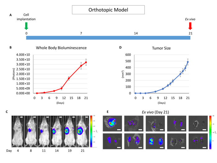
Figure 1: Overview of the orthotopic model without mastectomy. (A) This panel shows a time course of the orthotopic model utilizing the 4T1 syngeneic model. (B) This panel shows the whole-body bioluminescence of an orthotopic model which mainly reflects the primary tumor. The error bar indicates the standard error of the mean (n = 10). (C) This panel shows serial images of whole-body bioluminescence (of the same mouse). The scale bar indicates 1 cm. (D) This panel shows the actual tumor size of panel B, measured by calipers. The error bar indicates the standard error of the mean (n = 10). (E) This panel shows lung ex vivo bioluminescence images of 10 orthotopic model mice (n = 10). The scale bar indicates 1 cm. Please click here to view a larger version of this figure.
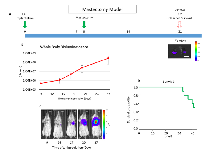
Figure 2: Overview of the mastectomy model. (A) This panel shows a time course of the mastectomy model after 4T1 orthotopic syngeneic implantation. The scale bar indicates 1 cm. (B) This panel shows the whole-body bioluminescence of the mastectomy model, which mainly reflects metastatic lesion. The error bar indicates the standard error of the mean (n = 10). (C) This panel shows serial images of whole-body bioluminescence (of the same mouse). Utilizing ex vivo imaging, it was confirmed that the signals were from lung metastasis, and no local recurrence was confirmed by whole-body imaging after lung removal. Scale bar = 1 cm. (D) This panel shows the Kaplan-Meier survival curve of the mastectomy model without any drug treatment. Mice were euthanized when they reached euthanasia criteria with any morbid condition. Please click here to view a larger version of this figure.
For the last decade, we have been establishing multiple murine cancer models, including breast cancer models3,7,13,16,20,21. Previously, we demonstrated that breast cancer cell orthotopic inoculation into the mammary gland tissue under direct vision produced a larger tumor with less size variability compared to injecting cells around the nipple without a surgical incision7. This model has been improved upon by utilizing a cell suspension in a gelatinous protein mixture. The shapes of the tumor generated by the gelatinous protein mixture-suspended cells were round with less variability, whereas those generated by PBS-suspended cells were scattered in the mammary gland3.
We further verified that the tumors generated by the surgical orthotopic implantation method presented here highly expressed the genes whose interaction networks are important in cancer progression compared to those generated by non-surgical or subcutaneous injection7. These findings imply that this model mimics human breast cancer better compared to non-surgical orthotopic or subcutaneous implantation7. We also demonstrated that this model is suitable to evaluate the immune-mediated regression of breast cancer using allogenic rejection3. However, skin incision can also cause inflammation around the tumor14. Therefore, depending on the hypothesis tested by the investigator, it is important to consider inflammation as a possible confounding factor.
Other than the neoadjuvant setting, the vast majority of systemic therapies are administered after the primary breast tumor is surgically removed11. To date, the majority of preclinical studies for new drug development evaluate drug response in the primary tumor, but not in the metastatic lesions1,18. This discrepancy between animal models and human treatment is important because the gene profile of the metastatic lesions is significantly different from those of the primary lesion22, which has important implications for cancer biology and treatment, especially in the era of targeted therapy22. Therefore, the efficacy of the drug for adjuvant therapy should be evaluated in the metastatic lesions, not merely in the primary tumor. We verified the timing of the lymph node and lung metastasis formation after orthotopic inoculation, utilizing the 4T1 breast cancer orthotopic model16. We also demonstrated that a resection of the primary tumor improved survival in the metastatic breast cancer utilizing the mastectomy model23. Thus, we tried to establish a mastectomy model after orthotopic cancer cell implantation. However, a long time was spent to establish this low local recurrence model. Because cancer cells are injected through surgical incision, cancer cells easily migrate into the wound. In addition, cancer cells invade tissues in direct contact with the tumor, such as the surrounding chest wall muscle and skin. The cancer cells also metastasize to axillary lymph nodes without forming a visible axillary mass at the time of the mastectomy. While removing the tumor may be an easier technique, doing so is not translatable to mastectomy in breast cancer treatment as it causes local recurrence13. Thus, this "radical mastectomy model" was established in order to remove all cancer cells from the anterior chest wall and axillary lymph node basin, as described in Figure 2. We confirmed lung metastasis by histology and lung ex vivo imaging, and there was no shining lesion present by whole-body imaging after lung removal. In this 4T1 orthotopic model, cancer cells eventually metastasize to the lungs, in all cases within 21 days. However, the timing of metastasis depends on the primary tumor size; thus, less primary tumor size variability is important for monitoring not only the primary tumor but also the metastatic tumor burden. By using the gelatinous protein mixture, tumor size variability can be minimized. However, the gelatinous protein mixture can also affect the tumor microenvironment. Therefore, the potential confounding factors associated with gelatinous protein mixture use should be considered, depending on the hypothesis of the investigator.
Utilizing these techniques and luciferase-expressing cancer cells allow for tumor burden quantification with less measurement error, even in non-palpable tumors. There are two reporter systems, bioluminescent and fluorescent, to visualize the signals in live animals. While it has been reported that the signal of both bioluminescence and fluorescence can be bright enough for quantification, when the target signal is very low, the background signal of fluorescence is higher, which reduces the accuracy of the assay. In contrast, bioluminescence is detected with greater accuracy at a lower signal with less background interference24. Accordingly, most investigators prefer to use bioluminescence reporter systems in live animal experiments24,25. However, tumor burden quantification by bioluminescence has its limitations. Luciferase-expressing cells shine when luciferin reaches the cancer cells by cardiovascular perfusion26. If the lesion has an area of hypoxia and/or hypoperfusion, luciferin is not delivered to the cells and the bioluminescence value could, then, be measured to be lower than the actual tumor burden. Indeed, we have experienced inappropriate bioluminescence quantification when the tumor size reached more than 1.5 cm. We assume that it is due to the described hypoxic and/or hypoperfused condition. Thus, when the tumor reaches a palpable size, the tumor burden is quantified by both bioluminescence and caliper measurement. An important limitation of bioluminescence quantification to consider is the black fur of mice. Even after the fur is removed, there is pigmentation on the skin27. While bioluminescence can be detected and quantified in mice with black fur, it is decreased and, therefore, it is important to compare mice of the same fur color to each other in order to reduce the confounding effects of fur color on bioluminescent quantification. The use of luciferase-expressing cells provides some unique modalities for study. For example, secretable luciferase can be used for tumor burden quantification by measuring blood or urine bioluminescence28. In the model presented here, direct tumor burden quantification of viable cancer cells in live animals with non-secretable luciferase is simple and straightforward, which does not require collecting blood or urine samples.
Similar to the models which introduced here, the production of a more efficient animal experiment by producing a higher success rate of tumorigenesis with less variability is key to prove the findings. In the present model, once an investigator has learned the technique, it is easy to achieve a 100% tumorigenesis rate. We also achieved higher tumorigenesis rates other than the models we have introduced; plus, we have established a colon cancer orthotopic model utilizing cell suspension in a gelatinous protein mixture, together with bioluminescence technology to quantify the tumor burden21. In conclusion, the animal model presented here provides a suitable murine model to conduct cancer study.
The authors have nothing to disclose.
This work was supported by NIH grant R01CA160688 and Susan G. Komen Foundation Investigator Initiated Research grant (IIR12222224) to K.T. Mice bioluminescence images were acquired by shared resource Translational Imaging Shared Resource at Roswell Park Comprehensive Cancer Center, which was supported by the Cancer Center Support Grant (P30CA01656) and Shared Instrumentation grant (S10OD016450).
Micro Dissection Scissors | Roboz | RS-5983 | For cancer cell inoculation and masstectomy |
Adson Forceps | Roboz | RS-5233 | For cancer cell inoculation and masstectomy |
Needle Holder | Roboz | RS-7830 | For cancer cell inoculation and masstectomy |
Mayo | Roboz | RS-6873 | For ex vivo |
5-0 silk sutures | Look | 774B | For cancer cell inoculation and masstectomy |
Dry sterilant (Germinator 500) | Braintree Scientific | GER 5287-120V | For cancer cell inoculation and masstectomy |
Clipper | Wahl | 9908-717 | For cancer cell inoculation and masstectomy |
Matrigel | Corning | 354234 | For cancer cell inoculation |
D-Luciferin, potassium salt | GOLD-Bio | LUCK-1K | For bioluminescence quantification |
Roswell Park Memorial Insitute 1640 | Gibco | 11875093 | For cell culture |
Fetal Bovine Serub | Gibco | 10437028 | For cell culture |
Trypsin-EDTA (0.25%) | Gibco | 25200056 | For cell culture |