The bone extracellular matrix (BEM) model for osteosarcoma (OS) is well established and shown here. It can be used as a suitable scaffold for mimicking primary tumor growth in vitro and providing an ideal model for studying the histologic and cytogenic heterogeneity of OS.
Osteosarcoma (OS) is the most common and a highly aggressive primary bone tumor. It is characterized with anatomic and histologic variations along with diagnostic or prognostic difficulties. OS comprises genotypically and phenotypically heterogeneous cancer cells. Bone microenvironment elements are proved to account for tumor heterogeneity and disease progression. Bone extracellular matrix (BEM) retains the microstructural matrices and biochemical components of native extracellular matrix. This tissue-specific niche provides a favorable and long-term scaffold for OS cell seeding and proliferation. This article provides a protocol for the preparation of BEM model and its further experimental application. OS cells can grow and differentiate into multiple phenotypes consistent with the histopathological complexity of OS clinical specimens. The model also allows visualization of diverse morphologies and their association with genetic alterations and underlying regulatory mechanisms. As homologous to human OS, this BEM-OS model can be developed and applied to the pathology and clinical research of OS.
Osteosarcoma (OS) usually occurs in actively growing areas, the metaphysis of long bones, during adolescence. More than 80% of the OS-affected sites have preference for the metaphysis of proximal tibia and proximal humerus as well as both distal and proximal femur, corresponding to the location of the growth plate1. OS comprises multiple cell subtypes with mesenchymal properties and considerable diversity in histologic features and grade. Evidences support mesenchymal stem cells (MSCs), osteoblasts committed precursors and pericytes as the cells of origin2,3,4,5. These cells can accumulate genetic or epigenetic alterations and give rise to OS under the influence of certain bone microenvironmental signals. Both intrinsic and extrinsic mechanisms result in the genomic instability and heterogeneity of OS, with multiple morphological and clinical phenotypes6,7. For individualized therapies or screening of new drugs, novel models need to be generated to against heterogeneity or other clinical disorders.
OS is an intra-osseous malignant solid tumor. The complexity and activity of surrounding microenvironment elements confer phenotypic and functional differences upon OS cells in different locations of a tumor. Bone extracellular matrix (BEM) provides a structural and biochemical scaffold for mineral deposition and bone remodeling. The organic portion of extracellular matrix (ECM) mainly consists of type I collagen secreted by osteoblastic lineage cells, while its mineralized portion is composed of calcium phosphate in the form of hydroxyapatite8. The dynamic role of ECM networks is to regulate cell adhesion, differentiation, cross-talk and tissue function maintenance9.
Demineralized BEM and ECM hydrogels have been successfully used in cell culture and can enhance cell proliferation10,11. Synthesized bone-like ECM can regulate the pool size, fate decisions and lineage progression of MSCs12,13,14. Moreover, results evidence its clinical significance to provide osteogenic activity by stimulating cellular processes during bone formation and regeneration15,16,17.
In this article, our group establishes a modified model and favorable alternative for three-dimensional long-term culture. OS cells injected into the tissue-derived BEM present a heterogeneously mesenchymal phenotype readily as compared to plastic two-dimensional cultures. BEM derived from site-specific homologous tissue show its dramatic advantage as being a native niche for OS cells in vitro and has great potential in OS theoretical and clinical research. This characterized BEM platform is simple but efficient for in vitro research and may be extended in modeling multiple cancers.
Animal care and use are conducted according to the National Institutes of Health Guide for the Care and Use of Laboratory Animals (NIH publication NO.80-23, revised in 1996) after approval from the Animal Ethics Committee of Sun Yat-sen University.
1. Bone preparation
2. Bone demineralization and decellularization
3. Cell seeding and culture
After demineralization and decellularization, BEM appears to be translucent with stronger resilience and tenacity compared to native mouse bone. A little muscle residue and the space of medullary cavity can be clearly observed (Figure 1A, B). To determine the effective decellularization of BEM, BEM is embedded in paraffin after fixation, and then sliced into 3–5 μm sections for hematoxylin-eosin (H&E) staining. The thorough removal of cell nuclei is shown by bright-field imaging. The natural porous structure and collagen network arrangement is well maintained in decellularized BEM (Figure 1C, D). Additionally, immunohistochemical (IHC) staining of predominant organic components of bone matrix, such as collagen I and collagen IV demonstrate no damage on ECM components in decellularized BEM compared to the native bone (Figure 1E). Therefore, BEM provides a suitable and promising scaffold with great biocompatibility for OS cell seeding and proliferation.
MNNG/HOS cells exhibit a highly atypical morphology with finely vacuolated cytoplasm, while MG-63 cells have fibroblast-like spindled shapes in monolayer culture (Figure 2A, B). The histological section from an OS patient displays significant cellular pleomorphism with rounded or polygonal cells, anisonucleosis and multiple mitoses (Figure 2C). To verify the viability and quality of the BEM model, both cell lines are injected into the medullary cavity of BEM and monitored via fluorescence imaging during the 14-day culture (Figure 3A, B). Histological sections with H&E staining reveal that OS cells attach to muscle residues and grow into thick piles or adhere along bone matrix and proliferate. Both periosteum and endosteum are infiltrated by the expansion of OS cells. Strikingly, the cell growth patterns of OS-BEM model differ from two-dimensional plate culture. As illustrated in Figure 3C, OS cells on the decellularized BEM show highly heterogeneous morphology similar to the cytopathologic features of an OS section. Some OS cells locating in cancellous bone and medullary cavity are spherical and partly spread out, whereas the cells resting along the periosteum and endosteum are extremely spread out into elongated cells accompanied by nuclear pleomorphism. Cell activity is determined using Ki67 immunostaining, which also shows great advantages in long-term cultures. Also, OS cells in BEM culture highly express bone matrix glycoprotein—secreted protein acidic and rich in cysteine (SPARC/osteonectin)—which is specific for osteoid matrix (Figure 3D).
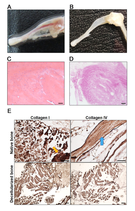
Figure 1: The structural characteristics of mouse decellularized BEM. (A, B) Overview of mouse native (A) and decellularized (B) bone. (C, D) Decellularization was assessed by H&E staining of mouse native tibia (C) and decellularized bone (D). Nuclei stained with hematoxylin could be observed in native mouse tibia, but not in the BEM. (E) IHC staining for collagen I and collagen IV to detect the main components of ECM that are preserved in mouse tibia after decellularization. Yellow arrow points out the abundant collagen I within cancellous bone and blue arrow points out the abundant collagen IV within compact bone. Scale bars = 50 μm. Please click here to view a larger version of this figure.
Figure 2: The cytomorphological characteristics of OS. (A, B) Human OS cell lines MNNG/HOS (A) and MG-63 (B) expanded in plastic flask culture. Scale bar = 100 μm. (C) Histopathologic section with H&E staining of OS patient. Scale bar = 50 μm. Please click here to view a larger version of this figure.
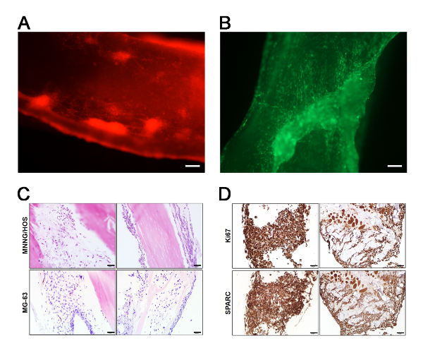
Figure 3: Characterization of OS cells in decellularized bone extracellular matrix model. (A, B) Representative mCherry expression (red) image of MNNG/HOS (A) and GFP expression (green) image of MG-63 (B) by fluorescence microscopy after seeding and culturing in BEM. Scale bar = 100 μm. (C) H&E analysis showing typical morphology of the injected MNNG/HOS and MG-63 cells after culturing in BEM. (D) IHC analysis on Ki67 and SPARC expression level of MNNG/HOS cells after culturing in BEM model for 14 days. The representative images are two sets of serial sections stained with Ki67 and SPARC. Scale bar = 50 μm. Please click here to view a larger version of this figure.
Generally, OS can be classified as osteoblastic, chondroblastic, and fibroblastic subtypes depending on its dominant histologic component. Its prognosis is dependent not only on histologic parameters but also on its anatomic site. It may occur inside the bones (in the intramedullary or intracortical compartment), on the surfaces of bones, and in extraosseous sites19. The emergence and heterogeneity of OS can be elucidated as a conjugation of oncogenic events and an adequate microenvironmental boost, followed by increasing development and migration to distant organs20,21,22,23. Mystery during OS evolution might be deciphered with a proper model to outline clinical implications targeting the OS environment and niche.
Cultivation either on plastic dishes or in flasks in vitro can hardly recapitulate the dynamic and intricate biological microenvironment. Great strides of various pre-clinical models (e.g., mouse, zebrafish and dog) mimicking the osteosarcoma have been declared and applied to pathogenesis investigation and drug development4,24,25,26,27,28. Still, researchers have concern for experimental animals due to their discomfort and suffering during experiments. In vitro three-dimensional models like our decellularized BEM model has the advantage due to its convenience, quick operation and cost saving; it provides long-term and easy maintenance of viable cells or tissues, and is also closer to the native biological situation than plastic culture. It has been used in our research demonstrating the phenotypic heterogeneity and regulatory mechanism of OS dedifferentiation with success29.
This protocol clarified the feasibility to generate a tissue-derived BEM from mouse and might be used for tissues from human, rat and dog. The most critical steps for successful establishment and application of BEM are: (i) complete removal of cell debris; (ii) maintenance of a sterile, healthy culture condition; (iii) manual dexterity and gentle pipetting during injection, transfer and culture of OS-BEM model.
Other reported protocols generally employ pressurization or a combination of chemical and enzymatic treatments, such as Triton X-100, sodium dodecyl sulfate (SDS) and DNase/RNase solution to achieve potent decellularization30,31. The cartilage tissue that undergoes decellularization with detergents has been shown to remove ECM components including glycosaminoglycans32. To recapitulate a more intact BEM to the greatest extent, a moderate yet powerful decellularization method is performed here to avoid the dissolution and damage of key components and native architecture of the bone environment.
However, it is not to be neglected that this OS-BEM model rested in a plate without flowing medium, consequently leading to an uneven distribution of oxygen and nutrients. Vascular network and other cell subtypes that help regulating the communication and interaction of microenvironmental signals and bone homeostasis need to be taken into consideration24, 33,34,35. Hopefully, this model will be combined with other high-tech engineering techniques to shed light on OS research and guide precision medicine.
The authors have nothing to disclose.
The authors value the support of Liuying Chen for her administrative assistance and Long Zhao for his excellent technical assistance during the construction of bone extracellular matrix scaffolds. This study is supported by grants from the National Natural Science Foundation of China (31871413).
15 mL centrifuge tube | Greiner | 188271 | |
50 mL centrifuge tube | Greiner | 227270 | |
6 cm cell culture dish | Greiner | 628160 | |
6-well plate | Greiner | 657160 | |
Ampicillin | Sigma-Aldrich | A9393 | |
C57-BL/6J mouse | Sun Yat-sen University Laboratory Animal Center | ||
CO2 incubator | SHEL LAB | SCO5A | |
Dibasic sodium phosphate | Guangzhou Chemical Reagent Factory | BE14-GR-500G | |
DMEM/F12 | Sigma-Aldrich | D0547 | |
Fetal bovine serum | Hyclone | SH30084.03 | |
Hemocytometer | BLAU | 717805 | |
Kanamycin | Sigma-Aldrich | PHR1487 | |
MG-63 | Chinese Academy of Science, Shanghai Cell Bank | Human osteosarcoma cell line | |
MNNG/HOS | Chinese Academy of Science, Shanghai Cell Bank | Human osteosarcoma cell line | |
Phenol red | Sigma-Aldrich | P4633 | A solution of phenol red is used as a pH indicator: its color exhibits a gradual transition from yellow to red over the pH range 6.6 to 8.0. |
Potassium chloride | Sangon Biotech | A100395 | |
Potassium Phosphate Monobasic | Sangon Biotech | A501211 | |
Sodium chloride | Sangon Biotech | A501218 |