1. Preparation of chitosan-PVA hydrogels
- Prepare 2% (w/w) chitosan and 10% (w/w) PVA solutions. Dissolve 0.2 g of chitosan in 10 mL of 0.1 M CH3COOH solution (previously filtered) at room temperature and maintain continuous mechanical stirring overnight. Dissolve 1 g of PVA in 10 mL of distilled water and stir at 80 °C for 1 h.
- Mix both solutions 1:1 using a magnetic stirrer until they are homogeneous at room temperature, and pour the mixtures on Petri dishes. Leave the samples for 2 h at atmospheric pressure to degas.
- Freeze the hydrogels at -4 °C, -20 °C or -80 °C for 20 h and 4 cycles (samples CP4-4, CP4-20 and CP4-80, respectively). Freeze another hydrogel at -80 °C for 20 h using 5 or 6 freezing cycles (samples CP5-80 and CP6-80). After the third freezing cycle, wash the hydrogels with deionized water. At the end, freeze-dry the hydrogels at -46 °C for 48 h and store for further characterization (methodology adapted from2).
2. FT-IR characterization
- Place a little piece (1 mm x 2 mm) of hydrogel in the FT-IR spectrometer in ATR mode. Take the FT-IR spectra from 4000 to 600 cm-1 (2 cm-1 of resolution and average of 32 scans).
3. Swelling assays
- Cut out discs (13 mm in diameter and 10 mm in height) from the hydrogel and weigh them. Incubate the discs in 50 mL of deionized water with shaking at 25 °C. Repeat three times.
- Every 30 min remove the sample from the medium, blotter to eliminate the excess of water, and weigh. Calculate the swelling degree using the equation 1 and calculate the equilibrium state of swelling,
 , at 24 h using the equation 2.
 )
)
Where is the weight of the dry hydrogel and
  is the weight of the wet hydrogel.
4. Electronic Microscopy
- Cover a little piece of hydrogel with a thin gold layer (30 s and 10 mA) in a sputter coater.
- Put the sample in a scanning electron microscope (SEM). Analyze the samples under vacuum at 20 kV and take the images with a 500x and 1500x magnification.
5. Porosimetry
- Place discs 15 mm in diameter weighing around 0.26 g into the penetrometer (a solid penetrometer, having a bulk volume of 0.3660 mL and 5.7831 mL of stem volume). Analyze the porosity and pore size by Mercury Intrusion Porosimetry (MIP).
- Conduct the experiment in the hysteresis mode (intrusion-extrusion). Measure the total intrusion volume (mL/g), total pore area (m2/g), pore diameter (µm), porosity (%), permeability (mDarcy) and tortuosity. Repeat twice.
6. Drug loading and release
- Before loading, prepare 4 L of 15 mg/L diflunisal solution and stir overnight. Confirm the concentration of the solution by UV-Vis spectroscopy (initial concentration). Indeed, swell 400 mg of freeze-dried samples of hydrogel in 6 mL of distilled water for 24 h.
- For loading, fill a flask with 50 mL of diflunisal solution and maintain at 25 °C with constant stirring. Submerge each swelled hydrogel in the flask.
- Take aliquots of remaining diflunisal solution (2 mL) at different times in order to determine the plateau region of the curve, for example: 3, 6, 24, 27, 30 and 48 h. After 24 h replace the solution with a fresh one.
- Measure the absorbance at 252 nm of each aliquot, and determine the concentration of diflunisal present in the solution, using a calibration curve of diflunisal. Calculate the amount of diflunisal retained in the hydrogel at 24 and 48 h, as the difference of initial and final concentrations, taking into account the total volume (56 mL).
- Determine the encapsulation efficiency (EE) using the equation 3.

- Freeze the loaded hydrogels at -80 °C and lyophilize them at -50 °C.
- Determine the encapsulation efficiency (EE) using the equation 3.
- For drug release, submerge 300 mg of freeze-dried diflunisal loaded hydrogels in 50 mL of phosphate buffer (pH 7.4) at 25 °C. Maintain constant stirring. Withdraw aliquots of 2 mL at different times and replace with fresh medium to keep a constant volume.
- Determinate the diflunisal released spectrophotometrically at 252 nm, according to a calibration curve.
- Deduce the predominant drug release mechanism in the hydrogels adjusting the drug release data corresponding to the first 60%, to the Korsmeyer-Peppas model (Equation 4), to obtain the kinetic (k) and the diffusion (n) constants. The n values indicate the mechanism of drug release24,25. Then, n values close to 0.5 are related to Fickian diffusion, meanwhile values of 0.5-1.0 for anomalous transport, where are involved diffusion and relaxation chains, and finally, values of 1.0 are related to case II transport.

- To confirm the results, use the Higuchi, First order, and Zero order mathematical models (Equations 5 to 7) and select the better fit.


where t represents the release time, Mt the amount of drug delivered at a given time, and M∞ the total amount of drug delivered at the end of the process.
Hydrogels preparation
Chitosan-PVA hydrogels were obtained at -4 °C, -20 °C and -80 °C with 4 freezing cycles and at -80 °C with 5 and 6 freezing cycles by the previously reported freeze-thawing method2. All hydrogels were homogeneous, semi-transparent, flexible and resistant against manipulation.
FT-IR characterization
The FT-IR spectra are shown in Figure 2. Seven characteristics signals of chitosan and PVA polymers were detected: at 3286 cm-1 the stretching vibration mode of PVA hydroxyl group (-OH) and at 2918 cm-1 the stretching vibration mode of -CH group26,27. The signals of amide groups, representative of chitosan structure, were found at 1652 cm-1 to the stretching vibration mode of C=O (amide I), at 1560 cm-1 to the flection vibration mode of N-H (amide II) and 1325 cm-1 to the vibration of amide III28,29,30. Other signals, at 1418 cm-1 to the flection vibration mode of C-H and at 1086 cm-1 to the stretching vibration mode of C-O groups, both of PVA, were detected27,31,32.
Electronic Microscopy
All CS-PVA hydrogels showed a highly porous surface (Figure 3, from left to right) and distinctive changes were observed according to the preparation conditions. Hydrogels prepared at -4 °C (CP4-4) presented larger pores than the hydrogels prepared at -80 °C (CP4-80). Moreover, the latter appears to have a more porous network. This effect may be due to the fact that, at lower temperature, the water crystal formation was faster and many small crystals emerged and were sublimated during the freeze-drying process, leaving void pores14,33. Meanwhile, the effect of the number of freezing cycles seems to promote more defined and circular pores in hydrogels CP6-80 (Figure 3, from top to bottom).
Porosimetry
Samples CP4-4, CP4-80 and CP6-80 presented more pronounced changes; in order to complement the information about morphology, they were analyzed by MIP (Table 1). The comparison between hydrogels CP4-4 and CP4-80 (Figure 3a) showed that, at a lower temperature of freezing, hydrogels developed a more porous network, which presented a large total intrusion volume and higher total pore area. However, hydrogels CP6-80 showed less permeability than CP4-80 (Figure 3b), probably due to their high tortuosity, which was also reflected in a lower total intrusion volume. Figure 3 presents the different pore sizes of these hydrogels. Two pore sizes were distinguished, one between 0.3-5.0 μm and other between 5.0-30 μm. In hydrogels CP4-80 and CP6-80, the porous network had a greater number of small pores than large ones, compared with CP4-4 hydrogel. These results were similar to those observed by SEM micrographs and suggested that, at lower temperature greater interactions between the PVA chains were favored and more crystalline zones were formed. In such a way, the formation of crystalline zones by PVA chains, was stimulated at low temperature.
Swelling assays
The swelling behavior of CS-PVA hydrogels can be seen in the Figure 4. They quickly absorbed large amounts of water; for the first 5 hours they retained 10x their weight, and after 20 hours they retain up to 15x their weight (equilibrium point). However, in relation to hydrogels prepared at the same number of freezing cycles, the hydrogel CP4-80 showed less swelling capacity in the first 5 hours as a consequence of the temperature that was used for its preparation (-80 °C). In the case of hydrogels prepared at different number of freezing cycles (CP4-80, CP5-80 and CP6-80) no differences were found at any time. Probably, the decreased swelling capacity observed in hydrogels prepared at -80 °C was caused by the small pore size of the hydrogel network.
Drug loading and release
To evaluate the capacity of CS-PVA hydrogels as drug delivery systems, the anti-inflammatory drug diflunisal was loaded in the network and subsequently released. The encapsulation efficiency (EE) in all these systems was around 70%; however, the CP4-80 hydrogel presented more slightly EE at 73% (Table 2). Meanwhile, the releasing kinetics of diflunisal from the CS-PVA hydrogels were maintained for about 30 h in all cases. The CP4-80 hydrogel released the highest amount of diflunisal (Figure 5). This may be due to the fact this hydrogel showed a more porous structure in comparison with the other two types of hydrogel. This feature allowed the small molecule of drug to easily enter in the hydrogel network and, then, to be released. Between CP4-80 and CP6-80 hydrogels not differences were observed during release times (Figure 6). No burst effect was observed in any of the CS-PVA hydrogels, which is promising for pharmaceutical applications. Mathematical models were used to determine the main release mechanism in CS-PVA hydrogels. The results were adjusted to different mathematical models (Table 3) and according to the n values, it was found that the Fick diffusion dominates the drug release process.
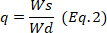
Figure 1: Chemical structure of PVA (a), chitosan (b) and diflunisal (c). Please click here to view a larger version of this figure.
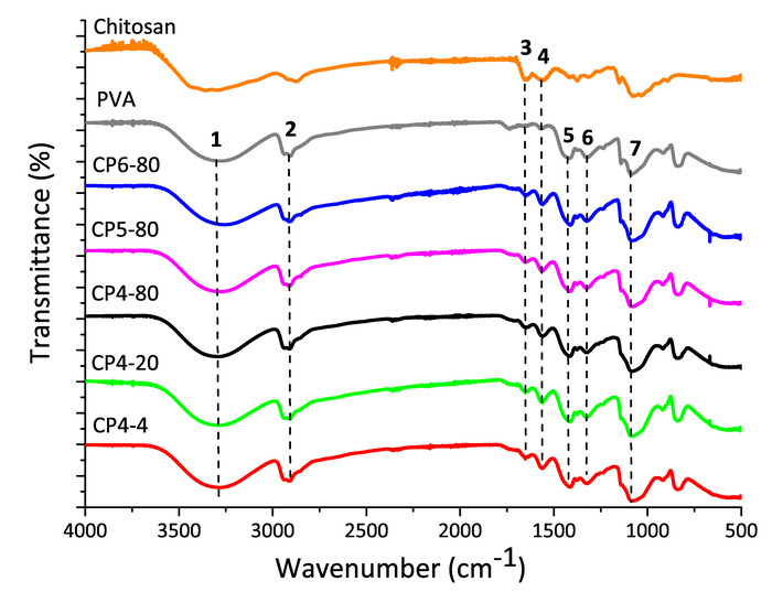
Figure 2: FT-IR spectra of pure chitosan and PVA and, chitosan-PVA hydrogels prepared at different conditions of freezing. Please click here to view a larger version of this figure.
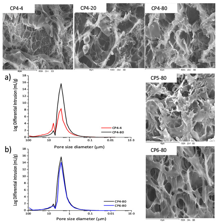
Figure 3: SEM micrographs of chitosan-PVA hydrogels at 1500x magnification. Pore size distributions of chitosan-PVA hydrogels: a) hydrogels prepared with 4 cycles of freezing and at -4 °C and -80 °C. b) Hydrogels prepared at -80 °C and, 4 and 6 cycles. Please click here to view a larger version of this figure.
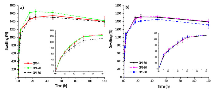
Figure 4: Swelling kinetics of chitosan-PVA hydrogels: a) hydrogels with 4 cycles of freezing and b) hydrogels prepared at -80 °C. Please click here to view a larger version of this figure.
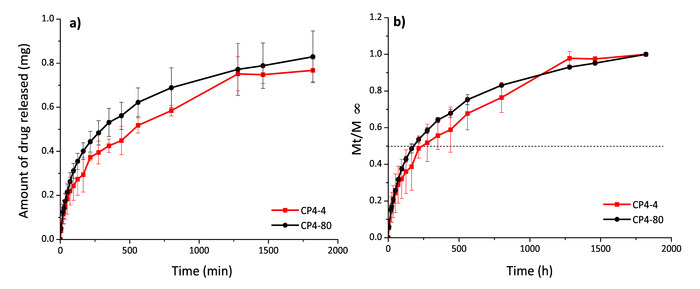
Figure 5: Diflunisal release profiles in mg (a) and Mt/ (b) for hydrogels CP4-4 and CP4-80. Please click here to view a larger version of this figure.
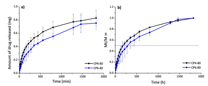
Figure 6: Diflunisal release profiles in mg (a) and Mt/ (b) for hydrogels CP4-80 and CP6-80. Please click here to view a larger version of this figure.
| Hydrogel | Total intrusion volume (mL/g) | Total pore area (m2/g) | Porosity (%) | Permeability (mdarcy) | Tortuosity |
| CP4-4 | 5.16 | 10.19 | 67.13 | 132.43 | 10.46 |
| CP4-80 | 7.36 | 15.14 | 85.95 | 151.16 | 5.83 |
| CP6-80 | 6.69 | 12.86 | 84.82 | 129.28 | 12.2 |
Table 1: Porosimetry parameters of the porous structure of chitosan-PVA hydrogels.
| Sample | Diflunisal loaded | Diflunisal released | |
| mg/g hydrogel | Encapsulation efficiency (%) | % released respect to loaded | |
| CP4-4 | 3.05± 0.09 | 71 | 79 ± 3.33 |
| CP4-80 | 3.22 ± 0.47 | 73 | 86 ± 0.4 |
| CP6-80 | 3.19 ± 0.05 | 68 | 80 ± 3.9 |
Table 2: Encapsulation and release efficiencies for chitosan-PVA hydrogels.
| Sample | Korsmeyer-Peppas | Higuchi | First Order | Zero Order | |||||
| kKP x 102 | n | R2 | kH x 102 | R2 | k1 x 102 | R2 | k0 x 102 | R2 | |
| (min-n) | (min-0.5) | (min-1) | (min-1) | ||||||
| CP4-4 | 4.3 ± 0.39 | 0.44 ± 0.02 | 0.99 | 3.1 ± 0.1 | 0.98 | 0.29 ± 0.03 | 0.803 | 0.18 ± 0.02 | 0.54 |
| CP4-80 | 3.6 ± 0.33 | 0.50 ± 0.02 | 0.99 | 3.7 ± 0.1 | 0.99 | 0.42 ± 0.03 | 0.894 | 0.27 ± 0.02 | 0.7 |
| CP6-80 | 2.3 ± 0.24 | 0.54 ± 0.02 | 0.99 | 2.9 ± 0.1 | 0.99 | 0.27 ± 0.02 | 0.925 | 0.17 ± 0.01 | 0.77 |
| k= kinetic constant; n= diffusion constant. | |||||||||
Table 3: Kinetic parameters of diflunisal release from chitosan-PVA hydrogels.
